Summary
Rev. bras. ginecol. obstet.. 2024;46:e-rbgo5
This study aims to correlate pelvic ultrasound with female puberty and evaluate the usual ultrasound parameters as diagnostic tests for the onset of puberty and, in particular, a less studied parameter: the Doppler evaluation of the uterine arteries.
Cross-sectional study with girls aged from one to less than eighteen years old, with normal pubertal development, who underwent pelvic ultrasound examination from November 2020 to December 2021. The presence of thelarche was the clinical criterion to distinguish pubescent from non-pubescent girls. The sonographic parameters were evaluated using the ROC curve and the cutoff point defined through the Youden index (J).
60 girls were included in the study. Uterine volume ≥ 2.45mL had a sensitivity of 93%, specificity of 90%, PPV of 90%, NPV of 93% and accuracy of 91% (AUC 0.972) for predicting the onset of puberty. Mean ovarian volume ≥ 1.48mL had a sensitivity of 96%, specificity of 90%, PPV of 90%, NPV of 97% and accuracy of 93% (AUC 0.966). Mean PI ≤ 2.75 had 100% sensitivity, 48% specificity, 62% PPV, 100% NPV and 72% accuracy (AUC 0.756) for predicting the onset of puberty.
Pelvic ultrasound proved to be an excellent tool for female pubertal assessment and uterine and ovarian volume, the best ultrasound parameters for detecting the onset of puberty. The PI of the uterine arteries, in this study, although useful in the pubertal evaluation, showed lower accuracy in relation to the uterine and ovarian volume.
Summary
Rev Bras Ginecol Obstet. 2024;46:e-rbgo1
To evaluate the association between clinical and imaging with surgical and pathological findings in patients with suspected neuroendocrine tumor of appendix and/or appendix endometriosis.
Retrospective descriptive study conducted at the Teaching and Research Institute of Hospital Israelita Albert Einstein, in which medical records and databases of patients with suspected neuroendocrine tumor of appendix and/or endometriosis of appendix were analyzed by imaging.
Twenty-eight patients were included, all of which had some type of appendix alteration on the ultrasound examination. The pathological outcome of the appendix found 25 (89.3%) lesions compatible with endometriosis and three (10.7%) neuroendocrine tumors. The clinical findings of imaging and surgery were compared with the result of pathological anatomy by means of relative frequency.
It was possible to observe a higher prevalence of appendix endometriosis when the patient presented more intense pain symptoms. The image observed on ultrasound obtained a high positive predictive value for appendicular endometriosis.
Summary
Rev Bras Ginecol Obstet. 2022;44(12):1134-1140
Gestational diabetes mellitus (GDM)is an entity with evolving conceptual nuances that deserve full consideration. Gestational diabetes leads to complications and adverse effects on the mother's and infants' health during and after pregnancy. Women also have a higher prevalence of urinary incontinence (UI) related to the hyperglycemic status during pregnancy. However, the exact pathophysiological mechanism is still uncertain. We conducted a narrative review discussing the impact of GDM on the women's pelvic floor and performed image assessment using three-dimensional ultrasonography to evaluate and predict future UI.
Summary
Rev Bras Ginecol Obstet. 2022;44(9):838-844
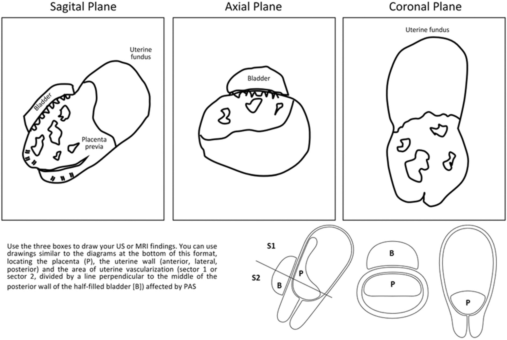
The immediate referral of patients with risk factors for placenta accreta spectrum (PAS) to specialized centers is recommended, thus favoring an early diagnosis and an interdisciplinary management. However, diagnostic errors are frequent, even in referral centers (RCs). We sought to evaluate the performance of the prenatal diagnosis for PAS in a Latin American hospital.
A retrospective descriptive study including patients referred due to the suspicion of PAS was conducted. Data from the prenatal imaging studies were compared with the final diagnoses (intraoperative and/or histological).
A total of 162 patients were included in the present study. The median gestational age at the time of the first PAS suspicious ultrasound was 29 weeks, but patients arrived at the PAS RC at 34 weeks. The frequency of false-positive results at referring hospitals was 68.5%. Sixty-nine patients underwent surgery based on the suspicion of PAS at 35 weeks, and there was a 28.9% false-positive rate at the RC. In 93 patients, the diagnosis of PAS was ruled out at the RC, with a 2.1% false-negative frequency.
The prenatal diagnosis of PAS is better at the RC. However, even in these centers, false-positive results are common; therefore, the intraoperative confirmation of the diagnosis of PAS is essential.
Summary
Rev Bras Ginecol Obstet. 2022;44(9):838-844
The immediate referral of patients with risk factors for placenta accreta spectrum (PAS) to specialized centers is recommended, thus favoring an early diagnosis and an interdisciplinary management. However, diagnostic errors are frequent, even in referral centers (RCs). We sought to evaluate the performance of the prenatal diagnosis for PAS in a Latin American hospital.
A retrospective descriptive study including patients referred due to the suspicion of PAS was conducted. Data from the prenatal imaging studies were compared with the final diagnoses (intraoperative and/or histological).
A total of 162 patients were included in the present study. The median gestational age at the time of the first PAS suspicious ultrasound was 29 weeks, but patients arrived at the PAS RC at 34 weeks. The frequency of false-positive results at referring hospitals was 68.5%. Sixty-nine patients underwent surgery based on the suspicion of PAS at 35 weeks, and there was a 28.9% false-positive rate at the RC. In 93 patients, the diagnosis of PAS was ruled out at the RC, with a 2.1% false-negative frequency.
The prenatal diagnosis of PAS is better at the RC. However, even in these centers, false-positive results are common; therefore, the intraoperative confirmation of the diagnosis of PAS is essential.
Summary
Rev Bras Ginecol Obstet. 2022;44(7):646-653
This study aims to describe the behavior of chromosomopathy screenings in euploid fetuses.
This is a prospective descriptive study with 566 patients at 11 to 14 weeks of gestation. The associations between ultrasound scans and serological variables were studied. For the quantitative variables we used the Spearman test; for the qualitative with quantitative variables the of Mann-Whitney U-test; and for qualitative variables, the X2 test was applied. Significance was set at p ≤ 0.05.
We have found that gestational age has correlation with ductus venosus, nuchal translucency, free fraction of β subunit of human chorionic gonadotropin, pregnancy-associated plasma protein-A and placental growth factor; there is also a correlation between history of miscarriages and nasal bone. Furthermore, we correlated body mass index with nuchal translucency, free fraction of β subunit of human chorionic gonadotropin, and pregnancy-associated plasma protein-A. Maternal age was associated with free fraction of β subunit of human chorionic gonadotropin and pregnancy-associated plasma protein-A.
Our study demonstrates for the first time the behavior of the biochemical and ultrasonographic markers of chromosomopathy screenings during the first trimester in euploid fetuses in Colombia. Our information is consistent with international reference values. Moreover, we have shown the correlation of different variables with maternal characteristics to determine the variables that could help with development of a screening process during the first trimester with high detection rates.
Summary
Rev Bras Ginecol Obstet. 2022;44(3):287-294
To evaluate the association between polycystic ovary syndrome (PCOS) and metabolic syndrome (MetS), adding liver assessment through elastography and ultrasound, for correlation with non-alcoholic fatty liver disease (NAFLD). Metabolic syndrome occurs in~43% of women with PCOS, and NAFLD is the hepatic expression of MetS.
One hundred women, 50 with PCOS and 50 controls, matched by age (18- 35 years) and body mass index (BMI) were included, restricted to patients with overweight and obesity grade 1, at the Assis Chateaubrian Maternity School, Universidade Federal do Ceará, Brazil. For the diagnosis of PCOS, we adopted the Rotterdam criteria, and for the diagnosis of MetS, the criteria of the National Cholesterol Education Program (NCEP/ATP III). Hepatic elastography and ultrasound were performed to assess liver stiffness and echotexture, respectively.
The average ages were 29.1 (±5.3) and 30.54 (±4.39) years, for the PCOS and the control group, respectively. Patients with PCOS had a risk 4 times higher of having MetS, odds ratio (95% confidence interval)=4.14, than those in the control group. Women with PCOS had higher average of abdominal circumference (100.9±9.08 cm vs 94.96±6.99 cm) and triglycerides (162±54.63 mg/dL vs 137.54±36.91mg/dL) and lower average of HDL cholesterol (45.66±6.88 mg/dL vs 49.78±7.05 mg/dL), with statistically significant difference. Hepatic steatosis was observed on ultrasound in women with PCOS; however, with no statistically significant difference. There was no change to NAFLD at elastography in any group.
Women with PCOS had 4-fold higher frequency of MetS andmore hepatic steatosis, with no statistically significant difference. There was no change in liver stiffness between the groups at elastography. The results can be extended only to populations of overweight and obesity grade 1, with PCOS or not. They cannot be generalized to other untested groups.
Summary
Rev Bras Ginecol Obstet. 2022;44(3):231-237
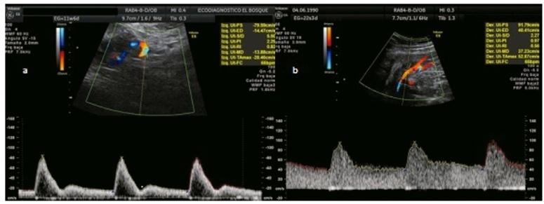
To analyze whether acetylsalicylic (ASA) intake modifies the mean uterine arteries pulsatility index (UtA-PI) at the 2nd or 3rd trimester in a cohort of pregnant women with abnormal mean UtA-PI at between 11 and 14 weeks of gestation.
This is a retrospective cohort study. Singleton pregnancies with abnormal mean UtA-PI at between 11 and 14 weeks of gestation were studied. The participants were divided into 3 groups: 1) If the participant did not take ASA during pregnancy; 2) If the participant took ASA before 14 weeks of gestation; and 3) If the participant took ASA after 14 weeks of gestation. The mean UtA-PI was evaluated at the 2nd and 3rd trimesters, and it was considered to improve when it decreased below the 95th percentile. The prevalence ratio (PR) and the number needed to treat (NNT) werecalculated.
A total of 72 participants with a mean UtA-PI>95th percentile at the 1st trimester of gestation were evaluated. Out of the 18 participants who took ASA, 8 participants started it before 14 weeks of gestation and 10 after. A total of 33.3% of these participants had improved the mean UtA-PI at the 2nd and 3rd trimesters of gestation, although it was not statistically significant (p=0.154). The prevalence ratio was 0.95 (95% confidence interval [CI]: 0.31-1.89), but between the 1st and 2nd trimesters of gestation, the PR was 0.92 (95%CI: 0.21-0.99) and it was statistically significant.
The present work demonstrates a modification of the mean UtA-PI in participants who took ASA compared with those who did not. It is important to check if ASA can modify the normal limits of uterine arteries because this could have an impact on surveillance.
Summary
Rev Bras Ginecol Obstet. 2022;44(3):231-237
To analyze whether acetylsalicylic (ASA) intake modifies the mean uterine arteries pulsatility index (UtA-PI) at the 2nd or 3rd trimester in a cohort of pregnant women with abnormal mean UtA-PI at between 11 and 14 weeks of gestation.
This is a retrospective cohort study. Singleton pregnancies with abnormal mean UtA-PI at between 11 and 14 weeks of gestation were studied. The participants were divided into 3 groups: 1) If the participant did not take ASA during pregnancy; 2) If the participant took ASA before 14 weeks of gestation; and 3) If the participant took ASA after 14 weeks of gestation. The mean UtA-PI was evaluated at the 2nd and 3rd trimesters, and it was considered to improve when it decreased below the 95th percentile. The prevalence ratio (PR) and the number needed to treat (NNT) werecalculated.
A total of 72 participants with a mean UtA-PI>95th percentile at the 1st trimester of gestation were evaluated. Out of the 18 participants who took ASA, 8 participants started it before 14 weeks of gestation and 10 after. A total of 33.3% of these participants had improved the mean UtA-PI at the 2nd and 3rd trimesters of gestation, although it was not statistically significant (p=0.154). The prevalence ratio was 0.95 (95% confidence interval [CI]: 0.31-1.89), but between the 1st and 2nd trimesters of gestation, the PR was 0.92 (95%CI: 0.21-0.99) and it was statistically significant.
The present work demonstrates a modification of the mean UtA-PI in participants who took ASA compared with those who did not. It is important to check if ASA can modify the normal limits of uterine arteries because this could have an impact on surveillance.
Summary
Rev Bras Ginecol Obstet. 2021;43(12):911-918
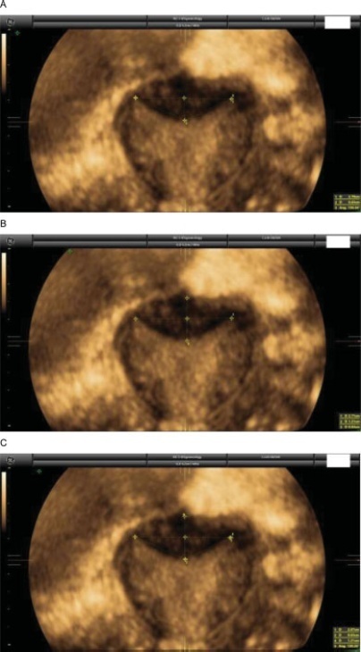
Currently, there are up to three different classifications for diagnosing septate uterus. The interobserver agreement among them has been poorly assessed.
A total of 50 three-dimensional (3D) volumes of a nonconsecutive series of women with suspected uterine malformation were used. Two nonexpert examiners evaluated a single 3D volume of the uterus of each woman, blinded to each other. The following measurements were performed: indentation depth, indentation angle, uterine fundal wall thickness, external fundal indentation, and indentation-to-wall-thickness (I:WT) ratio. Each observer had to assign a diagnosis in each case, according to the three classification systems (ESHRE/ESGE, ASRM, and CUME). The interobserver agreement regarding the ESHRE/ESGE, ASRM, and CUME classifications was assessed using the Cohen weighted kappa index (k). Agreement regarding the three classifications (ASRM versus ESHRE/ESGE, ASRM versus CUME, ESHRE/ESGE versus CUME) was also assessed.
The interobserver agreement between the 2 nonexpert examiners was good for the ESHRE/ESGE (k = 0.74; 95% confidence interval [CI]: 0.55–0.92) and very good for the ASRM and CUME classification systems (k = 0.95; 95%CI: 0.86–1.00; and k = 0.91; 95%CI: 0.79–1.00, respectively). Agreement between the ESHRE/ESGE and ASRM classifications was moderate for both examiners. Agreement between the ESHRE/ESGE and CUME classifications was moderate for examiner 1 and good for examiner 2. Agreement between the ASRM and CUME classifications was good for both examiners.
The three classifications have good (ESHRE/ESGE) or very good (ASRM and CUME) interobserver agreement. Agreement between the ASRM and CUME classifications was higher than that for the ESHRE/ESGE and ASRM and ESHRE/ESGE and CUME classifications.
Summary
Rev Bras Ginecol Obstet. 2021;43(12):911-918
Currently, there are up to three different classifications for diagnosing septate uterus. The interobserver agreement among them has been poorly assessed.
A total of 50 three-dimensional (3D) volumes of a nonconsecutive series of women with suspected uterine malformation were used. Two nonexpert examiners evaluated a single 3D volume of the uterus of each woman, blinded to each other. The following measurements were performed: indentation depth, indentation angle, uterine fundal wall thickness, external fundal indentation, and indentation-to-wall-thickness (I:WT) ratio. Each observer had to assign a diagnosis in each case, according to the three classification systems (ESHRE/ESGE, ASRM, and CUME). The interobserver agreement regarding the ESHRE/ESGE, ASRM, and CUME classifications was assessed using the Cohen weighted kappa index (k). Agreement regarding the three classifications (ASRM versus ESHRE/ESGE, ASRM versus CUME, ESHRE/ESGE versus CUME) was also assessed.
The interobserver agreement between the 2 nonexpert examiners was good for the ESHRE/ESGE (k = 0.74; 95% confidence interval [CI]: 0.55–0.92) and very good for the ASRM and CUME classification systems (k = 0.95; 95%CI: 0.86–1.00; and k = 0.91; 95%CI: 0.79–1.00, respectively). Agreement between the ESHRE/ESGE and ASRM classifications was moderate for both examiners. Agreement between the ESHRE/ESGE and CUME classifications was moderate for examiner 1 and good for examiner 2. Agreement between the ASRM and CUME classifications was good for both examiners.
The three classifications have good (ESHRE/ESGE) or very good (ASRM and CUME) interobserver agreement. Agreement between the ASRM and CUME classifications was higher than that for the ESHRE/ESGE and ASRM and ESHRE/ESGE and CUME classifications.
Search
Search in:
breast (42) breast cancer (42) breast neoplasms (95) Cesarean section (72) endometriosis (66) infertility (56) Maternal mortality (43) menopause (82) obesity (58) postpartum period (40) pregnancy (225) Pregnancy complications (99) Prenatal care (68) prenatal diagnosis (50) Prevalence (41) Quality of life (51) risk factors (94) ultrasonography (79) urinary incontinence (40) women's health (48)