Summary
Rev Bras Ginecol Obstet. 2019;41(12):703-709
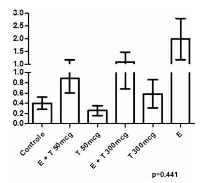
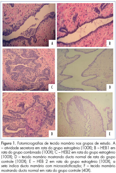
To investigate the action of testosterone (T), isolated or associated with estradiol benzoate (EB), on the proliferation markers and apoptosis of breasts of ovariectomized rats.
A total of 48 castrated female Wistar rats were divided into 6 groups, and each of them were submitted to one of the following treatments for 5 weeks: 1) control; 2) EB 50 mcg/day + T 50 mcg/day; 3) T 50mcg/day; 4) EB 50 mcg +T 300 mcg/day; 5) T 300 mcg/day; and 6) EB 50 mcg/day. After the treatment, the mammary tissue was submitted to a histological analysis and immunoexpression evaluation of proliferation markers (proliferating cell nuclear antigen, PCNA) and apoptosis (caspase-3).
There was a statistically significant difference among the groups regarding microcalcifications and secretory activity, with higher prevalence in the groups treated with EB. There was no difference among the groups regarding atrophy, but a higher prevalence of atrophy was found in the groups that received T versus those that received EB +T. There was a difference among the groups regarding the PCNA (p = 0.028), with higher expression in the group submitted to EB +T 300 mcg/day. Regarding caspase-3, there was no difference among the groups; however, in the group submitted to EB +T 300 mcg/day, the expression was higher than in the isolated T group.
Isolated T did not have a proliferative effect on the mammary tissue, contrary to EB. Testosterone in combination with EB may or may not decrease the proliferation, depending on the dose of T.
Summary
Rev Bras Ginecol Obstet. 2019;41(12):703-709
To investigate the action of testosterone (T), isolated or associated with estradiol benzoate (EB), on the proliferation markers and apoptosis of breasts of ovariectomized rats.
A total of 48 castrated female Wistar rats were divided into 6 groups, and each of them were submitted to one of the following treatments for 5 weeks: 1) control; 2) EB 50 mcg/day + T 50 mcg/day; 3) T 50mcg/day; 4) EB 50 mcg +T 300 mcg/day; 5) T 300 mcg/day; and 6) EB 50 mcg/day. After the treatment, the mammary tissue was submitted to a histological analysis and immunoexpression evaluation of proliferation markers (proliferating cell nuclear antigen, PCNA) and apoptosis (caspase-3).
There was a statistically significant difference among the groups regarding microcalcifications and secretory activity, with higher prevalence in the groups treated with EB. There was no difference among the groups regarding atrophy, but a higher prevalence of atrophy was found in the groups that received T versus those that received EB +T. There was a difference among the groups regarding the PCNA (p = 0.028), with higher expression in the group submitted to EB +T 300 mcg/day. Regarding caspase-3, there was no difference among the groups; however, in the group submitted to EB +T 300 mcg/day, the expression was higher than in the isolated T group.
Isolated T did not have a proliferative effect on the mammary tissue, contrary to EB. Testosterone in combination with EB may or may not decrease the proliferation, depending on the dose of T.
Summary
Rev Bras Ginecol Obstet. 2018;40(8):491-493
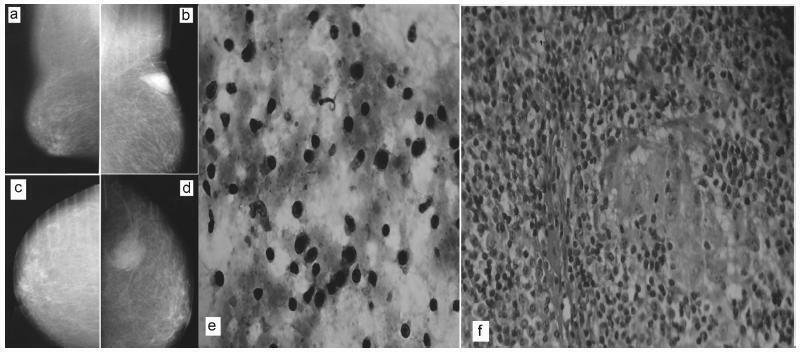
There are rare benign diseases that can mimic malignant breast neoplasms in the clinical exam and in mammography. We evaluated the contribution of an accessible procedure to most clinicians, the fine-needle aspiration cytology, to identify a rare mimicker of malignant breast neoplasms. A type 2 diabetic 85-year-old female presented with a 6-month history of a left breast lump. The physical exam and mammography were compatible with breast cancer. Nevertheless, after fine-needle aspiration cytology, the diagnosis was plasma cellmastitis. Once this rare diagnosis was established, the tumor was extirpated, and the final histologic diagnosis corroborated chronic plasma cellmastitis. The patient’s postoperative evolution was uneventful, and no other treatment was needed. Fine-needle aspiration cytology could be a valuable tool to identify rare mimickers of malignant breast neoplasms.
Summary
Rev Bras Ginecol Obstet. 2018;40(8):491-493
There are rare benign diseases that can mimic malignant breast neoplasms in the clinical exam and in mammography. We evaluated the contribution of an accessible procedure to most clinicians, the fine-needle aspiration cytology, to identify a rare mimicker of malignant breast neoplasms. A type 2 diabetic 85-year-old female presented with a 6-month history of a left breast lump. The physical exam and mammography were compatible with breast cancer. Nevertheless, after fine-needle aspiration cytology, the diagnosis was plasma cellmastitis. Once this rare diagnosis was established, the tumor was extirpated, and the final histologic diagnosis corroborated chronic plasma cellmastitis. The patient’s postoperative evolution was uneventful, and no other treatment was needed. Fine-needle aspiration cytology could be a valuable tool to identify rare mimickers of malignant breast neoplasms.
Summary
Rev Bras Ginecol Obstet. 2016;38(5):239-245
to evaluate the agreement between the clinical and pathological stagings of breast cancer based on clinical and molecular features.
this was a cross-sectional study, in which clinical, epidemiological and pathological data were collected from 226 patients who underwent surgery at the Prof. Dr. José Aristodemo Pinotti Women's Hospital (CAISM/Unicamp) from January 2008 to September 2010. Patients were staged clinically and pathologically, and were classified as: understaged, when the clinical staging was lower than the pathological staging; correctly staged, when the clinical staging was the same as the pathological one; and overstaged, when the clinical staging was greater than the pathological staging.
understaged patients were younger (52.2 years; p < 0.01) and more symptomatic at diagnosis (p = 0.04) when compared with correctly or overstaged patients. Clinicopathological surrogate subtype, menopausal status, parity, hormone replace therapy and histology were not associated with differences in staging. Women under 57 years of age were clinically understaged mainly due to underestimation of T ( tumor staging) (p < 0.001), as were the premenopausal women (p < 0.01). Patients whose diagnosis was made due to clinical complaints, and not by screening, were clinically understaged due to underestimation of N (lymph nodes staging) (p < 0.001).
the study shows that the clinicopathological surrogate subtype is not associated with differences in staging, while younger women diagnosed because of clinical complaints tend to have their breast tumors understaged during clinical evaluation.
Summary
Rev Bras Ginecol Obstet. 2016;38(2):112-116
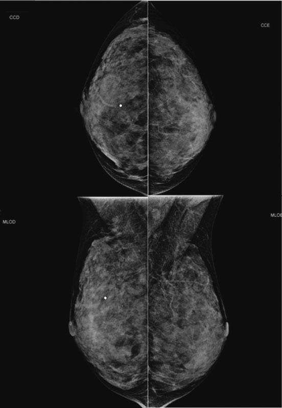
Lobular carcinoma in situ (LCIS) is associated with an increased risk of breast cancer and accounts for 1 to 2% of all breast cancers. LCIS diagnosis currently remains one of the major identifiable risk factors for subsequent breast cancer development. Imaging methods are becoming increasingly sensitive, and the consequent detection of small lesions and subtle abnormalities increases the chance of detection of in situ and invasive carcinomas, leading to a reduction in mortality. This report describes a case of a palpable complaint with abnormal imaging findings, including a solid LCIS mass.
Summary
Rev Bras Ginecol Obstet. 2016;38(2):112-116
Lobular carcinoma in situ (LCIS) is associated with an increased risk of breast cancer and accounts for 1 to 2% of all breast cancers. LCIS diagnosis currently remains one of the major identifiable risk factors for subsequent breast cancer development. Imaging methods are becoming increasingly sensitive, and the consequent detection of small lesions and subtle abnormalities increases the chance of detection of in situ and invasive carcinomas, leading to a reduction in mortality. This report describes a case of a palpable complaint with abnormal imaging findings, including a solid LCIS mass.
Summary
Rev Bras Ginecol Obstet. 2013;35(5):221-225
DOI 10.1590/S0100-72032013000500006
PURPOSE: We aimed to determine whether clinical examination could adequately ascertain the volume of tissue to be resected during breast-conserving surgery after neoadjuvant therapy. METHODS: We reviewed the clinical reports of 279 patients with histologically diagnosed invasive breast carcinomas treated with neoadjuvant therapy followed by surgery or with primary surgery alone. We estimated volumes of excised tissues, the volume of the tumor mass and the optimal volume required for excision based on 1 cm of clear margins. The actual excess of resected volume was estimated by calculating the resection ratio measured as the volume of the resected specimen divided by the optimal specimen volume. The study endpoints were to analyze the extent of tissue resection and to ascertain the effect of excess resected tissue on surgical margins in both groups of patients. RESULTS: The median tumor diameter was 2.0 and 1.5 cm in the surgery and neoadjuvant therapy groups, respectively. The median volume of resected mammary tissue was 64.3 cm³ in the primary surgery group and 90.7 cm³ in the neoadjuvant therapy group. The median resection ratios in the primary surgery and neoadjuvant therapy groups were 2.0 and 3.3, respectively (p<0.0001). Surgical margin data were similar in both groups. Comparison of the volume of resected mammary tissues with the tumor diameters showed a positive correlation in the primary surgery group and no correlation in the neoadjuvant therapy group. CONCLUSION: Surgeons tend to excise large volumes of tissue during breast-conserving surgery after neoadjuvant therapy, thereby resulting in a loss of the correlation between tumor diameter and volume of the excised specimen.
Summary
Rev Bras Ginecol Obstet. 2013;35(4):164-170
DOI 10.1590/S0100-72032013000400006
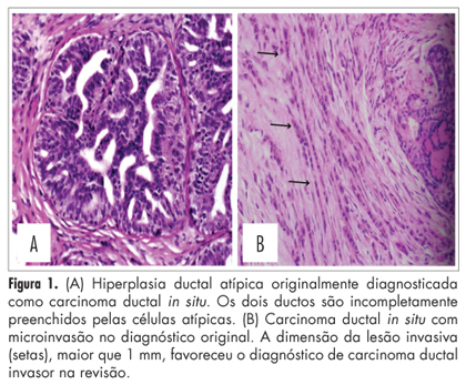
PURPOSE: To evaluate the agreement about the histopathological diagnosis of intraductal proliferative breast lesions between general pathologists and a specialist in breast pathology. METHODS: This was an observational, cross-sectional study of 209 lesions received in consultation at the Breast Pathology Laboratory of the School of Medicine, Federal University of Minas Gerais, from 2007 to 2011, comparing the original diagnosis and the review. We included only cases with a formal request for review and cases in which the original diagnosis or reviewer's diagnosis showed proliferative lesions, pure ductal carcinoma in situ, ductal carcinoma in situ associated with microinvasion or associated with invasive carcinoma. The kappa index and percent concordance were used in the statistical analyses. RESULTS: A moderate agreement was observed between the original histopathological diagnosis and the second opinion (kappa=0.5; percentual concordance=83%). After the review, the diagnosis of malignancy was confirmed in 140/163 cases (86%) and the diagnosis of benign lesions was confirmed in 34/46 cases (74%). Regarding specific diagnosis, we observed moderate agreement between the original diagnosis and the reviewer's diagnosis (136/209 cases; kappa=0.5; percent concordance=65%). The highest disagreement was observed in cases of ductal carcinoma in situ with microinvasion (6/6 cases; 100%). Important discordance was observed in cases of atypical ductal hyperplasia (16/30 cases; 53%) and ductal carcinoma in situ (25/75 cases; 33%). Regarding the histological grade of ductal carcinoma in situ, we observed good agreement between the original diagnosis and the review (29/39 cases; kappa=0.6, percent agreement=74%). CONCLUSION: Our data confirm that intraductal proliferative breast lesions, especially atypical ductal hyperplasia, ductal carcinoma in situ and ductal carcinoma in situ with microinvasion show relevant discrepancies in the histopathological diagnoses, which may induce errors in therapeutic decisions.
Summary
Rev Bras Ginecol Obstet. 2013;35(4):164-170
DOI 10.1590/S0100-72032013000400006
PURPOSE: To evaluate the agreement about the histopathological diagnosis of intraductal proliferative breast lesions between general pathologists and a specialist in breast pathology. METHODS: This was an observational, cross-sectional study of 209 lesions received in consultation at the Breast Pathology Laboratory of the School of Medicine, Federal University of Minas Gerais, from 2007 to 2011, comparing the original diagnosis and the review. We included only cases with a formal request for review and cases in which the original diagnosis or reviewer's diagnosis showed proliferative lesions, pure ductal carcinoma in situ, ductal carcinoma in situ associated with microinvasion or associated with invasive carcinoma. The kappa index and percent concordance were used in the statistical analyses. RESULTS: A moderate agreement was observed between the original histopathological diagnosis and the second opinion (kappa=0.5; percentual concordance=83%). After the review, the diagnosis of malignancy was confirmed in 140/163 cases (86%) and the diagnosis of benign lesions was confirmed in 34/46 cases (74%). Regarding specific diagnosis, we observed moderate agreement between the original diagnosis and the reviewer's diagnosis (136/209 cases; kappa=0.5; percent concordance=65%). The highest disagreement was observed in cases of ductal carcinoma in situ with microinvasion (6/6 cases; 100%). Important discordance was observed in cases of atypical ductal hyperplasia (16/30 cases; 53%) and ductal carcinoma in situ (25/75 cases; 33%). Regarding the histological grade of ductal carcinoma in situ, we observed good agreement between the original diagnosis and the review (29/39 cases; kappa=0.6, percent agreement=74%). CONCLUSION: Our data confirm that intraductal proliferative breast lesions, especially atypical ductal hyperplasia, ductal carcinoma in situ and ductal carcinoma in situ with microinvasion show relevant discrepancies in the histopathological diagnoses, which may induce errors in therapeutic decisions.
Summary
Rev Bras Ginecol Obstet. 2011;33(7):137-142
DOI 10.1590/S0100-72032011000700004
PURPOSE: To evaluate the efect of trimegestone on the histological changes of the mammary tissue of castrated rats. METHODS: Forty-five virgin female Wistar rats were used after oophorectomy. Sixty days after surgery, with hypoestrogenisms confirmed, the experimental rats were randomly assigned to three groups of 15 animals each, when then the specific treatment for each group was started. The control group (C) and experimental groups 1 and 2 respectively received 0.9% saline solution, 17-beta-estradiol and 17-beta-estradiol in combination with trimegestone for 60 consecutive days. After the end of treatment , the inguinal mammary glands were removed, stained with hematoxylin and eosin (HE) for morphometry and examined by immunohistochemistry for the quantification of anti-PCNA antibody in the mammary tissue, followed by euthanasia. The morphometric parameters evaluated were: epithelium cell-proliferation, secretor activity and mammary stroma changes. There were nine deaths during the experiment. The variables were submitted to statistical analysis adopting the 0.05 level of significance. RESULTS:Histological changes were observed in 16/36 rats, mild epithelial hyperplasia in 13/36, moderate epithelial hyperplasia in 3/36, with no cases of severe epithelial hyperplasia. Stromal fibrosis was found in 10/36 and secretory activity in 5/36 rats. All morphometric variables were significant in the estrogen group compared to control (p=0.0361), although there were no difference between the group receiving combined treatment and the controls (p=0.405). The immunohistochemical analysis showed no difference between groups. CONCLUSIONS:The hormones administered to castrated rats, i.e., 17 beta-estradiol alone or in combination with trimegestone, increased the proliferation of breast cells, but this effect appeared to be lower in the combined treatment, the same occurring regarding fibrosis of the mammary stroma.
Summary
Rev Bras Ginecol Obstet. 2010;32(5):241-246
DOI 10.1590/S0100-72032010000500007
PURPOSE: to assess the knowledge, attitude and practice of breast self-examination (BSE) of women from the municipality of São Luís (MA), Brazil, and associated socio-demographic variables. METHODS: prospective and cross-sectional study, with conglomerate sampling, in which 552 women from 14 census sections of São Luís were included during the period from January to September 2003. The knowledge, attitude and practice (dependent variables) were evaluated by means of analysis of the responses of the women as "adequate" or "inadequate". The main independent variables were: age, schooling, family income and marital and menopausal status. The χ2 test was used to determine the association between categorical variables and the measurement of the crude/adjusted Odds Ratio (OR) after multivariate analysis by means of logistic regression. RESULTS: although 1/3 of the studied population did not know about BSE, the group of women who were informed about it showed adequate knowledge (60.9%), practice (59.5%) and attitude (90%). The family history of breast cancer (8.9%) was not associated with better knowledge and practice. The media (63.6%) was found to be important in disseminating information about BSE. After multivariate analysis, women with a partner (OR=1.9) presented more adequate knowledge; women older than 50 years (OR=11.7) had a better attitude towards BSE; women with more than five years of schooling (OR=2) and with a partner (OR=1.7) were associated with a more correct practice of BSE. CONCLUSION: most of the patients know and practice the BSE in São Luís and their attitude towards the procedure is extremely positive. There was a great participation of the media in the dissemination of information concerning BSE.
Search
Search in:
breast (42) breast cancer (42) breast neoplasms (95) Cesarean section (72) endometriosis (66) infertility (56) Maternal mortality (43) menopause (82) obesity (58) postpartum period (40) pregnancy (225) Pregnancy complications (99) Prenatal care (68) prenatal diagnosis (50) Prevalence (41) Quality of life (51) risk factors (94) ultrasonography (79) urinary incontinence (40) women's health (48)