Summary
Rev Bras Ginecol Obstet. 2024;46:e-rbgo32
To ascertain how screening for preterm birth is performed among obstetricians working in public and private practice in a middle-income country.
Cross-sectional study of 265 obstetrician-gynecologists employed at public and private facilities. An online questionnaire was administered, with items designed to collect data on prematurity screening and prevention practices.
The mean age of respondents was 44.5 years; 78.5% were female, and 97.7% had completed a medical residency program. Universal screening (i.e., by ultrasound measurement of cervical length) was carried out by only 11.3% of respondents in public practice; 43% request transvaginal ultrasound if the manual exam is abnormal, and 74.6% request it in pregnant women with risk factors for preterm birth. Conversely, 60.7% of respondents in private practice performed universal screening. This difference in screening practices between public and private practice was highly significant (p < 0.001). Nearly all respondents (90.6%) reported prescribing vaginal progesterone for short cervix.
In the setting of this study, universal ultrasound screening to prevent preterm birth was used by just over half of doctors in private practice. In public facilities, screening was even less common. Use of vaginal progesterone in cervical shortening was highly prevalent. There is an unmet need for formal protocols for screening and prevention of preterm birth in middle-income settings.
Summary
Rev Bras Ginecol Obstet. 2024;46:e-rbgo6
BI-RADS® is a standardization system for breast imaging reports and results created by the American College of Radiology to initially address the lack of uniformity in mammography reporting. The system consists of a lexicon of descriptors, a reporting structure with final categories and recommended management, and a structure for data collection and auditing. It is accepted worldwide by all specialties involved in the care of breast diseases. Its implementation is related to the Mammography Quality Standards Act initiative in the United States (1992) and breast cancer screening. After its initial creation in 1993, four additional editions were published in 1995, 1998, 2003 and 2013. It is adopted in several countries around the world and has been translated into 6 languages. Successful breast cancer screening programs in high-income countries can be attributed in part to the widespread use of BI-RADS®. This success led to the development of similar classification systems for other organs (e.g., lung, liver, thyroid, ovaries, colon). In 1998, the structured report model was adopted in Brazil. This article highlights the pioneering and successful role of BI-RADS®, created by ACR 30 years ago, on the eve of publishing its sixth edition, which has evolved into a comprehensive quality assurance tool for multiple imaging modalities. And, especially, it contextualizes the importance of recognizing how we are using BI-RADS® in Brazil, from its implementation to the present day, with a focus on breast cancer screening.

Summary
Rev Bras Ginecol Obstet. 2023;45(12):790-795
To compare cytological and histological results from women > 64 years old who followed the Brazilian national cervical cancer screening guidelines with those who did not.
The present observational retrospective study analyzed 207 abnormal cervical smear results from women > 64 years old in a mid-sized city in Brazil over 14 years. All results were reported according to the Bethesda System. The women were divided into those who followed the screening guidelines and those who did not.
Atypical squamous cells of undetermined significance and low-grade squamous intraepithelial lesion cytology results were found in 128 (62.2%) cases. Of these, 112 (87.5%) had repeated cytology with positive results. The other 79 (38.1%) with abnormal results should have been referred to colposcopy and biopsy. Out of 41 (51.9%) biopsied women, 23 (29.1%) had a confirmed diagnosis of neoplasia or precursor lesion. In contrast, among the 78 (37.7%) biopsied patients, 40 (51.3%) followed the guideline recommendations, with 9 (22.5%) positive biopsies. Of the 38 (48.7%) women who did not follow the guidelines, there were 24 (63.1%) positive results. Women who did not follow the guidelines demonstrated higher chances of cancer and precursor lesions (odds ratio [OR]: 5.904; 95% confidence interval [CI]: 2.188–15.932; p = 0.0002).
Women > 64 years old who did not follow the national screening protocol showed significant differences in the frequency of abnormal results and severity of diagnosis compared with those who followed the protocol.
Summary
Rev Bras Ginecol Obstet. 2022;44(7):646-653
This study aims to describe the behavior of chromosomopathy screenings in euploid fetuses.
This is a prospective descriptive study with 566 patients at 11 to 14 weeks of gestation. The associations between ultrasound scans and serological variables were studied. For the quantitative variables we used the Spearman test; for the qualitative with quantitative variables the of Mann-Whitney U-test; and for qualitative variables, the X2 test was applied. Significance was set at p ≤ 0.05.
We have found that gestational age has correlation with ductus venosus, nuchal translucency, free fraction of β subunit of human chorionic gonadotropin, pregnancy-associated plasma protein-A and placental growth factor; there is also a correlation between history of miscarriages and nasal bone. Furthermore, we correlated body mass index with nuchal translucency, free fraction of β subunit of human chorionic gonadotropin, and pregnancy-associated plasma protein-A. Maternal age was associated with free fraction of β subunit of human chorionic gonadotropin and pregnancy-associated plasma protein-A.
Our study demonstrates for the first time the behavior of the biochemical and ultrasonographic markers of chromosomopathy screenings during the first trimester in euploid fetuses in Colombia. Our information is consistent with international reference values. Moreover, we have shown the correlation of different variables with maternal characteristics to determine the variables that could help with development of a screening process during the first trimester with high detection rates.
Summary
Rev Bras Ginecol Obstet. 2022;44(7):678-685
To determine the prevalence and possible variables associated with anal intraepithelial neoplasia and anal cancer in immunocompetent women with high-grade cervical intraepithelial neoplasia.
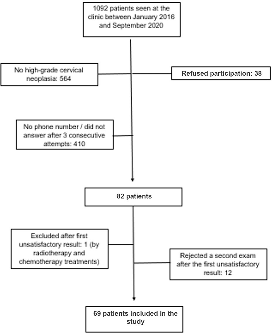
A cross-sectional study involving immunocompetent women with a histological diagnosis of high-grade cervical intraepithelial neoplasia and cervical cancer, conducted between January 2016 and September 2020. All women underwent anal cytology and answered a questionnaire on characterization and potential risk factors. Women with altered cytology were submitted to anoscopy and biopsy.
A total of 69 women were included in the study. Of these, 7 (10.1%) had abnormal anal cytology results: (high-grade lesion, atypical squamous cells of undetermined significance, and atypical squamous cells, cannot exclude high-grade lesions: 28,5% each; low grade lesion: 14,3%). Of the anoscopies, 3 (42.8%) showed alterations. Of the 2 (28,5% of all abnormal cytology results) biopsies performed, only 1 showed low-grade anal intraepithelial neoplasia. The average number of pregnancies, vaginal deliveries, and abortions was associated with abnormal anal cytology. However, the highest mean regarding the cesarean sections was associated with normal cytology.
The prevalence of anal intraepithelial neoplasia was compatible with data from recent studies, especially those conducted in Brazil. Opportunistic screening for anal intraepithelial neoplasia in this high-risk population should be considered. Anal cytology is suitable for this purpose, due to its low cost and feasibility in public health services.
Summary
Rev Bras Ginecol Obstet. 2022;44(7):678-685
To determine the prevalence and possible variables associated with anal intraepithelial neoplasia and anal cancer in immunocompetent women with high-grade cervical intraepithelial neoplasia.
A cross-sectional study involving immunocompetent women with a histological diagnosis of high-grade cervical intraepithelial neoplasia and cervical cancer, conducted between January 2016 and September 2020. All women underwent anal cytology and answered a questionnaire on characterization and potential risk factors. Women with altered cytology were submitted to anoscopy and biopsy.
A total of 69 women were included in the study. Of these, 7 (10.1%) had abnormal anal cytology results: (high-grade lesion, atypical squamous cells of undetermined significance, and atypical squamous cells, cannot exclude high-grade lesions: 28,5% each; low grade lesion: 14,3%). Of the anoscopies, 3 (42.8%) showed alterations. Of the 2 (28,5% of all abnormal cytology results) biopsies performed, only 1 showed low-grade anal intraepithelial neoplasia. The average number of pregnancies, vaginal deliveries, and abortions was associated with abnormal anal cytology. However, the highest mean regarding the cesarean sections was associated with normal cytology.
The prevalence of anal intraepithelial neoplasia was compatible with data from recent studies, especially those conducted in Brazil. Opportunistic screening for anal intraepithelial neoplasia in this high-risk population should be considered. Anal cytology is suitable for this purpose, due to its low cost and feasibility in public health services.
Summary
Rev Bras Ginecol Obstet. 2021;43(12):904-910
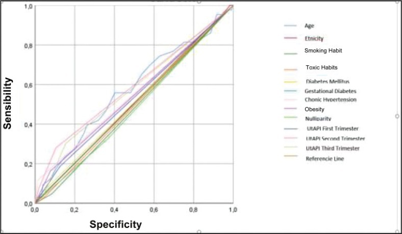
To evaluate the mean uterine artery pulsatility index (UtAPI) in each trimester of pregnancy as a predictor of early or late pre-eclampsia (PE) in Colombian pregnant women.
The UtAPI was measured in singleton pregnancies in each trimester. Uterine artery pulsatility index as predictor of PE was evaluated by odds ratio (OR), receiver operating characteristic (ROC) curves, and Kaplan-Meier diagram.
Analysis in the 1st and 3rd trimester showed that abnormal UtAPI was associated with early PE (OR: 5.99: 95% confidence interval [CI]: 1.64–21.13; and OR: 10.32; 95%CI: 2.75–42.49, respectively). Sensitivity and specificity were 71.4 and 79.6%, respectively, for developing PE (area under the curve [AUC]: 0.922). The Kaplan-Meier curve showed that a UtAPI of 0.76 (95%CI: 0.58–1.0) in the 1st trimester was associated with early PE, and a UtAPI of 0.73 (95%CI: 0.55–0.97) in the 3rd trimester was associated with late PE.
Uterine arteries proved to be a useful predictor tool in the 1st and 3rd trimesters for early PE and in the 3rd trimester for late PE in a pregnant population with high prevalence of PE.
Summary
Rev Bras Ginecol Obstet. 2021;43(12):904-910
To evaluate the mean uterine artery pulsatility index (UtAPI) in each trimester of pregnancy as a predictor of early or late pre-eclampsia (PE) in Colombian pregnant women.
The UtAPI was measured in singleton pregnancies in each trimester. Uterine artery pulsatility index as predictor of PE was evaluated by odds ratio (OR), receiver operating characteristic (ROC) curves, and Kaplan-Meier diagram.
Analysis in the 1st and 3rd trimester showed that abnormal UtAPI was associated with early PE (OR: 5.99: 95% confidence interval [CI]: 1.64–21.13; and OR: 10.32; 95%CI: 2.75–42.49, respectively). Sensitivity and specificity were 71.4 and 79.6%, respectively, for developing PE (area under the curve [AUC]: 0.922). The Kaplan-Meier curve showed that a UtAPI of 0.76 (95%CI: 0.58–1.0) in the 1st trimester was associated with early PE, and a UtAPI of 0.73 (95%CI: 0.55–0.97) in the 3rd trimester was associated with late PE.
Uterine arteries proved to be a useful predictor tool in the 1st and 3rd trimesters for early PE and in the 3rd trimester for late PE in a pregnant population with high prevalence of PE.
Summary
Rev Bras Ginecol Obstet. 2021;43(6):452-456
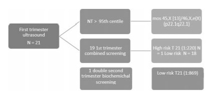
The objective of the present study was to determine the frequency of malformations and chromosomal abnormalities in a population of fetuses with an aberrant right subclavian artery (ARSA).
This is a 6-year retrospective study of fetuses with a prenatal diagnosis of ARSA conducted during the period between September 2013 and June 2019 at a fetal medicine unit. Data were collected from ultrasound, fetal echocardiograms, genetic studies, and neonatal records.
An ARSA was diagnosed in 22 fetuses. An ARSA was an isolated finding in 18 out of 22 cases (82%). Associated abnormal sonographic findings were found in 4 cases. All cases underwent invasive testing. In 1 of the cases, a chromosomal abnormality was detected (mos 45,X [13]/46,X,e(X) (p22.1q22.1)). No cases of congenital heart disease were found in any of these fetuses. There were two cases in which the postnatal evaluation revealed amalformation: one case of hypospadias and 1 case of cleft palate.
The presence of an isolated ARSA is benign and is not associated with chromosomal abnormalities. The finding of ARSA, however, warrants a detailed fetal ultrasound in order to exclude major fetal abnormalities and other soft markers.
Summary
Rev Bras Ginecol Obstet. 2021;43(6):452-456
The objective of the present study was to determine the frequency of malformations and chromosomal abnormalities in a population of fetuses with an aberrant right subclavian artery (ARSA).
This is a 6-year retrospective study of fetuses with a prenatal diagnosis of ARSA conducted during the period between September 2013 and June 2019 at a fetal medicine unit. Data were collected from ultrasound, fetal echocardiograms, genetic studies, and neonatal records.
An ARSA was diagnosed in 22 fetuses. An ARSA was an isolated finding in 18 out of 22 cases (82%). Associated abnormal sonographic findings were found in 4 cases. All cases underwent invasive testing. In 1 of the cases, a chromosomal abnormality was detected (mos 45,X [13]/46,X,e(X) (p22.1q22.1)). No cases of congenital heart disease were found in any of these fetuses. There were two cases in which the postnatal evaluation revealed amalformation: one case of hypospadias and 1 case of cleft palate.
The presence of an isolated ARSA is benign and is not associated with chromosomal abnormalities. The finding of ARSA, however, warrants a detailed fetal ultrasound in order to exclude major fetal abnormalities and other soft markers.
Summary
Rev Bras Ginecol Obstet. 2021;43(3):190-199
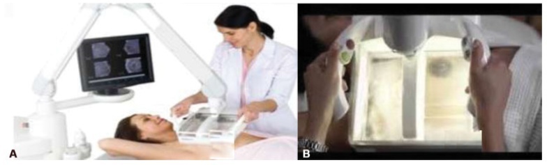
To compare hand-held breast ultrasound (HHBUS) and automated breast ultrasound (ABUS) as screening tool for cancer.
A cross-sectional study in patients with mammographically dense breasts was conducted, and both HHBUS and ABUS were performed. Hand-held breast ultrasound was acquired by radiologists and ABUS by mammography technicians and analyzed by breast radiologists. We evaluated the Breast Imaging Reporting and
(BI-RADS) classification of the exam and of the lesion, as well as the amount of time required to perform and read each exam. The statistical analysis employed was measures of central tendency and dispersion, frequencies, Student t test, and a univariate logistic regression, through the odds ratio and its respective 95% confidence interval, and with p<0.05 considered of statistical significance.
Atotal of 440 patientswere evaluated. Regarding lesions,HHBUS detected 15 (7.7%) BI-RADS 2, 175 (89.3%) BI-RADS 3, and 6 (3%) BI-RADS 4, with 3 being confirmed by biopsy as invasive ductal carcinomas (IDCs), and 3 false-positives. Automated breast ultrasound identified 12 (12.9%) BI-RADS 2, 75 (80.7%) BI-RADS 3, and 6 (6.4%) BI-RADS 4, including 3 lesions detected by HHBUS and confirmed as IDCs, in addition to 1 invasive lobular carcinoma and 2 high-risk lesions not detected by HHBUS. The amount of time required for the radiologist to read the ABUS was statistically inferior compared with the time required to read the HHBUS (p<0.001). The overall concordance was 80.9%. A total of 219 lesions were detected, from those 70 lesions by both methods, 126 only by HHBUS (84.9% not suspicious by ABUS) and 23 only by ABUS.
Compared with HHBUS, ABUS allowed adequate sonographic study in supplemental screening for breast cancer in heterogeneously dense and extremely dense breasts.
Summary
Rev Bras Ginecol Obstet. 2021;43(3):190-199
To compare hand-held breast ultrasound (HHBUS) and automated breast ultrasound (ABUS) as screening tool for cancer.
A cross-sectional study in patients with mammographically dense breasts was conducted, and both HHBUS and ABUS were performed. Hand-held breast ultrasound was acquired by radiologists and ABUS by mammography technicians and analyzed by breast radiologists. We evaluated the Breast Imaging Reporting and
(BI-RADS) classification of the exam and of the lesion, as well as the amount of time required to perform and read each exam. The statistical analysis employed was measures of central tendency and dispersion, frequencies, Student t test, and a univariate logistic regression, through the odds ratio and its respective 95% confidence interval, and with p<0.05 considered of statistical significance.
Atotal of 440 patientswere evaluated. Regarding lesions,HHBUS detected 15 (7.7%) BI-RADS 2, 175 (89.3%) BI-RADS 3, and 6 (3%) BI-RADS 4, with 3 being confirmed by biopsy as invasive ductal carcinomas (IDCs), and 3 false-positives. Automated breast ultrasound identified 12 (12.9%) BI-RADS 2, 75 (80.7%) BI-RADS 3, and 6 (6.4%) BI-RADS 4, including 3 lesions detected by HHBUS and confirmed as IDCs, in addition to 1 invasive lobular carcinoma and 2 high-risk lesions not detected by HHBUS. The amount of time required for the radiologist to read the ABUS was statistically inferior compared with the time required to read the HHBUS (p<0.001). The overall concordance was 80.9%. A total of 219 lesions were detected, from those 70 lesions by both methods, 126 only by HHBUS (84.9% not suspicious by ABUS) and 23 only by ABUS.
Compared with HHBUS, ABUS allowed adequate sonographic study in supplemental screening for breast cancer in heterogeneously dense and extremely dense breasts.
Search
Search in:
breast (42) breast cancer (42) breast neoplasms (95) Cesarean section (72) endometriosis (66) infertility (56) Maternal mortality (43) menopause (82) obesity (58) postpartum period (40) pregnancy (225) Pregnancy complications (99) Prenatal care (68) prenatal diagnosis (50) Prevalence (41) Quality of life (51) risk factors (94) ultrasonography (79) urinary incontinence (40) women's health (48)