Summary
Revista Brasileira de Ginecologia e Obstetrícia. 2008;30(11):561-565
DOI 10.1590/S0100-72032008001100006
PURPOSE: to evaluate the influence of age on the quality of semen in men submitted to spermatic analysis in a human reproduction service, in cases of conjugal infertility. METHODS: a retrospective study in which the spermiograms of all men in process of investigation for conjugal infertility in a service of assisted reproduction in the Northeast of Brazil were evaluated from September 2002 to December 2004. A number of 531 individuals submitted to 531 spermatic evaluations were included in the study. The following parameters have been analyzed: spermatic volume, concentration, motility and morphology. The men under investigation have been divided in groups, according to the results obtained in each of the variables studied. Seminal volume groups were divided in: hypospermia, normospermia and hyperspermia. Spermatic concentration groups were divided in: azoospermia, oligospermia, normospermia and polyspermia. Motility groups were divided in: normal motility and asthenospermia. Morphology groups were divided in: normal morphology and teratospermia. The t test has been used to compare the average age of patients in groups with normal and in groups with altered parameters. The program XLSTAT (p<0.05) has been used for the statistical analysis. RESULTS: the individuals studied presented an average of 37±7.9 years old, with an average of seminal volume of 3±1.4 mL, a spermatic concentration of 61.4±66.4 spermatozoids by mL of semen, a progressive motility of 44.7±19.4% of the total of spermatozoids and normal morphology of 11.2±6.6% of the spermatozoids. Average age among groups were similar, except for that of individuals with hypospermia, which was significantly higher than the one from men with normospermia (39.6±10.3 versus 36.5±7.3, p=0.001). CONCLUSIONS: age interferes in an inversely proportional way on the ejaculated volume, but does not influence spermatic concentration, motility and morphology.
Summary
Revista Brasileira de Ginecologia e Obstetrícia. 2008;30(11):566-572
DOI 10.1590/S0100-72032008001100007
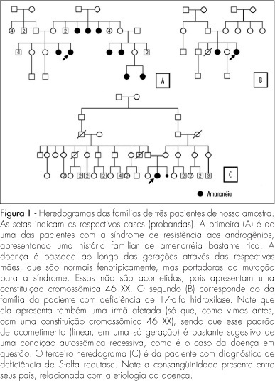
PURPOSE: to verify the prevalence and clinical characteristics of patients with primary amenorrhea and XY caryotype, evaluated in our Service, aiming at identifying findings which could help their recognition. METHODS: from January 1975 to November 2007, 104 patients with amenorrhea were evaluated. All the cases were analyzed by the caryotype by GTG bands. Among them, 21 (20.2%) presented a XY 46 constitution. Nevertheless, two of them were excluded from the study, because of incomplete data in their patient's chart. Most of the 19 patients who formed the sample had been referred to us by the gynecology clinics (63.2%). Their ages varied from 16 to 41 years old (an average of 22.1). Data were collected about their family and previous history, physical examination and results of complementary exams and the information was taken into consideration to determine the diagnosis. RESULTS: the predominant diagnosis was resistance to androgens syndrome (n=12; 63.2%); five patients (25.3%) presented XY pure gonadal dysgenesis (XY PGD), one (5.3%) 17 alpha-hydroxylase deficiency, and one (5.3%), 5 alpha-reductase deficiency. Clinical findings frequently found in these patients included abnormal development of secondary sexual characters (n=19), uterine agenesia with a blind vagina (n=14), family history of amenorrhea (n=8), and palpable gonads in the inguinal canal (n=5). Two of them presented a history of inguinal hernia. Systemic arterial hypertension was only diagnosed in the patient with 17 alpha-hydroxylase deficiency, and gonadal malignization, in the one with XY PGD. CONCLUSIONS: the rate of patients with XY caryotype (20%) was higher than the one described in the literature (3 to 11%). It is believed that this fact is related to the way patients are usually referred to our service. Some findings from the clinical history and from the physical examination should be evaluated as a routine in individuals with primary amenorrhea. This way, there would be a more precocious detection of XY 46 patients, and a better clinical management of them, as a consequence.
Summary
Revista Brasileira de Ginecologia e Obstetrícia. 2008;30(10):486-493
DOI 10.1590/S0100-72032008001000002
PURPOSE: to investigate factors accountable for macrosomia incidence in a study with mothers and progeny attended at a Basic Unity of Health in Rio de Janeiro, Brazil. METHODS: a prospective study, with 195 pairs of mothers and progeny, in which the dependent variable was macrosomia (weight at delivery >4,000 g - independent of the gestational age or of other demographic variables), and socioeconomic, previous pregnancies/gestation course, biochemical, behavioral and anthropometric, the independent variables. Statistical analysis has been done by multiple logistic regression. Relative risk (RR) values have been estimated, based on the simple form: RR=OR/ (1 - I0) + (I0 versus OR), in which I0 is the macrosomia incidence in non-exposed people. RESULTS: Macrosomia incidence was 6.7%, the highest value being found in the progeny of women >30 years old (12.8%), white (10.4%), with two or more children (16.7%), with male newborns (9.6%), with height >1,6 m (12.5%), with overweight or obesity as a nutritional pre-gestational state (13.6%), and with excessive gestational gain of weight (12.7%). The final model has shown that having two or more children (RR=3.7; CI95%=1.1-9.9), and having a male newborn (RR=7.5; CI95%=1.0-37.6) were the variables linked to the macrosomia occurrence. CONCLUSIONS: macrosomia incidence was higher than the one observed in Brazil as a whole, but inferior to the one reported in studies from developed countries. Having two or more children and a newborn male were the factors accountable for the occurrence of macrosomia.
Summary
Revista Brasileira de Ginecologia e Obstetrícia. 2008;30(10):494-498
DOI 10.1590/S0100-72032008001000003
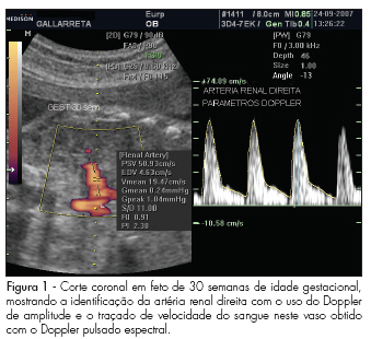
PURPOSE: to describe values found for the resistance index (RI), pulsatility index (PI) and the systole/diastole (S/D) ratio of fetal renal arteries in non-complicated gestations between the 22nd and the 38th week, and to evaluate whether those values vary along that period. METHODS: observational study, where 45 fetuses from non-complicated gestations have been evaluated in the 22nd, 26th, 30th and 38th weeks of gestational age. Doppler ultrasonography has been performed by the same observer, using a device with 4 to 7 MHz transducer. For the acquisition of the renal arteries velocity record, a 1 mm to 2 mm probe has been placed in the mean third of the renal artery for the evaluation through pulsed Doppler ultrasonography. The measurement of RI, PI and S/D ratio from three consecutive waves was performed with the automatic mode. To detect significant differences in the indexes' values along gestation, we have compared values obtained at the different gestational ages, through repeated measures ANOVA, followed by Tukey's post-hoc test. RESULTS: There were no significant differences between the right and left renal arteries, when the RI, IP and S/D ratio were compared. Nevertheless, a change in the values of these parameters has been observed between the 22nd week (RI=0.9 ± 0.02; PI=2.4 ± 0.02; S/D ratio=11.6 ± 2.2; mean ± standard deviation of the combined mean values of the right and left renal artery) and the 38th week (RI=0.8 ± 0.03; PI=2.1 ± 0.2; S/D ratio=8.7 ± 2.3) of gestation. CONCLUSIONS: the parameters evaluated (RI, PI and S/D ratio) have presented decreasing values between the 22nd and 38th, with no difference between the fetus's right and left sides.
Summary
Revista Brasileira de Ginecologia e Obstetrícia. 2008;30(10):499-503
DOI 10.1590/S0100-72032008001000004
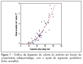
PURPOSE: to evaluate the embryo's volume (EV) between the seventh and the tenth gestational week, through tridimensional ultrasonography. METHODS: a transversal study with 63 normal pregnant women between the seventh and the tenth gestational week. The ultrasonographical exams have been performed with a volumetric abdominal transducer. Virtual Organ Computer-aided Analysis (VOCAL) has been used to calculate EV, with a rotation angle of 12º and a delimitation of 15 sequential slides. The average, median, standard deviation and maximum and minimum values have been calculated for the EV in all the gestational ages. A dispersion graphic has been drawn to assess the correlation between EV and the craniogluteal length (CGL), the adjustment being done by the determination coefficient (R²). To determine EV's reference intervals as a function of the CGL, the following formula was used: percentile=EV+K versus SD, with K=1.96. RESULTS: CGL has varied from 9.0 to 39.7 mm, with an average of 23.9 mm (±7.9 mm), while EV has varied from 0.1 to 7.6 cm³, with an average of 2.7 cm³ (±3.2 cm³). EV was highly correlated to CGL, the best adjustment being obtained with quadratic regression (EV=0.2-0.055 versus CGL+0.005 versus CGL²; R²=0.8). The average EV has varied from 0.1 (-0.3 to 0.5 cm³) to 6.7 cm³ (3.8 to 9.7 cm³) within the interval of 9 to 40 mm of CGL. EV has increased 67 times in this interval, while CGL, only 4.4 times. CONCLUSIONS: EV is a more sensitive parameter than CGL to evaluate embryo growth between the seventh and the tenth week of gestation.
Summary
Revista Brasileira de Ginecologia e Obstetrícia. 2008;30(10):504-510
DOI 10.1590/S0100-72032008001000005
PURPOSE: to translate from English into Portuguese, adapt culturally and validate the Female Sexual Function Index (FSFI). METHODS: knowing the objectives of this research, two Brazilian translators have prepared a version each from the FSFI into Portuguese. Both versions have then been retro-translated into English by two English translators. After harmonizing the differences, they have been pre-tested in a pilot study. The final versions from the FSFI and from another questionnaire, the Short-Form Health Survey, which had already been translated and published in Portuguese, have then been simultaneously administered to one hundred patients, to test the FSFI psychometric proprieties concerning reliability (internal consistency and testing-retesting) and construct validity. Retesting was done after four weeks from the first interview. RESULTS: the process of cultural adaptation has not altered the Portuguese version of the FSFI, as compared to the original. The FSFI standardized Cronbach alpha was 0.96, and the evaluation by domains has varied from 0.31 to 0.97. As a measure of test-retest confidentiality, it was applied the intra-class coefficient, which has been considered strong and identical (1.0). Pearson's correlation coefficient between the FSFI and the Short-Form Health Survey was positive, but weak in most of the interrelated domains, varying from 0.017 to 0.036. CONCLUSIONS: the FSFI English version has been translated into Portuguese and culturally adapted, being reliable to evaluate the sexual response of Brazilian women.
Summary
Revista Brasileira de Ginecologia e Obstetrícia. 2008;30(10):511-517
DOI 10.1590/S0100-72032008001000006
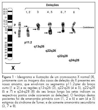
PURPOSE: to correlate the clinical manifestations of patients with amenorrhea and X chromosome abnormalities. METHODS: a retrospective analysis of the clinical and laboratorial findings of patients with amenorrhea and abnormalities of X chromosome, attended between January 1975 and November 2007 was performed. Their anthropometric measures were evaluated through standard growth tables, and, when present, minor and major anomalies were noted. The chromosomal study was performed through the GTG banded karyotype. RESULTS: from the total of 141 patients with amenorrhea, 16% presented numerical and 13% structural abnormalities of X chromosome. From these patients with X chromosome abnormalities (n=41), 35 had a complete clinical description. All presented hypergonadotrophic hypogonadism. Primary amenorrhea was observed in 24 patients, 91.7% of them with a Turner syndrome phenotype. Despite a case with Xq22-q28 deletion, all patients with this phenotype presented alterations involving Xp (one case with an additional cell lineage 46,XY). The two remaining patients with only primary amenorrhea had proximal deletions of Xq. Among the 11 patients with secondary amenorrhea, 54.5% presented a Turner phenotype (all with isolated or mosaic X chromosome monosomy). Patients with phenotype of isolated ovarian failure had only Xq deletions and X trisomy. CONCLUSIONS: the cytogenetic analysis must always be performed in women with ovarian failure of unknown cause, even in the absence of clinical dysmorphic features. This analysis is also extremely relevant in syndromic patients, because it can either confirm the diagnosis or identify patients in risk, like the cases involving a 46,XY lineage.
Summary
Revista Brasileira de Ginecologia e Obstetrícia. 2008;30(10):518-523
DOI 10.1590/S0100-72032008001000007
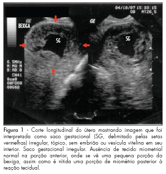
Ectopic pregnancy in a cesarean scar is the rarest form of ectopic pregnancy and probably the most dangerous one because of the risk of uterine rupture and massive hemorrhage. This condition must be distinguished from cervical pregnancy and spontaneous abortion in progress, so that the appropriate treatment can be immediately offered. Since the advent of endovaginal ultrasonography, ectopic pregnancy in a cesarean scar can be diagnosed early in pregnancy if the sonographer is familiarized with the diagnostic criteria of this situation, especially in women with previous cesarean scar. Here we describe a case of ectopic pregnancy in a cesarean scar in which the diagnosis was considerably late, with presentation of spontaneous regression.
