Summary
Rev Bras Ginecol Obstet. 2024;46:e-rbgo19
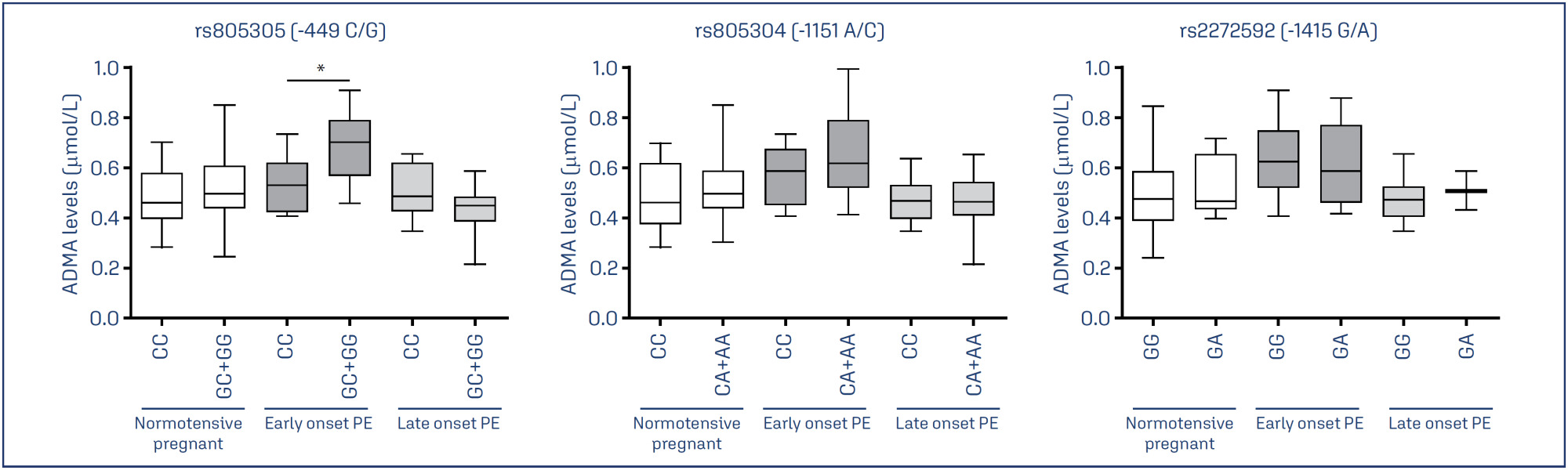
To examine whether the DDAH2 promoter polymorphisms -1415G/A (rs2272592), -1151A/C (rs805304) and -449G/C (rs805305), and their haplotypes, are associated with PE compared with normotensive pregnant women, and whether they affect ADMA levels in these groups.
A total of 208 pregnant women were included in the study and classified as early-onset (N=57) or late-onset PE (N =49), and as normotensive pregnant women (N = 102).
Pregnant with early-onset PE carrying the GC and GG genotypes for the DDAH2 -449G/C polymorphism had increased ADMA levels (P=0.01). No association of DDAH2 polymorphisms with PE in single-locus analysis was found. However, the G-C-G haplotype was associated with the risk for late-onset PE.
It is suggested that DDAH2 polymorphisms could affect ADMA levels in PE, and that DDAH2 haplotypes may affect the risk for PE.
Summary
Rev Bras Ginecol Obstet. 2024;46:e-rbgo19
To examine whether the DDAH2 promoter polymorphisms -1415G/A (rs2272592), -1151A/C (rs805304) and -449G/C (rs805305), and their haplotypes, are associated with PE compared with normotensive pregnant women, and whether they affect ADMA levels in these groups.
A total of 208 pregnant women were included in the study and classified as early-onset (N=57) or late-onset PE (N =49), and as normotensive pregnant women (N = 102).
Pregnant with early-onset PE carrying the GC and GG genotypes for the DDAH2 -449G/C polymorphism had increased ADMA levels (P=0.01). No association of DDAH2 polymorphisms with PE in single-locus analysis was found. However, the G-C-G haplotype was associated with the risk for late-onset PE.
It is suggested that DDAH2 polymorphisms could affect ADMA levels in PE, and that DDAH2 haplotypes may affect the risk for PE.
Summary
Rev Bras Ginecol Obstet. 2015;37(5):203-207
DOI 10.1590/SO100-720320150005293
To determine the frequency of Human Papillomavirus (HPV) in the placenta, in the
colostrum and in the umbilical cord blood of parturient women and their newborns
assisted at the Clinic of Gynecology and Obstetrics of the University Hospital of
Rio Grande (RS), Brazil.
Biopsies were collected from 150 placentas on the maternal side, 150 on the fetal
side, 138 samples of umbilical cord blood and 118 of the colostrum. The placenta
biopsies were collected from the central and peripheral portions. DNA was
extracted according to the manufacturer's protocol and to a reference found in the
literature. HPV was detected by the nested polymerase chain reaction (PCR-Nested)
using primers MY09/11 and GP5/GP6. Genotyping was performed by direct sequencing.
The participants responded to a self-applied questionnaire with demographic and
clinical data, in order to characterize the sample.
HPV was detected in 4% (6/150) of cases on the mother's side of the placentas, in
3.3% (5/150) on the fetal side, in 2.2% (3/138) in umbilical cord blood and in
0.84% (1/118) in colostrum samples. The vertical transmission rate was 50%. HPV-6
was the low-risk genotype found (60%) and the high-risk genotypes were HPV-16 and
HPV-18 (20% each).
These results suggest that HPV can infect the placenta, the colostrum and the
umbilical cord blood.
Summary
Rev Bras Ginecol Obstet. 2015;37(2):94-99
DOI 10.1590/SO100-720320150005206
The aim of this study was to compare the performance of two human papillomavirus (HPV) genotyping techniques, Linear Array and PapilloCheck, in women with high-grade squamous intraepithelial lesion (HSIL).
A total of 88 women with cytological diagnosis of HSIL were recruited at 2 reference centers in cervical pathology in Salvador, Bahia, Brazil, from July 2006 to January 2009. After the cytological diagnosis of HSIL, cervix cells were collected to determine the HPV genotype and a biopsy was obtained under colposcopic vision for histopathological analysis. After the confirmation of CIN2+ by histopathology, HPV genotyping was performed on 41 women by the Linear Array and PapilloCheck methods.
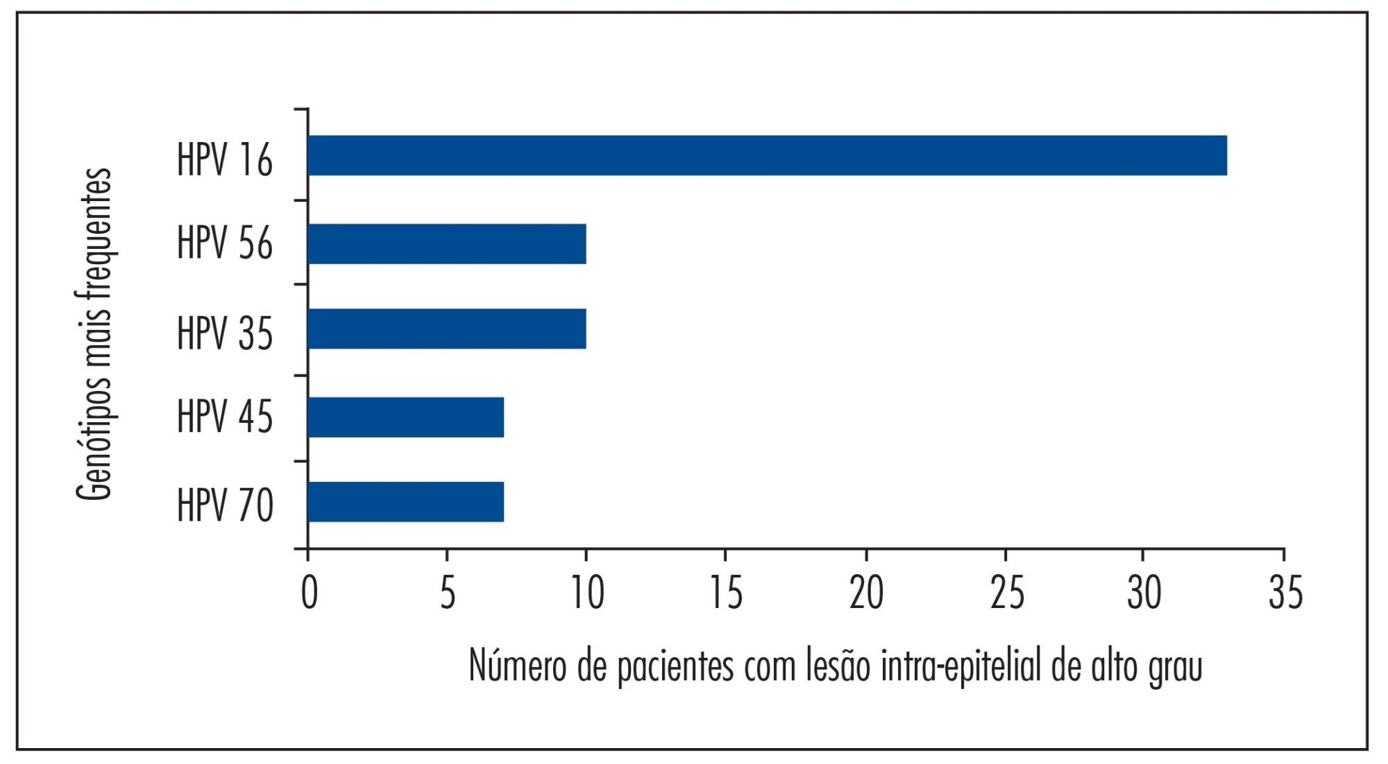
Both tests showed an overall concordance rate for HPV detection of 97.2% (35/36). Of the 36 valid samples, 35 (97.2%) were positive in both tests and 1 (2.8%) was discordant, with the Linear Array indicating the presence of multiple types. The most prevalent HPV genotypes detected by the Linear Array technique were HPV 16, HPV 56, HPV 35, HPV 45, and HPV 70; and those detected by the PapilloCheck technique were HPV 16, HPV 56, HPV 11, HPV 35, and HPV 42. A similar rate of infection with multiple HPV types was observed with the two tests (72.5% with the Linear Array and 75.0% with the PapilloCheck).
Linear Array genotyping assay and PapilloCheck showed equivalent performance for the detection of oncogenic HPV types in women with HSIL, with PapilloCheck having the advantage of being a method that avoids subjectivity when reading the HPV genotypes.
Summary
Rev Bras Ginecol Obstet. 2015;37(2):94-99
DOI 10.1590/SO100-720320150005206
The aim of this study was to compare the performance of two human papillomavirus (HPV) genotyping techniques, Linear Array and PapilloCheck, in women with high-grade squamous intraepithelial lesion (HSIL).
A total of 88 women with cytological diagnosis of HSIL were recruited at 2 reference centers in cervical pathology in Salvador, Bahia, Brazil, from July 2006 to January 2009. After the cytological diagnosis of HSIL, cervix cells were collected to determine the HPV genotype and a biopsy was obtained under colposcopic vision for histopathological analysis. After the confirmation of CIN2+ by histopathology, HPV genotyping was performed on 41 women by the Linear Array and PapilloCheck methods.
Both tests showed an overall concordance rate for HPV detection of 97.2% (35/36). Of the 36 valid samples, 35 (97.2%) were positive in both tests and 1 (2.8%) was discordant, with the Linear Array indicating the presence of multiple types. The most prevalent HPV genotypes detected by the Linear Array technique were HPV 16, HPV 56, HPV 35, HPV 45, and HPV 70; and those detected by the PapilloCheck technique were HPV 16, HPV 56, HPV 11, HPV 35, and HPV 42. A similar rate of infection with multiple HPV types was observed with the two tests (72.5% with the Linear Array and 75.0% with the PapilloCheck).
Linear Array genotyping assay and PapilloCheck showed equivalent performance for the detection of oncogenic HPV types in women with HSIL, with PapilloCheck having the advantage of being a method that avoids subjectivity when reading the HPV genotypes.
Summary
Rev Bras Ginecol Obstet. 2014;36(9):416-422
DOI 10.1590/SO100-720320140004995
The aim of this study was to evaluate the human papillomavirus genotypes and the frequency of multiple human papillomavirus infections, as well as to assess the association between human papillomavirus genotype, cyto-histopathological abnormalities and age range.
A retrospective cross-sectional study was carried out between June 2010 and October 2013 in Salvador, Bahia, Brazil. We analyzed 351 results of positive human papillomavirus genotyping performed using the PapilloCheck(r) test, designed to detect 24 human papillomavirus types. The cyto-histopathological abnormalities were classified as negative (negative cytology and histopathology), low-grade lesions (cytologic low-grade squamous intraepithelial lesion diagnosis or histopathologic cervical intraepithelial neoplasia 1 or vaginal intraepithelial neoplasia 1 diagnosis) and high-grade lesions (cytologic high-grade squamous intraepithelial lesion diagnosis or histopathologic cervical intraepithelial neoplasia 2+ or vaginal intraepithelial neoplasia 2+ diagnosis).
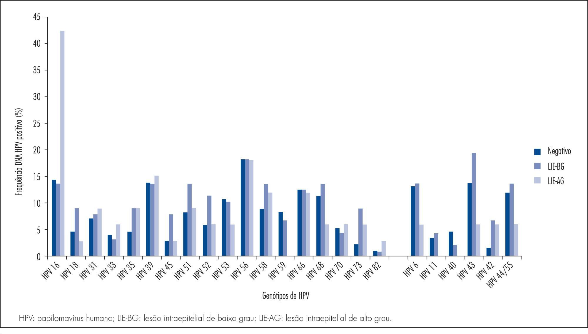
The most frequently detected high risk human papillomavirus genotype was HPBV 16, with 18.5%, 95% confidence interval (95%CI) 14.6-23.0, followed by HPV 56 (14%; 95%CI 10.5-18.0) and HPV 39 (13.4%; 95%CI 9.5-16.8). HPV 18 (5.4%; 95%CI 3.3-8.3) was among the least frequent types. Among the low risk types, HPV 42 (15.7%; 95%CI 12.0-20.0), HPV 6 (11.4%; 95%CI 8.3-15.2) and HPV 44/55 (11.1%; 95%CI 8.0-14.9) were the most frequent, while HPV 11 (2.8%; 95%CI 1.4-5.2) was the least common. The proportion of HPV 16-positive women increased with severity of cyto-histopathological abnormalities: 13.8% (12/87) in low-grade lesion and 42.4% (14/33) in high-grade lesion. There was association between low- or high-grade cyto-histopathological lesion and the high risk genotypes, HPV16, HPV 52, HPV 73 and HPV 82, and the low risk type, HPV 43. Women under 30 years showed a significantly higher frequency of HPV 16 (22.2 versus 12.9%, p =0.01), HPV 42 (19.7 versus 10.9%, p=0.01) and HPV 45 (6.6 versus 1.4%, p=0.01), and multiple human papillomavirus infections (58.1 versus 47.4%, p=0.04).
We observed variability of human papillomavirus genotype distribution in women from the state of Bahia. HPV 16 was the most frequently detected high risk human papillomavirus, as also reported for other geographic areas of Brazil and for the world in general. HPV 56 and HPV 39 were the second and the third most common genotypes, whereas HPV 18 was among the least frequent types. HPV 42, 6 and 44/55 were the most frequently detected low risk human papillomavirus, and HPV 11 was the least common.
Summary
Rev Bras Ginecol Obstet. 2014;36(9):416-422
DOI 10.1590/SO100-720320140004995
The aim of this study was to evaluate the human papillomavirus genotypes and the frequency of multiple human papillomavirus infections, as well as to assess the association between human papillomavirus genotype, cyto-histopathological abnormalities and age range.
A retrospective cross-sectional study was carried out between June 2010 and October 2013 in Salvador, Bahia, Brazil. We analyzed 351 results of positive human papillomavirus genotyping performed using the PapilloCheck(r) test, designed to detect 24 human papillomavirus types. The cyto-histopathological abnormalities were classified as negative (negative cytology and histopathology), low-grade lesions (cytologic low-grade squamous intraepithelial lesion diagnosis or histopathologic cervical intraepithelial neoplasia 1 or vaginal intraepithelial neoplasia 1 diagnosis) and high-grade lesions (cytologic high-grade squamous intraepithelial lesion diagnosis or histopathologic cervical intraepithelial neoplasia 2+ or vaginal intraepithelial neoplasia 2+ diagnosis).
The most frequently detected high risk human papillomavirus genotype was HPBV 16, with 18.5%, 95% confidence interval (95%CI) 14.6-23.0, followed by HPV 56 (14%; 95%CI 10.5-18.0) and HPV 39 (13.4%; 95%CI 9.5-16.8). HPV 18 (5.4%; 95%CI 3.3-8.3) was among the least frequent types. Among the low risk types, HPV 42 (15.7%; 95%CI 12.0-20.0), HPV 6 (11.4%; 95%CI 8.3-15.2) and HPV 44/55 (11.1%; 95%CI 8.0-14.9) were the most frequent, while HPV 11 (2.8%; 95%CI 1.4-5.2) was the least common. The proportion of HPV 16-positive women increased with severity of cyto-histopathological abnormalities: 13.8% (12/87) in low-grade lesion and 42.4% (14/33) in high-grade lesion. There was association between low- or high-grade cyto-histopathological lesion and the high risk genotypes, HPV16, HPV 52, HPV 73 and HPV 82, and the low risk type, HPV 43. Women under 30 years showed a significantly higher frequency of HPV 16 (22.2 versus 12.9%, p =0.01), HPV 42 (19.7 versus 10.9%, p=0.01) and HPV 45 (6.6 versus 1.4%, p=0.01), and multiple human papillomavirus infections (58.1 versus 47.4%, p=0.04).
We observed variability of human papillomavirus genotype distribution in women from the state of Bahia. HPV 16 was the most frequently detected high risk human papillomavirus, as also reported for other geographic areas of Brazil and for the world in general. HPV 56 and HPV 39 were the second and the third most common genotypes, whereas HPV 18 was among the least frequent types. HPV 42, 6 and 44/55 were the most frequently detected low risk human papillomavirus, and HPV 11 was the least common.
Summary
Rev Bras Ginecol Obstet. 2011;33(10):315-320
DOI 10.1590/S0100-72032011001000008
PURPOSE: to compare three methods for the detection of HPV infection and to determine the prevalence of the genotypes found. METHODS: a total of 120 cervical scrape samples from patients with cervical intraepithelial neoplasia were analyzed by the conventional polymerase chain reaction using the MY09/11 and GP05+/06+ primers, and by the Nested polymerase chain reaction. The samples were subjected to DNA amplification with the GH20 and PC04 primers (β-globin) to verify DNA quality and also by polymerase chain reaction and Nested polymerase chain reaction. The amplicons were visualized in 1.2% agarose gel stained with Blue Green Loading Dye I. Positive samples also were sequenced using the automatic DNA sequencer "MegaBACE 1000". The Χ2 and Fisher tests were used for statistical analysis with the level of significance set at 5%. RESULTS: fifteen samples were eliminated from the study because they failed to amplify the β-globin gene. Of the remaining samples, 40% (42/105) were positive using primers MY09/11, 98% (103/105) using primers GP05+/06+, and 92% (97/105) using Nested-PCR. With the MY09/11 and GP05+/06+ techniques, it was possible to obtain 100% HPV-positive samples. In this study, the prevalence of the genotypes found was 57, 23, 5, 4 and 3% for HPV genotypes 16, 18, 31, 33 and 56, respectively. HPVs 67 and 83 were present in 2%, and genotypes 6, 11, 58 and candHPV85 were present in 1% each. The prevalence of the more common genotypes (HPV 16 and 18) in this study agrees with that reported worldwide (IC95%=0.4657-0.8976). CONCLUSIONS: to obtain more reliable results, it is necessary the use of more than one primer system to detect HPV infections. We believe that the three techniques studied are important and suitable for the clinical diagnosis of HPV, when they are appropriately combined.
Summary
Rev Bras Ginecol Obstet. 2011;33(2):65-69
DOI 10.1590/S0100-72032011000200002
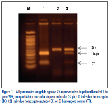
PURPOSE: to evaluate the frequency of VDR gene polymorphism Fok1 in infertile women with endometriosis and Control and its relation to the disease. METHODS: a case-control study that included 147 infertile women with endometriosis and 154 fertile women without endometriosis as Control. Fok1 polymorphism (rs10735810, T2C), which promotes a T/C exchange in exon 2 of the VDR gene, was identified by the polymerase chain reaction-restriction fragment length polymorphism (PCR-RFLP), that involves the combination of amplification by PCR and digestion with restriction endonuclease. The χ2 test was used to compare allele and genotype frequencies between groups. All p-values were two-tailed and a p-value < 0.05 was considered statistically significant. RESULTS: the TT, TC and CC genotype frequencies of VDR Fok1 polymorphism were 44.2%, 46.9% and 8.9% in infertile women with endometriosis and 41.6%, 50% and 8.4% in the Control Group. No significant difference was found (p=0.8), even when the patients were subdivided according to the stage of endometriosis (p=0.3 for minimal and mild endometriosis and p=0.2 for moderate and severe endometriosis). Alleles T and C were present, respectively, in 67.6% and 32.3% of infertile women with endometriosis (p=0.8), in 63.5% and 36.5% of women with minimal/mild endometriosis (p=0.5), in 72.5% and 27.5% of women with moderate/severe endometriosis (p=0.2), and in 66.6% and 33.4% of the Control Group. No statistically significant difference was found among any groups and the Control. CONCLUSION: the results suggest that VDR gene polymorphism Fok1 does not confer genetic susceptibility to endometriosis-associated infertility in the Brazilian population.
Summary
Rev Bras Ginecol Obstet. 2011;33(2):65-69
DOI 10.1590/S0100-72032011000200002
PURPOSE: to evaluate the frequency of VDR gene polymorphism Fok1 in infertile women with endometriosis and Control and its relation to the disease. METHODS: a case-control study that included 147 infertile women with endometriosis and 154 fertile women without endometriosis as Control. Fok1 polymorphism (rs10735810, T2C), which promotes a T/C exchange in exon 2 of the VDR gene, was identified by the polymerase chain reaction-restriction fragment length polymorphism (PCR-RFLP), that involves the combination of amplification by PCR and digestion with restriction endonuclease. The χ2 test was used to compare allele and genotype frequencies between groups. All p-values were two-tailed and a p-value < 0.05 was considered statistically significant. RESULTS: the TT, TC and CC genotype frequencies of VDR Fok1 polymorphism were 44.2%, 46.9% and 8.9% in infertile women with endometriosis and 41.6%, 50% and 8.4% in the Control Group. No significant difference was found (p=0.8), even when the patients were subdivided according to the stage of endometriosis (p=0.3 for minimal and mild endometriosis and p=0.2 for moderate and severe endometriosis). Alleles T and C were present, respectively, in 67.6% and 32.3% of infertile women with endometriosis (p=0.8), in 63.5% and 36.5% of women with minimal/mild endometriosis (p=0.5), in 72.5% and 27.5% of women with moderate/severe endometriosis (p=0.2), and in 66.6% and 33.4% of the Control Group. No statistically significant difference was found among any groups and the Control. CONCLUSION: the results suggest that VDR gene polymorphism Fok1 does not confer genetic susceptibility to endometriosis-associated infertility in the Brazilian population.
Summary
Rev Bras Ginecol Obstet. 2010;32(10):476-485
DOI 10.1590/S0100-72032010001000002
PURPOSE: to analyze the characteristics of viral infection and the risk factors for high-grade squamous intraepithelial lesion and cervical carcinoma in women with cervical HPV infection. METHODS: a case-control study was conducted on women with cervical HPV at a Gynecology reference service enrolled at the Public Health System, located in Recife, Northeastern Brazil. The groups of cases (72 women with high-grade squamous intraepithelial lesion or cervical cancer) and controls (176 women with normal Pap smear or benign alterations) were investigated for six viral genotypes (HPV 16, 18, 31, 33, 6, 11) in ecto- and endocervical material using MY09/MY11 primers. The independent variables were ranked in three levels of determination: distal (sociodemographic), intermediate (behavioral) and proximal (previous Pap smear). The homogeneity of proportions was tested (χ2), unadjusted Odds Ratios (OR) were obtained and hierarchical logistic regression was applied to the final model, with adjustment of the effect of each variable to the outcome based on the variables in the same and previous levels of causality. RESULTS: the viral genotype of cervical infection was identified in 76.6% of the 248 women participating in the study. High-risk HPV genotypes (83.4% of cases and 67.1% of controls) were predominant, especially HPV 16 and 31. The distal risk factors identified were: living in a rural area (OR=2.71, 95%CI: 1.18-6.23), less than three years of study (OR=3.97, 95%CI: 2.09-7.54) and family income below two minimum wages (OR=3.30, 95%CI: 1.04-10.51); intermediate: four or more pregnancies (OR=2.00, 95%CI: 1.06-3.76); and proximal: absence of a previous Pap smear (OR=9.74, 95%CI: 2.48-38.28). CONCLUSIONS: genotypes 16 and 31 of cervical HPV infection are predominant among women assisted by the Public Health System in Northeastern Brazil. Socioeconomic and reproductive factors, as well as the absence of cytological screening, represent risk factors for the progression of infection to high-grade squamous intraepithelial lesion and cervical cancer.
Summary
Rev Bras Ginecol Obstet. 2009;31(12):609-614
DOI 10.1590/S0100-72032009001200006
PURPOSE: to describe the genetic diversity of HIV-1 isolates from serum positive women followed up at a reference center. METHODS: transversal study, including 96 women with two ELISA serological tests and a Western Blot confirmatory test. The viral charge was determined by the b-DNA kit, and the counting of T CD4 and T CD8 lymphocytes, by the Excalibur flow cytometry, from the samples of peripheral blood. The extraction and purification of pro-viral DNA was performed by the polymerase (PCR) chain reaction, using the QIAamp Blood kit (Qiagen Inc., Chatsworth, CA, U.S.A.). Sequencing of the pol region was done in 52 isolates with the 3100 Genetic Analyzer (Applied Biosystems Inc., Foster City, CA), and the genotyping was assessed by the Rega Subtyping Tool. The resistance pattern to anti-retrovirals (ARV) was inferred by the algorithm from the Stanford HIV Resistance data bank. Participants' clinical stages were defined as A, B or C, according to the criteria established by the Center for Diseases Control (CDC). For statistical analysis, the χ2 test was used for the categorical variables and the Student's t test, for the numerical variables. RESULTS: The average age of the sample, the disease and treatment average duration were respectively: 33.7 years old, 3.8 and 2.5 years. The viral charge average was log10 2.3 copies/mL; the T CD4 e T CD8 lymphocytes, 494.9 cells/µL and 1126.4 cells/µL. Concerning the clinical stage, 30 women were in stage A, 47 in B and 19 in C. Sequencing from the 52 isolates found 33 of B subtype, 4 of F, 1 of C and 14 of BF recombinant. The analysis of resistance to ARV has shown 39 (75.0%) susceptible isolates, 13 (25.0%) resistant to reversal transcriptase inhibitors (RTIN), and 3 (5.7%) resistant to protease inhibitor (PI). CONCLUSIONS: There has been a large variety of HIV-1 and a high percentage of isolates resistant to ARV in the studied sample.
Search
Search in:
breast (42) breast cancer (42) breast neoplasms (95) Cesarean section (72) endometriosis (66) infertility (56) Maternal mortality (43) menopause (82) obesity (58) postpartum period (40) pregnancy (225) Pregnancy complications (99) Prenatal care (68) prenatal diagnosis (50) Prevalence (41) Quality of life (51) risk factors (94) ultrasonography (79) urinary incontinence (40) women's health (48)