Summary
Rev Bras Ginecol Obstet. 2006;28(2):75-80
DOI 10.1590/S0100-72032006000200002
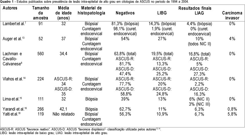
PURPOSE: to determine the prevalence of high-grade squamous intraepithelial lesions (HSIL) and cancer in women with cytological diagnosis of persistent ASCUS (atypical squamous cells of undetermined significance) for 6 months in the last 7 years. We also assessed if age could be a predictive factor for presence of HSIL/cancer in this group. METHODS: we included 215 cases of non-pregnant and HIV-seronegative women with cytological diagnosis of persistent ASCUS (unespecific) with at least 6 months of interval between smears. This cytological diagnosis was compared to histological diagnosis obtained by biopsy (large loop excision of the transformation zone) or cone biopsies, and considered negative when colposcopy was satisfactory without lesions or, when unsatisfactory, no lesion was detected after at least one cytological and colposcopic follow-up. RESULTS: among the 215 cases, 49.3% had negative results (CI 95%: 42.6-55.9). The prevalence of histological confirmed low-grade squamous intraepithelial lesion was 38.6% (CI 95%: 32.1- 45.1) and HSIL was 10.7% (CI 95%: 6.5-14.8). Cases of cancer were found in 1.4% of patients (CI 95%: 0-2.9). We could not find a significant difference between the prevalence of HSIL/cancer according to age group using the cutoff point of 35 years. CONCLUSION: HSIL/cancer prevalence observed in this study has shown the risk of finding this kind of lesions in about 12% of women assisted in our public health system with two cytological diagnosis of ASCUS. A higher probability of HSIL/cancer in the different age groups was not found but this result was limited by our small sample size.
Summary
Rev Bras Ginecol Obstet. 2006;28(2):75-80
DOI 10.1590/S0100-72032006000200002
PURPOSE: to determine the prevalence of high-grade squamous intraepithelial lesions (HSIL) and cancer in women with cytological diagnosis of persistent ASCUS (atypical squamous cells of undetermined significance) for 6 months in the last 7 years. We also assessed if age could be a predictive factor for presence of HSIL/cancer in this group. METHODS: we included 215 cases of non-pregnant and HIV-seronegative women with cytological diagnosis of persistent ASCUS (unespecific) with at least 6 months of interval between smears. This cytological diagnosis was compared to histological diagnosis obtained by biopsy (large loop excision of the transformation zone) or cone biopsies, and considered negative when colposcopy was satisfactory without lesions or, when unsatisfactory, no lesion was detected after at least one cytological and colposcopic follow-up. RESULTS: among the 215 cases, 49.3% had negative results (CI 95%: 42.6-55.9). The prevalence of histological confirmed low-grade squamous intraepithelial lesion was 38.6% (CI 95%: 32.1- 45.1) and HSIL was 10.7% (CI 95%: 6.5-14.8). Cases of cancer were found in 1.4% of patients (CI 95%: 0-2.9). We could not find a significant difference between the prevalence of HSIL/cancer according to age group using the cutoff point of 35 years. CONCLUSION: HSIL/cancer prevalence observed in this study has shown the risk of finding this kind of lesions in about 12% of women assisted in our public health system with two cytological diagnosis of ASCUS. A higher probability of HSIL/cancer in the different age groups was not found but this result was limited by our small sample size.
Summary
Rev Bras Ginecol Obstet. 2006;28(1):24-31
DOI 10.1590/S0100-72032006000100005
PURPOSE: to verify the coverage and factors associated with Papanicolaou (Pap) testing in Londrina (PR), Brazil. METHODS: this is a cross-sectional study, carried out in 2004, in microareas of five Basic Health Units (BHU) of Londrina. One or two microareas from each BHU were selected and a list of all women aged 20-59 years resident in these places, was made through search in the Basic Attention Information System, the women being then visited and interviewed. Those with a Pap test in the last three years were considered as having an updated examination, and the remaining as delayed. The association of some factors with the examination situation was investigated. Data analysis was performed using Epi-Info 6.04d. RESULTS: Pap smear coverage among the 513 participants of the study was 80.7%, ranging from 71.5% to 88.4%. Delay in taking the test was higher (p<0.05) among women who worked only at home (22.4% as compared with 14.3% among those who worked outside), and among those who belonged to D/E social classes (24.9%) as compared to C (17.5%) and A/B (8.3%) classes. The proportion who ignored the next test date was higher (p<0.01) among those who had the last Papanicolaou testing at a BHU (14.7%), as compared to those who had been attended privately or by a health insurance company (5.8%). CONCLUSION: the coverage of Pap smear in the studied areas can be considered satisfactory, although there is a need of improving compliance with Pap test, mainly among women who are the poorest and who work only at home.
Summary
Rev Bras Ginecol Obstet. 2005;27(12):744-749
DOI 10.1590/S0100-72032005001200007
PURPOSE: to assess to what extent the surgical staging differs from the clinical staging among cases of advanced uterine cervix carcinoma, and also to assess the percentage of cases with positive para-aortic ganglia in this group of patients. METHODS: this is a descriptive prospective study in which 36 patients with histological diagnosis of uterine cervix carcinoma considered locally advanced were included (stages IB2, IIB, IIIA and B, and IVA). The cases were submitted to clinical staging, according to FIGO criteria. All patients were to be treated with neoadjuvant chemotherapy. Age ranged from 40 to 73 years, with a mean of 56.2±7.9. The procedure started with pelvic lymphadenectomy followed by para-aortic lymphadenectomy, in case the pelvic lymph nodes were positive on surgical examination. Examination of the abdominal cavity and lymphadenectomy were done either through laparotomy or laparoscopy, chosen at random. In each case, the clinical staging was compared to the surgical staging, considered the gold standard. RESULTS: in the clinical staging (CS), 7 cases were classified as IB2 (tumors larger than 4 cm), 22 cases as CSII and 7 cases as CSIII. The surgical assessment changed the clinical staging as follows: the stage was decreased in six cases, and increased in 13. There was agreement only in 18 cases (50%). The para-aortic lymph nodes were affected in six cases. CONCLUSIONS: clinical staging of locally advanced uterine cervix carcinoma is incorrect in most of the cases. Such inconsistency may lead to excessive treatment in some cases, but about one fourth of the patients with positive para-aortic ganglia would not be adequately treated with the current standard treatment radiotherapy with chemosensitization, which aims at the local regional control of the pelvic disease.
Summary
Rev Bras Ginecol Obstet. 2005;27(11):656-660
DOI 10.1590/S0100-72032005001100004
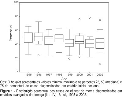
PURPOSE: to analyze time trends in the clinical staging at the moment of diagnosis in patients with breast and cervix cancer based on data produced by the Brazilian Public Health System (SUS). METHODS: in the first part of this study we identified the published documents describing clinical staging of patients at the moment of diagnosis. Considering their scarcity and poor representativity we conducted the second part of this study through an active search for information. A form was sent via regular mail to all cancer centers in the country (n=173) requesting information about the tumor site and stage at diagnosis by year, in the period of 1995-2002. The statistical analysis was performed using the "R" statistical package. The results are reported as percentage and boxplots. RESULTS: in the first part of the study (1990-1994) we described data from 18 hospitals concerning 7,458 patients with breast cancer and 7,216 patients with cervix cancer. The median of the percentage of cancers diagnosed at an advanced stage (stages III or IV) was 52.6 and 56.8%, respectively. In the second part of the study (1995-2002) data were collected from 89 cancer hospitals and 7 chemotherapy or radiotherapy clinics. There was a total of 43,442 cases of breast cancer and 29,263 of cervix cancer. The response rate based on the potential contact list was 55%. The median percentage of patients in advanced stage was 45.3% for breast cancer and 42.5% for cervix cancer. CONCLUSIONS: few studies have examined the time trends in staging of cancer at diagnosis in Brazilian hospitals. Data obtained from Hospital Cancer Registries showed that in the last decade there was a reduction in the percentage of cervix and breast cancer at the advanced stage. This reduction can be due to an improvement in early detection of these cancers.
Summary
Rev Bras Ginecol Obstet. 2005;27(11):656-660
DOI 10.1590/S0100-72032005001100004
PURPOSE: to analyze time trends in the clinical staging at the moment of diagnosis in patients with breast and cervix cancer based on data produced by the Brazilian Public Health System (SUS). METHODS: in the first part of this study we identified the published documents describing clinical staging of patients at the moment of diagnosis. Considering their scarcity and poor representativity we conducted the second part of this study through an active search for information. A form was sent via regular mail to all cancer centers in the country (n=173) requesting information about the tumor site and stage at diagnosis by year, in the period of 1995-2002. The statistical analysis was performed using the "R" statistical package. The results are reported as percentage and boxplots. RESULTS: in the first part of the study (1990-1994) we described data from 18 hospitals concerning 7,458 patients with breast cancer and 7,216 patients with cervix cancer. The median of the percentage of cancers diagnosed at an advanced stage (stages III or IV) was 52.6 and 56.8%, respectively. In the second part of the study (1995-2002) data were collected from 89 cancer hospitals and 7 chemotherapy or radiotherapy clinics. There was a total of 43,442 cases of breast cancer and 29,263 of cervix cancer. The response rate based on the potential contact list was 55%. The median percentage of patients in advanced stage was 45.3% for breast cancer and 42.5% for cervix cancer. CONCLUSIONS: few studies have examined the time trends in staging of cancer at diagnosis in Brazilian hospitals. Data obtained from Hospital Cancer Registries showed that in the last decade there was a reduction in the percentage of cervix and breast cancer at the advanced stage. This reduction can be due to an improvement in early detection of these cancers.
Summary
Rev Bras Ginecol Obstet. 2005;27(10):607-612
DOI 10.1590/S0100-72032005001000007
PURPOSE: to test the hypothesis that gene TP53 codon 72 polymorphism is a risk factor for premalignant and malignant cervical lesions associated or not with human papillomavirus (HPV). METHODS: uterine cervical samples were collected for HPV DNA and TP53 codon 72 polymorphism tests from 155 patients who underwent cervical biopsy. Three groups were formed according to histological diagnosis: low-grade squamous intraepithelial lesion (LSIL), high-grade squamous intraepithelial lesion (HSIL) and cervical carcinoma. Subjects without cytological and histological displasic changes were considered controls. To verify the association between the gene TP53 codon 72 polymorphism and the groups, the chi2 test was applied. Confidence interval was considered significant at 95% (alpha=0.05). RESULTS: forty subjects were found to present cervical carcinoma, 18 had HSIL, 24 had LSIL and 73 were grouped as controls. The genotype Arg/Arg p53 was found in 60% of the patients with cancer, in 50.0% of the cases with HSIL, 45.8% with LSIL, and in 45.2% of the controls. No significant differences were identified in the frequencies of p53 genotype between all groups, independently of the presence of HPV (chi2: 3.7; p=0.716). CONCLUSIONS: our data do not support hypothesis that the gene TP53 codon 72 polymorphism is important for the development of pre-malignant and malignant cervical lesions associated or not with HPV.
Summary
Rev Bras Ginecol Obstet. 2005;27(8):485-492
DOI 10.1590/S0100-72032005000800009
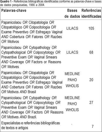
PURPOSE: to present an overview of the coverage of the Pap smear in Brazil, emphasizing the determinant factors associated with failure of women to submit to the test. METHODS: the literature was reviewed using the LILACS (Latin-American and Caribbean Literature in Sciences of the Health), MEDLINE - 1966 to 2004 (International Literature in Sciences of the Health), PAHO (Collection of the Library of the Pan-American Organization of Health), and WHOLIS (System of Information of the Library of OMS) databases. The review was enlarged through the search of bibliographical references of relevant studies, request for published and unpublished studies by specialists, and other sources. Articles that fulfilled the following criteria were selected: to be a cross-sectional study, carried out in Brazil, including information about periodicity of the Pap test (some time in life or in the last three years) and/or containing information about factors associated with failure of women to submit to the test. Duplicates and articles without summary were excluded. A total of 13 articles fulfilling these criteria were selected. RESULTS: there are few studies on the coverage of Pap smear in Brazil. Most of them are concentrated in the big cities of the South and Southeast regions of the country. Besides the shortage, little methodological standardization exists in relation to the sampling and profile of the investigated women, which turns difficult the comparison among them. These methodological differences must have contributed to the great variability found in the coverage. However, in spite of all of the problems, a trend of time series increase is observed in the percentage of women who had at least one Pap smear in life. The two studies accomplished in the eighties showed coverage of 55.0 and 68.9% some time in life, while a household survey carried out in 2002 and 2003 presented values that varied from 73.4 to 92.9%; however, two studies of national inclusion presented estimates below 70.0% in the last three years. On the other hand, some variables were associated with the women's failure to submit to the Pap smear: low socioeconomic level, low education, low family income, and to belong to the younger age groups. CONCLUSION: the data here presented point to regional inequalities in the coverage of the Pap smear in the Brazilian female population and to the need of intervention targeted to those factors associated with women's failure to submit to the Pap smear.
Summary
Rev Bras Ginecol Obstet. 2005;27(8):485-492
DOI 10.1590/S0100-72032005000800009
PURPOSE: to present an overview of the coverage of the Pap smear in Brazil, emphasizing the determinant factors associated with failure of women to submit to the test. METHODS: the literature was reviewed using the LILACS (Latin-American and Caribbean Literature in Sciences of the Health), MEDLINE - 1966 to 2004 (International Literature in Sciences of the Health), PAHO (Collection of the Library of the Pan-American Organization of Health), and WHOLIS (System of Information of the Library of OMS) databases. The review was enlarged through the search of bibliographical references of relevant studies, request for published and unpublished studies by specialists, and other sources. Articles that fulfilled the following criteria were selected: to be a cross-sectional study, carried out in Brazil, including information about periodicity of the Pap test (some time in life or in the last three years) and/or containing information about factors associated with failure of women to submit to the test. Duplicates and articles without summary were excluded. A total of 13 articles fulfilling these criteria were selected. RESULTS: there are few studies on the coverage of Pap smear in Brazil. Most of them are concentrated in the big cities of the South and Southeast regions of the country. Besides the shortage, little methodological standardization exists in relation to the sampling and profile of the investigated women, which turns difficult the comparison among them. These methodological differences must have contributed to the great variability found in the coverage. However, in spite of all of the problems, a trend of time series increase is observed in the percentage of women who had at least one Pap smear in life. The two studies accomplished in the eighties showed coverage of 55.0 and 68.9% some time in life, while a household survey carried out in 2002 and 2003 presented values that varied from 73.4 to 92.9%; however, two studies of national inclusion presented estimates below 70.0% in the last three years. On the other hand, some variables were associated with the women's failure to submit to the Pap smear: low socioeconomic level, low education, low family income, and to belong to the younger age groups. CONCLUSION: the data here presented point to regional inequalities in the coverage of the Pap smear in the Brazilian female population and to the need of intervention targeted to those factors associated with women's failure to submit to the Pap smear.
Summary
Rev Bras Ginecol Obstet. 2005;27(5):243-247
DOI 10.1590/S0100-72032005000500003
PURPOSE: to evaluate the association between p53 and Ki-67 expression in the tumor and clinicopathological features in patients with carcinoma of the cervix. METHODS: samples were taken from the tumor of 36 patients with stage IB (FIGO) cervical carcinoma submitted to radical hysterectomy. Tissue samples were taken from the tumor, fixed in formalin and embedded in paraffin. The specimens were analyzed by histopathology (hematoxylin and eosin) and immunohistochemically evaluated using monoclonal antibodies for p53 and Ki-67. Data were analyzed statistically by the chi2 test to evaluate eventual differences between the groups. RESULTS: the age of the patients ranged from 27 to 73 years (48.7±10.4 years). Clinical stage (FIGO) was IB1 in 27 cases (75%) and IB2 in 9 cases (25%). A positive tumoral expression of the p53 protein was found in half of the cases. In relation to the Ki-67 expression, a high cell proliferation index was shown in 73.3% of the cases. There was no association between tumoral p53 and Ki-67 expression with age (p=0.091 and 0.900), clinical stage (p=0.054 and 0.667), histological classification (p=0.674 and 0.674), grade of differentiation (p=0.070 and 0.282), presence of lymphatic vascular space invasion (p=0.248 and 0.667), parametrial involvement (p=0.729 and 0.763) and pelvic lymph node metastasis (p=0.729 and 0.636, respectively). CONCLUSIONS: tumoral expression of p53 and Ki-67 was not associated with the clinicopathological features in patients with stage IB carcinoma of the cervix.
Search
Search in:
breast (42) breast cancer (42) breast neoplasms (95) Cesarean section (72) endometriosis (66) infertility (56) Maternal mortality (43) menopause (82) obesity (58) postpartum period (40) pregnancy (225) Pregnancy complications (99) Prenatal care (68) prenatal diagnosis (50) Prevalence (41) Quality of life (51) risk factors (94) ultrasonography (79) urinary incontinence (40) women's health (48)