Summary
Revista Brasileira de Ginecologia e Obstetrícia. 2002;24(2):101-106
DOI 10.1590/S0100-72032002000200005
Purpose: to analyze the prevalence of gonorrhea, Chlamydia, syphilis and HIV among patients attending a family planning clinic regarding presence of STD symptoms and risk behaviors. Methods: women between the ages of 18 and 30 years who attended a public family planning clinic in Brazil were tested for gonorrhea and Chlamydia using the urine-based DNA amplification test (LCR, Abbott), and a blood test for syphilis (VDRL) and HIV. All participants were asked questions about their health care seeking behavior, the presence of STD symptoms, and about the STD risk behaviors. Results: Chlamydia was found in 11.4%, syphilis in 2%, gonorrhea in 0.5% and HIV was confirmed positive in 3%. Approximately 61% of the women who were infected with Chlamydia had no symptoms. Women who never used condoms had much higher risks for STD than women who used them always or most of the time. Although not statistically significant, there was a trend for women who never used any contraceptive to have a higher risk for STD than women who used some method of contraception (p=0.09). However, when examining separately each contraceptive, none of them alone offered protection against STD. Very few women reported problems related to the use of alcohol or illegal drugs. But among those who did report such use, the risk for STD was very high, particularly regarding marijuana use. Conclusions: the most significant findings in our study were the high STD rates among a population of women generally reporting low-risk health behaviors. Based upon our findings it is crucial to offer STD/HIV screening to all women under 30 years who visit public family planning clinics. Without screening all women, more than half of the infected women will never be identified or treated. Given the new sensitive and specific technology available to screen for Chlamydia, gonorrhea, and HIV, and the ease of collecting urine specimens for diagnosis, more efforts should be directed to surveillance of populations at risk, so that current clinical practice may reflect the true risk of the populations.
Summary
Revista Brasileira de Ginecologia e Obstetrícia. 2002;24(2):107-112
DOI 10.1590/S0100-72032002000200006
Purpose: to analyze the effects of prenatal and perinatal complications and the neurological development of surviving twins when the other had died in utero. Methods: fourteen cases of twin pregnancies where one of the twins had died during the pregnancy were analyzed. These patients gave birth between 1988 and 1994 and were subsequently followed-up by the Department of Obstetrics, Pathology Division, at the Hospital das Clínicas, Faculty of Medicine of Ribeirão Preto, University of São Paulo. Data from prenatal and perinatal records as well as findings from the deceased twins' autopsies were analyzed. In 1996, requests were made for the children to have a neurological examination as part of the study. The examination included developmental assessment and pathological signs in the motor, sensory and sensitivy areas and superior cortical functions such as praxis and agnosia. Results: ten of the fourteen contacted subjects complied with the request for neurological examination. Of the ten examined children only one had abnormal neurological findings, presenting a light degree of spastic paresis of the left leg. The pregnancy evaluation showed five cases of monochorionic placenta and one case of monoamnionic pregnancy; six of the fourteen cases reached full-term. In six cases (42.8%) one of the fetus died during the second trimester and in the other they died during the third trimester. Only one newborn, who had Apgar 0 at the first minute, developed neurological sequelae. Conclusion: the neurological problem of one fetus may be a consequence of the perinatal complications that this fetus developed. The other newborns did not develop sequelae, possibly because of the conservatory management, trying to make the pregnancy reach 32 weeks or more, thus decreasing the complications of preterm delivery.
Summary
Revista Brasileira de Ginecologia e Obstetrícia. 2002;24(2):113-120
DOI 10.1590/S0100-72032002000200007
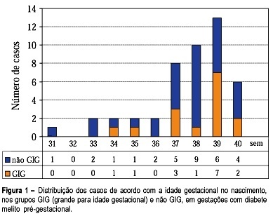
Purpose: to study fetal surveillance examinations in pregnancies complicated by pregestational diabetes mellitus, and to correlate them with large for gestational age (LGA) newborns. Methods: Between March 1999 and June 2001, 46 singleton pregnancies with pregestational diabetes mellitus without fetal anomalies were followed prospectively. From the 28th gestational week on, the following examinations were performed weekly: fetal biophysical profile, amniotic fluid index (AFI), and dopplervelocimetry of umbilical and middle cerebral arteries. The newborns with birthweight above the 90th percentile according to local standard values were characterized as LGA infants. Fisher's exact test and Student's t test were used for statistical analysis. Results: The mean gestational age at delivery was 37.6 weeks and 15 (32.6%) newborns were LGA. LGA fetuses showed significant increase in the AFI mean performed in the 32nd (16.5 cm, p=0.02), 33rd (16.7 cm, p=0.03), 34th (17.0 cm, p=0.02), 35th (17.9 cm, p=0.000), 36th (15.8 cm, p=0.03) and 37th (17.5 cm, p=0.003) weeks. Non-LGA fetuses presented the following mean AFI values: 13.5cm (32nd week), 13.1cm (33th week), 13.4 (34th week), 12.8 (35th week), 12.5 (36th week) and 12.8cm (37th week). AFI values equal to or above 18.0 cm were associated with the occurrence of LGA infants, when detected at the following gestational ages: 34th (60%, p=0.03), 35th (71.4%, p=0.01), 36th (80%, p=0.02) and 37th (66.7%, p=0.04) week. Non-LGA infants presented the following proportion of AFI values equal to or above 18.0 cm: 40.0% (34th week), 28.6% (35th week), 20.0% (36th week), and 33.3% (37th week). Conclusions: abnormal increase in AFI, mainly with values equal to or above 18.0 cm, is related to LGA infants at delivery. The maternal treatment should be adjusted to achieve the best result for maternal-fetal control, according to the AFI values during pregnancy.

Summary
Revista Brasileira de Ginecologia e Obstetrícia. 2002;24(2):121-127
DOI 10.1590/S0100-72032002000200008
Purpose: to estimate the performance of ultrasound to detect gestations at risk for fetal chromosomal abnormalities. Methods: four hundred and thirty-six patients selected for the study had undergone ultrasound examination and fetal karyotyping, between March 1993 and March 1998. Two hundred and seventy-seven patients had fetal karyotype for fetal malformation detected on ultrasound and 158 for parental anxiety with normal ultrasound examination. Ultrasound sensitivity and specificity were calculated using fetal karyotype as gold standard. The relative risk for each chromosomal abnormality was calculated according to the altered system on ultrasound examination and the risks of the presence of one or more abnormalities on ultrasound, using the Epi-Info 6.0 software package for statistical analysis. Results: the relative risks for chromosomal abnormalities were 89 for face malformations, 53 for abdominal wall and cardiovascular, 49.6 for neck, 44.6 for extremities, 42.4 for lung, 32.7 for gastrointestinal tract, 27.4 for central nervous system and 23.0 for urinary tract malformations. The relative risk for fetal chromosomal anomalies for genital, thorax, spine and muscle and/or skeletal malformations was not appropriate for calculation because they occurred at very low frequencies. An isolated malformation detected by ultrasound is associated with a 7.8 times higher relative risk for chromosomal anomalies than none, and associated morphologic malformations have a 33.8 times higher relative risk for chromosomal abnormalities. Conclusion: ultrasound has good performance to detect gestations at risk for chromosomal abnormalities.
Summary
Revista Brasileira de Ginecologia e Obstetrícia. 2002;24(2):81-86
DOI 10.1590/S0100-72032002000200002
Purpose: to evaluate the predictive capacity of the sentinel lymph node (SLN) in relation to the axillary lymph node status in patients with initial invasive breast carcinoma submitted or not to neoadjuvant chemotherapy. Method: a prospective study was performed in 112 patients divided into two groups. The first group comprised 70 patients who had not received previous chemotherapy (Group I) and the second consisted of 42 patients who were submitted to neoadjuvant chemotherapy in three cycles of AC (adriamycin + cyclophosphamide) (Group II). Regarding chemotherapy, we observed partial response >50% in 21 patients, being complete in three of them, and <50% in 19 patients; in two patients progression of the disease occurred. A peritumoral injection of 99mTc dextran was applied with the help of stereotaxy in 29 patients with nonpalpable tumors, 16 of Group I and 13 of Group II. The radioactive accumulation shown by scintigraphy guided the biopsy of the axillary SLN with the help of a probe. The anatomopathologic study of SLN was based initially on a single section. When the LSN was free, it was submitted to serial sections at 50 mum intervals, stained with HE. Results: SLN was identified in 108 patients. No identification has been obtained in four patients, all with nonpalpable lesions (3 patients of Group I and 1 of Group II). The method's accuracy in predicting the axillary lymph node status was 100% in patients who did not receive neoadjuvant chemotherapy and 93% in those to whom this kind of treatment was administered. This difference proved to be statistically significant. Conclusions: the present study allowed us to conclude that in all patients who did not receive previous chemotherapy treatment, the SLN study was effective in predicting the axillary lymph node status. The high rate of false-negative results in the group of patients submitted to neoadjuvant chemotherapy seems to invalidate the use of SLN study these patients.
Summary
Revista Brasileira de Ginecologia e Obstetrícia. 2002;24(2):87-91
DOI 10.1590/S0100-72032002000200003
Purpose: to analyze the prevalence of urogynecological symptoms and their relationship with final urodynamic diagnosis, and to compare the clinical sign of stress urinary incontinence with urodynamic diagnosis. Methods: a total of 114 patients were included in a retrospective study from June 2000 to January 2001. All patients were evaluated through medical interview, physical examination and urodynamic study. They were classified according to clinical symptom, presence of clinical sign of urine loss and urodynamic study. The data analysis was performed using a test to determine sensitivity, specificity, and positive and negative predictive values. Results: the mean age was 51 years (19-80), 61 patients (53.5%) were in menacme and 53 (46.5%) in postmenopausal stage. Ten (18.8%) were using hormone replacement therapy and 25 (21.9%) had been submitted to surgery for incontinence. The isolated clinical symptom of urine loss was reported in 41 (36.0%) patients, the isolated urgency/urgency-incontinence in 13 (11.4%) and mixed symptoms in 60 (52.6%). In the urodynamic study, of all patients with symptom of isolated urine loss, 34 (83%) had stress urinary incontinence (SUI), no patient had detrusor instability (DI), 2 (4.9%) had mixed incontinence (MI) and 5 (12.1%) had a normal result. Of all patients with isolated urgency/urgency-incontinence, in the urodynamic study, none had SUI, 5 (38.5%) had ID, 1 (7.7%) had MI and 7 (53.8%) had a normal result. Of the patients with mixed symptoms, we identified, on the urodynamic evaluation, 25 (41.6%) who had SUI, 10 (16.7%) ID, 10 (16.7%) MI and 15 (25.0%) a normal result. The clinical sign of urine loss was identified in 50 (43.9%) patients. A total of 35 (70%) had SUI on urodynamic study, 6 (12%) had SUI and another diagnosis and 9 (18%) did not have SUI. Urine loss was absent in 64 (56.1%) women. Of those 23 (35.9%) had SUI on urodynamic study, 7 (11%) had SUI and another diagnosis and 34 (53.1%) did not have SUI. Conclusions: clinical history and physical examination are important in the management of urinary incontinence, although they should not be used as the only diagnostic method. Objective tests are available and should be used together with clinical data.
Summary
Revista Brasileira de Ginecologia e Obstetrícia. 2002;24(2):93-99
DOI 10.1590/S0100-72032002000200004
Purpose: to evaluate the correlation between the laparoscopic aspects and the stromal histologic findings of peritoneal endometriosis in order to understand the evolutive theory of endometriosis. Methods: sixty-seven women were submitted to laparoscopy for pelvic pain, infertility, ovarian tumor and other pathologies. A peritoneal biopsy was taken from the typical (puckered black) and atypical endometriotic implants. The different aspects of endometriosis were classified as follows: red lesions (Group V), black lesions (Group N) and white lesions (Group B). The histological sections were examined according to a standardized protocol. The histologic parameters used were: depth of the lesion, presence of hemosiderin, vascularization of the stroma and fibrotic tissue in stroma. Results: regarding lesion depth, there were significant differences between the groups. Red lesions were located consistently on the surface of the peritoneum (100%) and black lesions were superficial in 55.6%, intermediate in 38.9% and deep in 5.5%. White lesions were superficial in 28%, intermediate in 68% and deep in 4%. The presence of hemosiderin showed equivalent results in the 3 groups. The large stromal vascularization was present in the red lesions (60%), which a statistically significant difference compared to the other groups. Fibrotic tissue was present in 70.6% of the white lesions (group B), a fact that was significantly different when compared to groups V and N. Conclusion: the parameters analyzed in this study confirmed the importance of the evolutive theory of endometriosis.