Summary
Revista Brasileira de Ginecologia e Obstetrícia. 2011;33(7):137-142
DOI 10.1590/S0100-72032011000700004
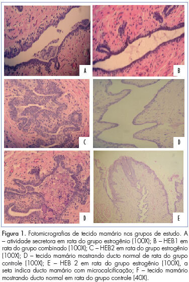
PURPOSE: To evaluate the efect of trimegestone on the histological changes of the mammary tissue of castrated rats. METHODS: Forty-five virgin female Wistar rats were used after oophorectomy. Sixty days after surgery, with hypoestrogenisms confirmed, the experimental rats were randomly assigned to three groups of 15 animals each, when then the specific treatment for each group was started. The control group (C) and experimental groups 1 and 2 respectively received 0.9% saline solution, 17-beta-estradiol and 17-beta-estradiol in combination with trimegestone for 60 consecutive days. After the end of treatment , the inguinal mammary glands were removed, stained with hematoxylin and eosin (HE) for morphometry and examined by immunohistochemistry for the quantification of anti-PCNA antibody in the mammary tissue, followed by euthanasia. The morphometric parameters evaluated were: epithelium cell-proliferation, secretor activity and mammary stroma changes. There were nine deaths during the experiment. The variables were submitted to statistical analysis adopting the 0.05 level of significance. RESULTS:Histological changes were observed in 16/36 rats, mild epithelial hyperplasia in 13/36, moderate epithelial hyperplasia in 3/36, with no cases of severe epithelial hyperplasia. Stromal fibrosis was found in 10/36 and secretory activity in 5/36 rats. All morphometric variables were significant in the estrogen group compared to control (p=0.0361), although there were no difference between the group receiving combined treatment and the controls (p=0.405). The immunohistochemical analysis showed no difference between groups. CONCLUSIONS:The hormones administered to castrated rats, i.e., 17 beta-estradiol alone or in combination with trimegestone, increased the proliferation of breast cells, but this effect appeared to be lower in the combined treatment, the same occurring regarding fibrosis of the mammary stroma.

Summary
Revista Brasileira de Ginecologia e Obstetrícia. 2011;33(7):143-149
DOI 10.1590/S0100-72032011000700005
PURPOSE: To determine the main contraceptive methods adopted by users of the public and private health sectors in the city of Aracaju (SE), Brazil, with a secondary focus on orientations for their use and reasons for interruption. METHODS: A cross-sectional study was conducted on 210 women, 110 from the public service and 100 from the private sector. The data were collected by applying a questionnaire to sexually active patients who agreed to sign a consent form. The software Statistical Package for Social Sciences (SPSS) version 15.0 was used for statistical analysis, with the ![]() test for categorical variables and the Student's t-test for independent samples. RESULTS: The overall prevalence of contraceptive use in this study was 83.3%. The main methods used in the public and private sectors, were the hormonal (41 and 24%, p=0.008) and permanent (20 and 26%, p=0.1) ones, respectively. The rate of condom use was 17.3% in the public sector and 12% in the private sector, with no significant difference (p=0.12). Medical orientation about the correct use of the method chosen and/or indicated was provided to 37.3% of users from the public sector and to 48% of users from the private sector. Discontinuation of the use of contraceptive methods was 14.5% in the public sector and 12.0% in the private sector, mainly because of side effects and the desire to become pregnant. CONCLUSIONS: The main contraceptive methods adopted by users of the public and private sectors were hormonal contraception and permanent contraception. It is important to highlights the low frequency of use of male condoms in the two groups studied.
test for categorical variables and the Student's t-test for independent samples. RESULTS: The overall prevalence of contraceptive use in this study was 83.3%. The main methods used in the public and private sectors, were the hormonal (41 and 24%, p=0.008) and permanent (20 and 26%, p=0.1) ones, respectively. The rate of condom use was 17.3% in the public sector and 12% in the private sector, with no significant difference (p=0.12). Medical orientation about the correct use of the method chosen and/or indicated was provided to 37.3% of users from the public sector and to 48% of users from the private sector. Discontinuation of the use of contraceptive methods was 14.5% in the public sector and 12.0% in the private sector, mainly because of side effects and the desire to become pregnant. CONCLUSIONS: The main contraceptive methods adopted by users of the public and private sectors were hormonal contraception and permanent contraception. It is important to highlights the low frequency of use of male condoms in the two groups studied.
Summary
Revista Brasileira de Ginecologia e Obstetrícia. 2011;33(7):150-157
DOI 10.1590/S0100-72032011000700006
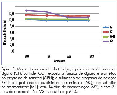
PURPOSE: the purpose of this study was to evaluate mortality, weight and body length, and the gastrocnemius muscle of the offspring of pregnant rats submitted to a swimming program associated with second-hand smoke. METHODS: twenty-four rats were divided into four groups: GF (exposed to cigarette smoke), GC (control), GFN (submitted to the swimming program and exposed to cigarette smoke), and GN (submitted to the swimming program). The mortality, weight and length of the offspring were measured at four time points. The gastrocnemius muscle of the pups was obtained for evaluation of muscle development. RESULTS: the average number of offspring was lower for GF (10.2) and GFN (10.3) and higher for GN (12.8). At birth, only GFN showed significantly lower weight (p=0.016) and length (p=0.02), whereas during lactation the groups exposed to cigarette smoke showed significantly lower weight. GFN had delayed muscle development compared to GC (p=0.03). CONCLUSIONS: Passive smoking during pregnancy and lactation negatively influenced number, weight and body length of offspring from birth to weaning and muscle development, and the swimming program positively influenced these variables at birth, although it did not provide the same benefits during lactation; and their association negatively affected these measures.

Summary
Revista Brasileira de Ginecologia e Obstetrícia. 2011;33(7):158-163
DOI 10.1590/S0100-72032011000700007
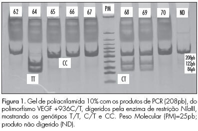
PURPOSE: To identify genetic polymorphisms of endothelial growth factor (VEGF), positions +936C/T and -2578C/A, in women with pre-eclampsia. METHODS: This was a cross-sectional study conducted on 80 women divided into two groups: pre-eclampsia and control. The sample was characterized using a pre-structured interview and data transcribed from the medical records. DNA extraction, amplification of sequences by the Polymerase Chain Reaction (PCR) with specific primers and polymorphism analysis of Restriction Fragment Length Polymorphism (RFLP) were performed to identify polymorphisms. The statistical analysis was performedin a descriptive manner and using the ![]() test. The multiple logistic regression model was used to determine the effect of polymorphisms on pre-eclampsia. RESULTS:Ahigher frequency of the T allele of theVEGF +936C/T polymorphism was observedin patients with pre-eclampsia, but with no significant difference. The presence of allele A of the VEGF -2578C/A was significantly higher in the control group. CONCLUSIONS:No significant association was observed between VEGF +936C/Tpolymorphism andpre-eclampsia. For the VEGF -2578C/A polymorphism a significant differencewas observed between thecontrol and pre-eclampsia group, with allele A being the most frequent in the control, suggesting the possibility that carriers of allele A have lower susceptibility to the development of pre-eclampsia.
test. The multiple logistic regression model was used to determine the effect of polymorphisms on pre-eclampsia. RESULTS:Ahigher frequency of the T allele of theVEGF +936C/T polymorphism was observedin patients with pre-eclampsia, but with no significant difference. The presence of allele A of the VEGF -2578C/A was significantly higher in the control group. CONCLUSIONS:No significant association was observed between VEGF +936C/Tpolymorphism andpre-eclampsia. For the VEGF -2578C/A polymorphism a significant differencewas observed between thecontrol and pre-eclampsia group, with allele A being the most frequent in the control, suggesting the possibility that carriers of allele A have lower susceptibility to the development of pre-eclampsia.

Summary
Revista Brasileira de Ginecologia e Obstetrícia. 2011;33(7):164-169
DOI 10.1590/S0100-72032011000700008
PURPOSE: to analyze the gait propulsion force and relate it to changes in the dimensions of the feet and to the influence on the quality of life of pregnant women. METHODS: two groups were studied, a control (C) one consisting of 20 non-pregnant women and a group of 13 pregnant women investigated during the three gestational trimesters (Gfirst, Gsecond, Gthird). The groups were subjected to an initial assessment; evaluation of gait propulsion force using the force platform (Bertec); measurement of foot length and width; assessment of perimetry by the figure eight method; and assessment of quality of life using the World Health Organization Quality of Life Instrument Bref (Whoqol-bref). The Mann-Whitney test was used to evaluate differences between group C and Gfirst, the Friedman test was used to determine differences between Gfirst, Gsecond and Gthird, and the Wilcoxon test was applied to significant cases. The level of significance was set at 5%. RESULTS: There was an increase in body mass (10.5 kg) and ankle edema (2.4 cm) during pregnancy. There was a decrease of gait propulsion force (10% of body mass) and an increase of mediolateral sway (10% of body mass) compared to Control Group. There was a reduced quality of life among pregnnat women, especially in the physical domain. CONCLUSIONS: Gait disorders occur during pregnancy, which can increase the risk of falls and musculoskeletal discomfort, which may affect the quality of life of pregnant wome
Summary
Revista Brasileira de Ginecologia e Obstetrícia. 2011;33(5):231-237
DOI 10.1590/S0100-72032011000500005
PURPOSE: To investigate the relationship between quality of life and spinal fracture in women aged over 60 living in Southern Brazil. METHODS: A case-control study was conducted with the application of the WHOQOL-bref questionnaire to 100 women living in the city of Chapecó (SC), aged over 60, postmenopausal, white or Caucasian, with no important cognitive impairment or a history of diseases known to affect bone metabolism, or malignant neoplasias. The population was divided into two groups depending on the presence or absence of fractures in the spine radiography. We analyzed variables related to the current and previous medical history, life habits and family history of fractures, and the domains and facets that compose the WHOQOL-bref. All participants were informed about the objectives and methodologies adopted and gave written informed consent to participate in the study. RESULTS: The mean age of the women in the fracture group was older than that of women with fractures (p<0.05). Also women with fractures tended to belong to a higher social class, to have more years of study, a higher family income, and a greater use of alcoholic drinks (p<0.05). In the evaluation of the WHOQOL-bref domains, the fracture group had the highest average in the psychological field (..=63.6± 3.0) and the lowest in the environment field (..=9.3±58.8). In the group without fracture, the highest average also occurred in the psychological domain (..=67.2± 9.3) and the lowest in the field of social relations (..=57.5±7.7). Statistical analysis showed no significant correlation between the averages of the facets that make up the areas between the groups with and without fractures. CONCLUSIONS: This study suggests that there is no impairment of quality of life among older women with vertebral fractures, but the relation between QL and time of occurrence and severity of the fractures should be better evaluated. Both groups had higher scores in the psychological domain, showing that the respondents rely on personal beliefs, spirituality and religion, accept their physical appearance while maintaining self-esteem and the ability to think, to learn and to concentrate despite the presence of this disease. There was no statistically significant difference between groups or between domains in the same group.
Summary
Revista Brasileira de Ginecologia e Obstetrícia. 2011;33(6):271-275
DOI 10.1590/S0100-72032011000600002
PURPOSE: to investigate the association between gene polymorphism of the progesterone receptor (PROGINS) and the risk of premature birth. METHODS: In this case-control study, 57 women with previous premature delivery (Case Group) and 57 patients with delivery at term in the current pregnancy and no history of preterm delivery (Control Group) were selected. A 10 mL amount of peripheral blood was collected by venipuncture and genomic DNA was extracted followed by the polymerase chain reaction (PCR) under specific conditions for this polymorphism and 2% agarose gel electrophoresis. The bands were visualized with an ultraviolet light transilluminator. Genotype and allele PROGINS frequencies were compared between the two groups by the χ2 test, with the level of significance set at value p<0.05. The Odds Ratio (OR) was also used, with 95% confidence intervals. RESULTS: PROGINS genotypic frequencies were 75.4% T1/T1, 22.8% T1/T2 and 1.8% T2/T2 in the Group with Preterm Delivery and 80.7% T1/T1, 19.3% T1/T2 and 0% T2/T2 in the term Delivery Group. There were no differences between groups when genotype and allele frequencies were analyzed: p=0.4 (OR=0.7) and p=0.4 (OR=0.7). CONCLUSIONS: the present study suggests that the presence of PROGINS polymorphism in our population does not constitute a risk factor for premature birth.
Summary
Revista Brasileira de Ginecologia e Obstetrícia. 2011;33(6):276-280
DOI 10.1590/S0100-72032011000600003
PURPOSE: To evaluate the effectiveness of misoprostol administered vaginally for uterine evacuation in interrupted early pregnancies and the time between the administration and emptying correlated with gestational age. METHODS: Clinical trial with 41 patients with pregnancies interrupted between the 7th and the 12th gestational weeks. The mean age was 27.3 (±6.1) years. Mean parity was 2.2 (±1.2) deliveries. The average number of previous abortions was 0.2 (± 0.5). Misoprostol was administered vaginally in a single 800 µg dose and transvaginal ultrasound was performed after 24 hours. Abortion was considered complete when the anteroposterior diameter of the endometrial cavity measured <15 mm. Patients whose diameter remained was larger than 15 mm underwent uterine curettage. Two groups (<8 and >8 weeks of gestational age) were compared using the binomial test and Student's t test regarding outcome: frequency of complete abortion and the interval between administration of misoprostol and abortion (in minutes). The level of significance was 5%. RESULTS: The mean gestational age at diagnosis was 8.5 weeks (SD=1.5). The intervals between administration of misoprostol and uterine contractions and between the administration and abortion were 322.5±97.0 minutes and 772.5±201.0 minutes, respectively. There was complete abortion in 80.3%. The success rate was 96.2% for the first group and 53.3% for the second (p<0.01). We observed a statistically significant difference in time between administration and uterine evacuation (676.2±178.9 vs. 939.5±105.7 minutes, p<0.01). The side effects observed were hyperthermia (12.1%), nausea (7.3%), diarrhea or breast pain (2.4%). No case of genital infection was observed. CONCLUSIONS: The use of vaginal misoprostol is a safe and effective alternative to curettage for interrupted early pregnancies, being better in pregnancies up to the 8th week. The time interval until emptying was lower in pregnancies that were interrupted earlier.