Summary
Revista Brasileira de Ginecologia e Obstetrícia. 2008;30(3):113-120
DOI 10.1590/S0100-72032008005000001
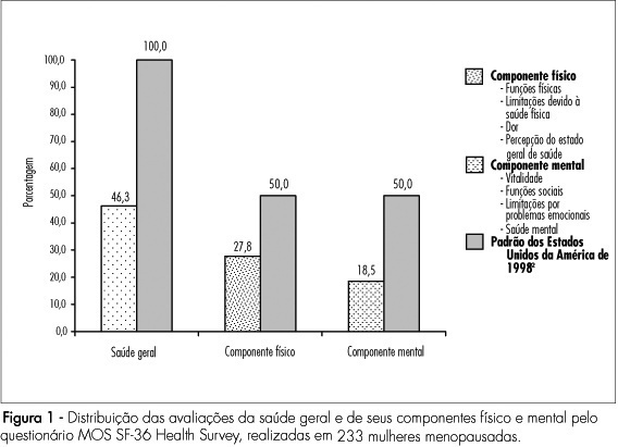
PURPOSE: to evaluate quality of life of climacteric women attended at a school hospital in Recife, Pernambuco, Brazil, adopting the Medical Outcome Study 36-item Short Form Health Survey (MOS SF-36 Health Survey) and the Women's Health Questionnaire (WHQ), as well as the modified Blatt-Kupperman index. METHODS: according to a descriptive, transversal study, 233 women, assisted from February to June 2006, were evaluated. Within a convenience sample, the inclusion criteria were age from 40 to 65 years old and agreement in participating of the research, excluding previous history of bilateral oophorectomy, hormonal therapy in the last semester and uncontrolled illnesses. The sample size was calculated admitting a prevalence of climacteric symptoms of 4% and a precision of 2.5%. The variables were: general health and physical and mental components based on the MOS SF-36 Health Survey; quality of health based on the WHQ and climacteric symptoms according to the modified Blatt-Kupperman index. Data were analyzed by the Statistical Package for Social Sciences, version 13.0 software. RESULTS: the quality of life was classified as bad. Based on the MOS SF-36 Health Survey, there was more damage in the mental component (18.53 versus 27.77% for physical components), higher losses in social functions (80.28%) and limitations for emotional problems (78.61%). According to WHQ, there were limitations due to sleep disturbances (69.77%), somatic (69.15%) and vasomotor symptoms (68.80%), considering regular sexual function and menstrual symptoms. Estrogenic deficiency symptoms were found in 53% of the women. The increase of hypoestrogenism symptoms were followed by worsening of general and menopausal health. CONCLUSIONS: it seemed reasonable to assume that menopause, for the researched women, was really configured as a biopsychosocial event, more than organic, derived predominantly from estrogenic deficiency.
Summary
Revista Brasileira de Ginecologia e Obstetrícia. 2008;30(3):107-112
DOI 10.1590/S0100-72032008005000005
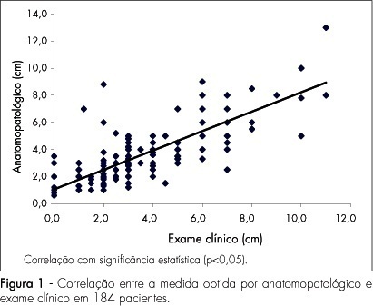
PURPOSE: to evaluate which method is the best to determine pre-surgically the size of breast cancer: clinical examination, mammography or ultrasonography, using as a reference the anatomopathological exam. METHODS: this study has included 184 patients with palpable-or-not breast lesions, detected by mammography and ultrasonography, that were submitted to surgical resection of the tumor, with histopathological diagnosis of breast cancer. The same examiner evaluated clinically the largest tumoral diameter, through clinical examination, mammography and ultrasonography, and the measurements obtained by each method were correlated with the maximum diameter obtained by the anatomopathological exam. The comparative analysis has been done by Pearson's correlation coefficient (r). RESULTS: Pearson's correlation coefficient between the anatomopathological and the clinical exams was 0.8; between the anatomopathological exam and the mammography, 0.7; and between anatomopathological exam and ultrasonography 0.7 (p<0.05). Pearson's correlation coefficients among the methods evaluated were also calculated and r=0.7 was obtained between clinical exam and mammography, r=0.8 between clinical examination and utrasonograhy, and r=0.8 between mammography and ultrasonography (p<0.05). CONCLUSIONS: clinical examination, mammography and ultrasonography have presented high correlation with the anatomopathological measures, besides high correlations among themselves, what seems to show that they may be used as equivalent methods in the pre-surgical evaluation of the breast tumoral size. Nevertheless, due to specific limitations of each method, clinical examination, mammography and ultrasonography should be seen as complementary to each other, in order to obtain a more accurate measurement of the breast cancer tumor.
Summary
Revista Brasileira de Ginecologia e Obstetrícia. 2008;30(3):142-148
DOI 10.1590/S0100-72032008005000004
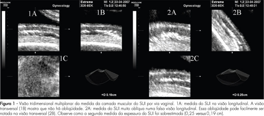
PURPOSE: to compare the intra and interobserver reproducibility of the total thickness measurement of the inferior uterine segment (IUS), through the abdominal route, and of the muscle layer measurement, through the vaginal route, using bi and tridimensional ultrasonography. METHODS: the IUS thickness measurement of 30 women, between the 36th and 39th weeks of gestation with previous caesarean section, done by two observers, was studied. Abdominal ultrasonography with the patient in both supine and lithotomy position was performed. In the sagittal section, the IUS was identified and four bidimensional images and two tridimensional blocks of the total thickness were collected through the abdominal route, and the same for the muscle layer, through the vaginal route. Tridimensional acquisitions were manipulated in the multiplanar mode. The time was measured with a chronometer. Reproducibility was evaluated by the computation of the absolute difference between measurements, the ratio of differences smaller than 1 mm, the intraclass coefficient (ICC), and the Bland and Altman's concordance limits. RESULTS: the average bidimensional measurement of IUS thickness was 7.4 mm through the abdominal and 2.7 mm through the vaginal route, and the tridimensional measurement was 6.9 mm through the abdominal and 5.1 mm through the vaginal route. Intra- and interobserver reproducibility of vaginal versus abdominal route: smaller absolute difference (0.2-0.4 mm versus 0.8-1.5 mm), greater ratio of differences (85.8-97.8% versus 48.7-72,8%), with p<0,0001, higher ICC (0.8-0.9 versus 0.6-0.8) and lower concordance limits (-0.9 to 1.5 versus -3.8 to 4 mm) for the vaginal route. Tri versus bidimensional ultrasonography: lower absolute difference (0.2-1.4 versus 0.4-1.5 mm), higher ratio of differences (57.7-97.8% versus 48.7-91.7%) with p>0.05[A1] and similar lower concordance limits (-38 to 3.4 versus -3.6 to 4 mm) for tridimensional ultrasonography and ICC (0.6-0.9 versus 0.7-0.9). CONCLUSIONS: from the above, we came to the conclusion that the measurement of the IUS muscle layer, through the vaginal route using tridimensional ultrasonography is more reproducible. Nevertheless, our results do not indicate that this measurement shows any clinical evidence to predict uterine tear, as that was not the aim of this study. The only work that has correlated the UIS thickness with risk of uterine tear, without interfering in the obstetrician behavior or anticipating delivery, was done by bidimensional abdominal measurements of the total thickness.
Summary
Revista Brasileira de Ginecologia e Obstetrícia. 2007;29(11):580-587
DOI 10.1590/S0100-72032007001100006
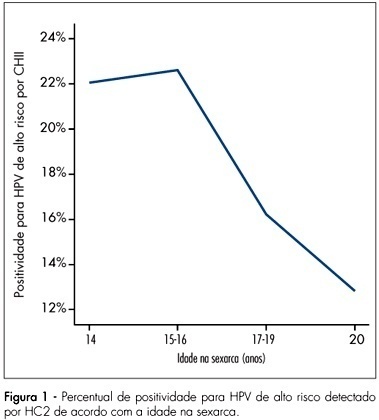
PURPOSE: to investigate women’s age at their first sexual intercourse and its correlation with their present age, human papillomavirus (HPV) infection and cytological abnormalities at Pap smear. METHODS: women from the general population were invited to be screened for cervical cancer and pre-malignant lesions. After answering a behavior questionnaire, they were submitted to screening with cervical cytology and high-risk HPV testing with Hybrid Capture 2 (HC2). This report is part of the Latin American Screening (LAMS) study, that comprises centers from Brazil and Argentina, and the data presented herein refer to the Brazilian women evaluated at the cities of Porto Alegre, São Paulo and Campinas. RESULTS: from 8,649 women that answered the questionnaire, 8,641 reported previous sexual activity and were included in this analysis. The mean age at the interview was 38.1±11.0 years and the mean age at the first sexual intercourse was 18.5±4.0 years. The age at the first sexual intercourse increased along with the age at the interview, i.e., younger women reported they had begun their sexual life earlier than older women (p<0.001). From the total of women who had already begun having sexual intercourse, 3,643 patients were tested for high-risk HPV infection and 17.3% of them had positive results. In all the centers, it became clear that the women with the first sexual intercourse at ages below the mean age of all the population interviewed presented higher rates of HPV infection (20.2%) than the women with the first sexual intercourse at ages above the mean (12.5%) - Odds Ratio (OR) 1.8 (IC95% 1.5-2.2;p<0,001). According to the cytology, the women with first sexual intercourse at ages under the mean, presented higher percentage of abnormal cytology > or = ASC-US (6.7%) than the women with the first sexual intercourse at ages above the mean (4.3%) - OR 1.6 (IC95% 1.3-2.;p<0.001). CONCLUSIONS: the high-risk HPV infection and cytological abnormalities identified during the asymptomatic population screening were significantly associated to the women’s age at the first sexual intercourse. Additionally, we have also identified that the women’s age at the first sexual intercourse has decreased during the last decades, suggesting an important contribution to the increase of HPV infection and the subsequent cervical lesions.
Summary
Revista Brasileira de Ginecologia e Obstetrícia. 2007;29(11):575-579
DOI 10.1590/S0100-72032007001100005
PURPOSE: the villoglandular adenocarcinoma (VGA) of the cervix has been identified as a variant of cervical adenocarcinoma that occurs in young women, which has an excellent prognosis. Considering the scarcity of studies related to the subject, we report six cases of VGA of the cervix. METHODS: we followed the development of six cases of VGA in the period from 1995 to 2006 at Hospital São Lucas of Pontifícia Universidade Católica do Rio Grande do Sul (PUC-RS). We collected clinical and histologic information of the patients and submitted all the surgical specimens to histological review. RESULTS: mean age at diagnosis was 43.5 years (range 27-61 years). Four patients were submitted to Wertheim-Meigs radical hysterectomy and bilateral pelvic lymphadenectomy, one to conization and subsequent radiotherapy and one to pelvic lymphadenectomy followed by radiotherapy. All the patients were alive and well at the time of this writing, without evidence of recurrence. CONCLUSIONS: the implications of therapy are discussed. We propose here the inclusion of the study of the pattern of lymphovascular involvement in determining the diagnosis of VGA. Thus, in referring to this diagnosis, we will be able to opt, with caution, for conservative therapy, except for particularities of each case.
Summary
Revista Brasileira de Ginecologia e Obstetrícia. 2007;29(11):568-574
DOI 10.1590/S0100-72032007001100004
PURPOSE: to evaluate the histological differentiation pattern in superficial peritoneum lesions and in deeply infiltrating endometriosis (DIE) in utero-sacral ligament, bowel (rectum and sigmoid colon) and rectovaginal septum. METHODS: this prospective non-randomized study included 139 patients. Of the total, 234 biopsies were obtained (179 with DIE - Deeply Group - and 55 superficial endometriosis - Superficial Group). From the 179 DIE lesions (Depply Group), 15 were obtained from rectovaginal septum, 72 from rectosigmoid nodules and 92 from utero-sacral ligament. Biopsies were classified in well-differentiated glandular pattern, undifferentiated glandular, mixed glandular differentiation and pure stromal disease, based on specific morphological classification. RESULTS: in the Depply Group (DIE), 33.5% of the biopsies showed undifferentiated glandular pattern and 46.9% mixed glandular pattern. In the Superficial Group, there was the predominance of the well-differentiated glandular pattern (41.8%). Comparing specifically the different localizations of the biopsies of DIE lesions (Deeply Group), a predominance of mixed pattern in bowel nodules (61.1%) was noted. CONCLUSIONS: it was possible to conclude that there is a predominance of well-differentiated glandular pattern in superficial endometriosis, a predominance of mixed undifferentiated in deeply pelvic endometriosis and, specifically studying endometriosis from the rectum and sigmoid colon, there was a predominance of the mixed pattern.
Summary
Revista Brasileira de Ginecologia e Obstetrícia. 2007;29(11):561-567
DOI 10.1590/S0100-72032007001100003
PURPOSE: to verify the association of abortion, recurrent fetal loss, miscarriage and severe pre-eclampsia with the presence of hereditary thrombophilias and antiphospholipid antibodies in pregnant women. METHODS: observational and transverse study of 48 pregnant women with past medical record of miscarriage, repeated abortion and fetal loss story (AB Group) and severe pre-eclampsia (PE Group), attended to in the High Risk Pregnancy Ambulatory of the Faculdade de Medicina (Famed) from the Universidade Federal de Mato Grosso do Sul (UFMS) from November 2006 to July 2007. The pregnant women of both groups were screened for the presence of antiphospholipid antibodies (anticardiolipin IgG and IgM, lupic anticoagulant and anti-beta2-glycoprotein I) and hereditary thrombophilias (protein C and S deficiency, antithrombin deficiency, hyperhomocysteinemia and factor V Leiden mutation). The laboratorial screening was performed during the pregnancy. The parametric data (maternal age and parity) were analyzed with Student’s tau test. The non-parametric data (presence/absence of hereditary thrombophilias and antiphospholipid antibodies, presence/absence of pre-eclampsia, fetal loss, miscarriage and repeated abortion) were analyzed with Fisher’s exact test in contingency tables. It was considered significant the association with p value <0.05. RESULTS: out of the 48 pregnant women, 31 (65%) were included in AB Group and 17 (35%) in PE Group. There was no significant difference between maternal age and parity within the groups. There was significant statistical association between recurrent fetal loss, recurrent abortions and previous miscarriages and maternal hereditary thrombophilias (p<0.05). There was no statistical association between the AB Group and the presence of antiphospholipid antibodies. Neither there were associations of the PE Group with maternal hereditary thrombophilias and the presence of antiphospholipid antibodies. CONCLUSIONS: the data obtained suggest routine laboratorial investigation for hereditary thrombophilias in pregnant women with previous obstetrical story of recurrent fetal loss, repeated abortion and miscarriage.