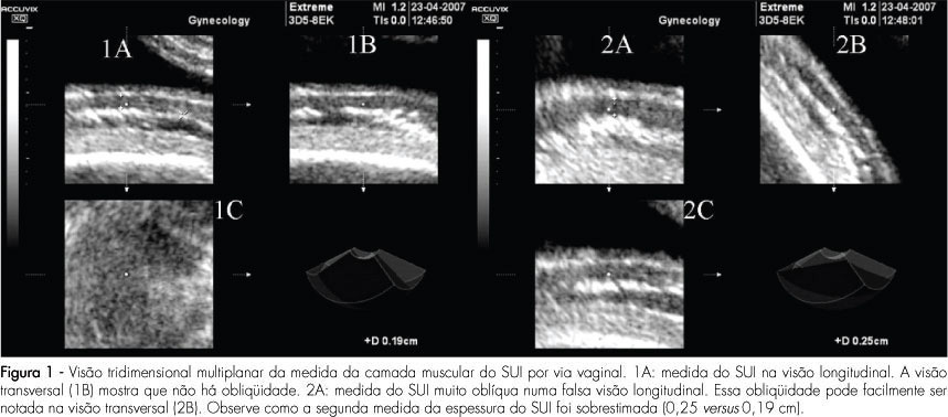
PURPOSE: to compare the intra and interobserver reproducibility of the total thickness measurement of the inferior uterine segment (IUS), through the abdominal route, and of the muscle layer measurement, through the vaginal route, using bi and tridimensional ultrasonography. METHODS: the IUS thickness measurement of 30 women, between the 36th and 39th weeks of gestation with previous caesarean section, done by two observers, was studied. Abdominal ultrasonography with the patient in both supine and lithotomy position was performed. In the sagittal section, the IUS was identified and four bidimensional images and two tridimensional blocks of the total thickness were collected through the abdominal route, and the same for the muscle layer, through the vaginal route. Tridimensional acquisitions were manipulated in the multiplanar mode. The time was measured with a chronometer. Reproducibility was evaluated by the computation of the absolute difference between measurements, the ratio of differences smaller than 1 mm, the intraclass coefficient (ICC), and the Bland and Altman’s concordance limits. RESULTS: the average bidimensional measurement of IUS thickness was 7.4 mm through the abdominal and 2.7 mm through the vaginal route, and the tridimensional measurement was 6.9 mm through the abdominal and 5.1 mm through the vaginal route. Intra- and interobserver reproducibility of vaginal versus abdominal route: smaller absolute difference (0.2-0.4 mm versus 0.8-1.5 mm), greater ratio of differences (85.8-97.8% versus 48.7-72,8%), with p<0,0001, higher ICC (0.8-0.9 versus 0.6-0.8) and lower concordance limits (-0.9 to 1.5 versus -3.8 to 4 mm) for the vaginal route. Tri versus bidimensional ultrasonography: lower absolute difference (0.2-1.4 versus 0.4-1.5 mm), higher ratio of differences (57.7-97.8% versus 48.7-91.7%) with p>0.05[A1] and similar lower concordance limits (-38 to 3.4 versus -3.6 to 4 mm) for tridimensional ultrasonography and ICC (0.6-0.9 versus 0.7-0.9). CONCLUSIONS: from the above, we came to the conclusion that the measurement of the IUS muscle layer, through the vaginal route using tridimensional ultrasonography is more reproducible. Nevertheless, our results do not indicate that this measurement shows any clinical evidence to predict uterine tear, as that was not the aim of this study. The only work that has correlated the UIS thickness with risk of uterine tear, without interfering in the obstetrician behavior or anticipating delivery, was done by bidimensional abdominal measurements of the total thickness.
Search
Search in:
Search
Search in:


Comments