Summary
Revista Brasileira de Ginecologia e Obstetrícia. 2012;34(5):221-227
DOI 10.1590/S0100-72032012000500006
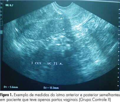
PURPOSE: To evaluate the thickness of the lower uterine segment by transvaginal ultrasound in a group of non-pregnant women and to describe the morphologic findings in the scar of those submitted to cesarean section. METHODS: A retrospective study of 155 transvaginal ultrasound images obtained from premenopausal and non-pregnant women, conducted between January 2008 and November 2011. the subjects were divided into three groups: women who were never pregnant (Control Group I), women with previous vaginal deliveries (Control Group II) and women with previous cesarean section (Observation Group). We excluded women with a retroverted uterus, intrauterine device users, pregnant women and those with less than one year of tsince the last obstetrical event. The data were analyzed statistically with Statistica®, version 8.0 software. ANOVA and LSD were used to compare the groups regarding quantitative variables and the Student's t-test was used to compare the thickness of the anterior and posterior isthmus. The Spearman correlation coefficient was calculated to estimate the association between quantitative variables. P values <0.05 were considered statistically significant. RESULTS: There was significant difference between the thickness of the anterior and posterior isthmus only in the group of women with previous cesarean section. Comparing the groups two by two, no significant differences between the thickness of the anterior and posterior isthmus were observed in the Control Groups, but this difference was significant when we compared the Observation Group with each Control Group. In the Observation Group, no correlation was found between the thickness of the isthmus and the number of previous cesarean deliveries or the time elapsed since the last birth. A niche was found in the cesarean scar in 30.6% of the women in the Observation Group, 93% of whom complained of post-menstrual bleeding. CONCLUSION: The relationship between the thickness of the anterior and posterior wall of the lower uterine segment by transvaginal ultrasound is a suitable method for the evaluation of the uterine lower segment in women with previous cesarean sections.
Summary
Revista Brasileira de Ginecologia e Obstetrícia. 2012;34(5):221-227
DOI 10.1590/S0100-72032012000500006
PURPOSE: To evaluate the thickness of the lower uterine segment by transvaginal ultrasound in a group of non-pregnant women and to describe the morphologic findings in the scar of those submitted to cesarean section. METHODS: A retrospective study of 155 transvaginal ultrasound images obtained from premenopausal and non-pregnant women, conducted between January 2008 and November 2011. the subjects were divided into three groups: women who were never pregnant (Control Group I), women with previous vaginal deliveries (Control Group II) and women with previous cesarean section (Observation Group). We excluded women with a retroverted uterus, intrauterine device users, pregnant women and those with less than one year of tsince the last obstetrical event. The data were analyzed statistically with Statistica®, version 8.0 software. ANOVA and LSD were used to compare the groups regarding quantitative variables and the Student's t-test was used to compare the thickness of the anterior and posterior isthmus. The Spearman correlation coefficient was calculated to estimate the association between quantitative variables. P values <0.05 were considered statistically significant. RESULTS: There was significant difference between the thickness of the anterior and posterior isthmus only in the group of women with previous cesarean section. Comparing the groups two by two, no significant differences between the thickness of the anterior and posterior isthmus were observed in the Control Groups, but this difference was significant when we compared the Observation Group with each Control Group. In the Observation Group, no correlation was found between the thickness of the isthmus and the number of previous cesarean deliveries or the time elapsed since the last birth. A niche was found in the cesarean scar in 30.6% of the women in the Observation Group, 93% of whom complained of post-menstrual bleeding. CONCLUSION: The relationship between the thickness of the anterior and posterior wall of the lower uterine segment by transvaginal ultrasound is a suitable method for the evaluation of the uterine lower segment in women with previous cesarean sections.
Summary
Revista Brasileira de Ginecologia e Obstetrícia. 2012;34(3):133-138
DOI 10.1590/S0100-72032012000300008
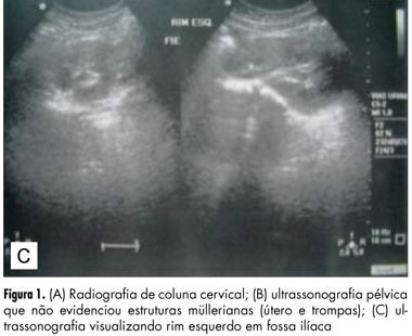
The atypical and more severe form of Mayer-Rokitansky-Kuster-Hauser syndrome (MRKH) or MRKH type II is also known as MURCS association, an acronym meaning aplasia/hypoplasia of Müllerian ducts (MU), congenital renal dysplasia (R) and cervico-thoracic dysplasia (CS). It affects female patients with normal karyotype and ovarian function, evolving to primary amenorrhea. It has an incidence of 1:50,000, but it is underestimated due to late diagnosis and undefined etiology. We describe the cases of a child and an adolescent in order to predict the diagnosis even in childhood, before the onset of amenorrhea. Patients had in common renal malformation, agenesis or hypoplasia of Müllerian derivatives and vertebral anomalies, establishing the diagnosis of MURCS. The relevance of this paper is to show the importance of further investigation when some of pathologic signs are present, researching correlated abnormalities in order to establish an early diagnosis and consequently to provide guidance to the patients and their families about the best way to conduct the case, including genetic counseling.
Summary
Revista Brasileira de Ginecologia e Obstetrícia. 2012;34(3):133-138
DOI 10.1590/S0100-72032012000300008
The atypical and more severe form of Mayer-Rokitansky-Kuster-Hauser syndrome (MRKH) or MRKH type II is also known as MURCS association, an acronym meaning aplasia/hypoplasia of Müllerian ducts (MU), congenital renal dysplasia (R) and cervico-thoracic dysplasia (CS). It affects female patients with normal karyotype and ovarian function, evolving to primary amenorrhea. It has an incidence of 1:50,000, but it is underestimated due to late diagnosis and undefined etiology. We describe the cases of a child and an adolescent in order to predict the diagnosis even in childhood, before the onset of amenorrhea. Patients had in common renal malformation, agenesis or hypoplasia of Müllerian derivatives and vertebral anomalies, establishing the diagnosis of MURCS. The relevance of this paper is to show the importance of further investigation when some of pathologic signs are present, researching correlated abnormalities in order to establish an early diagnosis and consequently to provide guidance to the patients and their families about the best way to conduct the case, including genetic counseling.
Summary
Revista Brasileira de Ginecologia e Obstetrícia. 2011;33(10):292-296
DOI 10.1590/S0100-72032011001000004
PURPOSE: To analyze the effectiveness and occurrence of complications, in addition to hospitalization time and blood losses. METHODS: Thirty patients were assigned alternatively and consecutively to one of two groups (15 to the Curettage Group and 15 to the Manual Vacuum Aspiration Group). The following variables were analyzed: effectiveness of the method, occurrence of complications, time before the procedure, time of execution of the procedure, time after the procedure, and total time of hospital permanence, in addition to hematocrit and hemoglobin, which were measured before and after the procedure. Patients were evaluated clinically 10 to 14 days after the procedure. Parametric and nonparametric tests were used for statistical analysis, with the level of significance set at p>0.05. RESULTS: Both methods were efficient and no complications were recorded. Blood losses were similar in the two groups, but the hospitalization time was significantly shorter for the Manual Vacuum Aspiration Group (p=0.03). CONCLUSION: Manual vacuum aspiration is as efficient and safe as uterine curettage, with the advantage of requiring shorter hospitalization, which increases the resolution of the method, improving the quality of care for these patients.
Summary
Revista Brasileira de Ginecologia e Obstetrícia. 2009;31(9):453-460
DOI 10.1590/S0100-72032009000900006
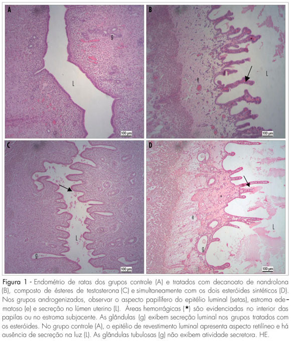
PURPOSE: to evaluate the effects of the administration of two synthetic steroids in the uterus morphology and in the reproductive parameters of adult female rats. METHODS: divided into four experimental groups: control (C; physiological solution); treated with nandrolone decanoate (DN; 7.5 mg/kg of body weight); with a testosterone esters compound (T; 7.5 mg/kg); and simultaneously with DN and T (7.5 mg/kg of each steroid), in a single intraperitoneal weekly dose, for eight weeks. Five females of each group were sacrificed and the uterine horns were collected, weighted and prepared for histological and morphometrical evaluation. The remaining rats were mated with normal male rats for reproductive parameters evaluation, composing the groups treated during the pre-gestational period. Another group of 20 female rats were treated during the gestational period (7th-14th days). For data analysis, the Kruskal-Wallis non-parametric variance analysis was used, followed by the test of Dunn or of Student-Newman-Keus (5% significance level). RESULTS: there was a significant body weight increase in the androgenized females (ND: 305±50; T: 280±35; ND+T: 275±30 versus C: 255±22 g; p<0.05). Uterine weight was not affected by the steroidal treatment (ND: 0.6±0.2; T: 0.4±0.04; ND+T: 0.7±0.1 versus C: 0.4±0.09 g). All the androgenized females presented estral acyclicity and endometrium characterized by papilliferous luminal lining, oedematous stroma with hemorrhagic areas and secretory activity. There were changes in the morphometrical thickness parameters of the luminal epithelium, myometrium and perimetrium in the androgenized groups. None of the female rats got pregnant when treated with steroids in the pre-gestational period and the treatment during organogenesis affected negatively the reproductive parameters. CONCLUSIONS: steroidal agents alter the uterine structure and impair fertility and gestational outcome in female rats.
Summary
Revista Brasileira de Ginecologia e Obstetrícia. 2009;31(9):453-460
DOI 10.1590/S0100-72032009000900006
PURPOSE: to evaluate the effects of the administration of two synthetic steroids in the uterus morphology and in the reproductive parameters of adult female rats. METHODS: divided into four experimental groups: control (C; physiological solution); treated with nandrolone decanoate (DN; 7.5 mg/kg of body weight); with a testosterone esters compound (T; 7.5 mg/kg); and simultaneously with DN and T (7.5 mg/kg of each steroid), in a single intraperitoneal weekly dose, for eight weeks. Five females of each group were sacrificed and the uterine horns were collected, weighted and prepared for histological and morphometrical evaluation. The remaining rats were mated with normal male rats for reproductive parameters evaluation, composing the groups treated during the pre-gestational period. Another group of 20 female rats were treated during the gestational period (7th-14th days). For data analysis, the Kruskal-Wallis non-parametric variance analysis was used, followed by the test of Dunn or of Student-Newman-Keus (5% significance level). RESULTS: there was a significant body weight increase in the androgenized females (ND: 305±50; T: 280±35; ND+T: 275±30 versus C: 255±22 g; p<0.05). Uterine weight was not affected by the steroidal treatment (ND: 0.6±0.2; T: 0.4±0.04; ND+T: 0.7±0.1 versus C: 0.4±0.09 g). All the androgenized females presented estral acyclicity and endometrium characterized by papilliferous luminal lining, oedematous stroma with hemorrhagic areas and secretory activity. There were changes in the morphometrical thickness parameters of the luminal epithelium, myometrium and perimetrium in the androgenized groups. None of the female rats got pregnant when treated with steroids in the pre-gestational period and the treatment during organogenesis affected negatively the reproductive parameters. CONCLUSIONS: steroidal agents alter the uterine structure and impair fertility and gestational outcome in female rats.
Summary
Revista Brasileira de Ginecologia e Obstetrícia. 2009;31(2):82-88
DOI 10.1590/S0100-72032009000200006
PURPOSE: to evaluate the effect of magnesium sulphate on the pulsatility index (PI) of the uterine, umbilical and fetal middle cerebral arteries, according to the persistency or not of the bilateral protodiastolic notch of the uterine arteries in severe pre-eclampsia. METHODS: a cohort study including 40 pregnant women with severe pre-eclampsia, 23 of them presenting bilateral protodiastolic notch, and 17, unilateral/absent notch. The patients were submitted to Doppler velocimetry before and 20 minutes after the intravenous administration of 6 g of magnesium sulphate. The examination was carried out with the patient in semi-Fowler position, the sonograms being obtained during fetal inactivity, in apnea and absent uterine contraction periods. All the exams were performed by two researchers, the average being considered as the final result. Wilcoxon's test was used to compare the PI, before and after magnesium sulphate in both groups. The difference between the two measurements (before and after magnesium sulphate) was compared between the groups (bilateral incision and unilateral/absent incision) using the Mann-Whitney test. RESULTS: there was a significant increase in the maternal heart rate (MHR) and decrease in the maternal blood pressure, and in the PI medians of the two uterine arteries and in the fetal middle cerebral artery, after magnesium sulphate in both groups. There was a significant decrease in the PI of the left uterine artery and in the umbilical artery, only in the protodiastolic unilateral/absent notch group. Nevertheless, it was not found any significant difference regarding the PI of the right uterine artery, or the cerebral/umbilical relationship, before and after magnesium sulphate in each group. No difference between the groups was found, before and after magnesium sulphate, for any of the studied outcomes. CONCLUSIONS: after the intravenous administration of 6 g of magnesium sulphate to patients with severe pre-eclampsia, a decrease in blood pressure and in the PI of the uterine, umbilical and fetal middle cerebral arteries occurs, besides the increase in the MHR, not influenced by the presence of bilateral protodiastolic notch in the uterine arteries.
Summary
Revista Brasileira de Ginecologia e Obstetrícia. 2008;30(3):142-148
DOI 10.1590/S0100-72032008005000004
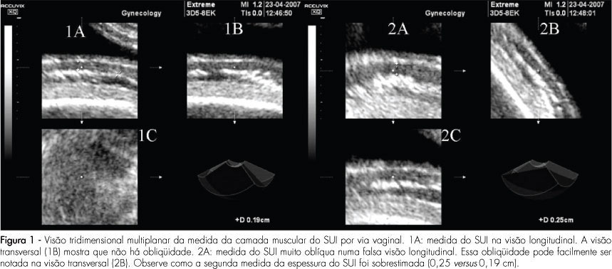
PURPOSE: to compare the intra and interobserver reproducibility of the total thickness measurement of the inferior uterine segment (IUS), through the abdominal route, and of the muscle layer measurement, through the vaginal route, using bi and tridimensional ultrasonography. METHODS: the IUS thickness measurement of 30 women, between the 36th and 39th weeks of gestation with previous caesarean section, done by two observers, was studied. Abdominal ultrasonography with the patient in both supine and lithotomy position was performed. In the sagittal section, the IUS was identified and four bidimensional images and two tridimensional blocks of the total thickness were collected through the abdominal route, and the same for the muscle layer, through the vaginal route. Tridimensional acquisitions were manipulated in the multiplanar mode. The time was measured with a chronometer. Reproducibility was evaluated by the computation of the absolute difference between measurements, the ratio of differences smaller than 1 mm, the intraclass coefficient (ICC), and the Bland and Altman's concordance limits. RESULTS: the average bidimensional measurement of IUS thickness was 7.4 mm through the abdominal and 2.7 mm through the vaginal route, and the tridimensional measurement was 6.9 mm through the abdominal and 5.1 mm through the vaginal route. Intra- and interobserver reproducibility of vaginal versus abdominal route: smaller absolute difference (0.2-0.4 mm versus 0.8-1.5 mm), greater ratio of differences (85.8-97.8% versus 48.7-72,8%), with p<0,0001, higher ICC (0.8-0.9 versus 0.6-0.8) and lower concordance limits (-0.9 to 1.5 versus -3.8 to 4 mm) for the vaginal route. Tri versus bidimensional ultrasonography: lower absolute difference (0.2-1.4 versus 0.4-1.5 mm), higher ratio of differences (57.7-97.8% versus 48.7-91.7%) with p>0.05[A1] and similar lower concordance limits (-38 to 3.4 versus -3.6 to 4 mm) for tridimensional ultrasonography and ICC (0.6-0.9 versus 0.7-0.9). CONCLUSIONS: from the above, we came to the conclusion that the measurement of the IUS muscle layer, through the vaginal route using tridimensional ultrasonography is more reproducible. Nevertheless, our results do not indicate that this measurement shows any clinical evidence to predict uterine tear, as that was not the aim of this study. The only work that has correlated the UIS thickness with risk of uterine tear, without interfering in the obstetrician behavior or anticipating delivery, was done by bidimensional abdominal measurements of the total thickness.
Summary
Revista Brasileira de Ginecologia e Obstetrícia. 2008;30(3):142-148
DOI 10.1590/S0100-72032008005000004
PURPOSE: to compare the intra and interobserver reproducibility of the total thickness measurement of the inferior uterine segment (IUS), through the abdominal route, and of the muscle layer measurement, through the vaginal route, using bi and tridimensional ultrasonography. METHODS: the IUS thickness measurement of 30 women, between the 36th and 39th weeks of gestation with previous caesarean section, done by two observers, was studied. Abdominal ultrasonography with the patient in both supine and lithotomy position was performed. In the sagittal section, the IUS was identified and four bidimensional images and two tridimensional blocks of the total thickness were collected through the abdominal route, and the same for the muscle layer, through the vaginal route. Tridimensional acquisitions were manipulated in the multiplanar mode. The time was measured with a chronometer. Reproducibility was evaluated by the computation of the absolute difference between measurements, the ratio of differences smaller than 1 mm, the intraclass coefficient (ICC), and the Bland and Altman's concordance limits. RESULTS: the average bidimensional measurement of IUS thickness was 7.4 mm through the abdominal and 2.7 mm through the vaginal route, and the tridimensional measurement was 6.9 mm through the abdominal and 5.1 mm through the vaginal route. Intra- and interobserver reproducibility of vaginal versus abdominal route: smaller absolute difference (0.2-0.4 mm versus 0.8-1.5 mm), greater ratio of differences (85.8-97.8% versus 48.7-72,8%), with p<0,0001, higher ICC (0.8-0.9 versus 0.6-0.8) and lower concordance limits (-0.9 to 1.5 versus -3.8 to 4 mm) for the vaginal route. Tri versus bidimensional ultrasonography: lower absolute difference (0.2-1.4 versus 0.4-1.5 mm), higher ratio of differences (57.7-97.8% versus 48.7-91.7%) with p>0.05[A1] and similar lower concordance limits (-38 to 3.4 versus -3.6 to 4 mm) for tridimensional ultrasonography and ICC (0.6-0.9 versus 0.7-0.9). CONCLUSIONS: from the above, we came to the conclusion that the measurement of the IUS muscle layer, through the vaginal route using tridimensional ultrasonography is more reproducible. Nevertheless, our results do not indicate that this measurement shows any clinical evidence to predict uterine tear, as that was not the aim of this study. The only work that has correlated the UIS thickness with risk of uterine tear, without interfering in the obstetrician behavior or anticipating delivery, was done by bidimensional abdominal measurements of the total thickness.
Summary
Revista Brasileira de Ginecologia e Obstetrícia. 2007;29(11):588-592
DOI 10.1590/S0100-72032007001100007
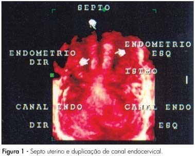
This report describes an unusual case of spontaneous pregnancy in a patient with Müllerian anomaly. The patient was a 34-years old, white, nulligravida, with regular menstrual cycles, and suspected uterine septum observed during a routine ultrasonographic examination. The gynecological examination revealed a complete longitudinal vaginal septum and two uterine cervices. Three-dimensional pelvic ultrasonography showed cervix duplication, uterine septum from isthmus to endometrial cavity and absence of uterine body division, compatible with complete uterine septum and true dual cervices. She returned after one month and reported unprotected sexual intercourse and delayed menstrual period. She was pregnant, had a good pregnancy evolution, and delivered a healthy term baby girl, by cesarean section, at 37 weeks of pregnancy. This report describes a case of normal-term pregnancy in a patient with a rare anomaly (vaginal septum and two cervices) who became spontaneously pregnant without treatment.
Summary
Revista Brasileira de Ginecologia e Obstetrícia. 2007;29(11):588-592
DOI 10.1590/S0100-72032007001100007
This report describes an unusual case of spontaneous pregnancy in a patient with Müllerian anomaly. The patient was a 34-years old, white, nulligravida, with regular menstrual cycles, and suspected uterine septum observed during a routine ultrasonographic examination. The gynecological examination revealed a complete longitudinal vaginal septum and two uterine cervices. Three-dimensional pelvic ultrasonography showed cervix duplication, uterine septum from isthmus to endometrial cavity and absence of uterine body division, compatible with complete uterine septum and true dual cervices. She returned after one month and reported unprotected sexual intercourse and delayed menstrual period. She was pregnant, had a good pregnancy evolution, and delivered a healthy term baby girl, by cesarean section, at 37 weeks of pregnancy. This report describes a case of normal-term pregnancy in a patient with a rare anomaly (vaginal septum and two cervices) who became spontaneously pregnant without treatment.
Summary
Revista Brasileira de Ginecologia e Obstetrícia. 2006;28(7):410-415
DOI 10.1590/S0100-72032006000700006
PURPOSE: to verify, through pelvic ultrasound, the existence of changes in the internal genitalia of girls with central precocious puberty, submitted to treatment with gonadotrophin-releasing hormone (GnRH) analogs. METHODS: pelvic ultrasound was performed in 18 girls with idiopathic central precocious puberty, before and after three months of onset of the treatment with GnRH analogs, to investigate the impact of the therapy on the internal genitalia. Ovarian and uterine volumes, uterine longitudinal length, relation between the longitudinal diameter of the uterine corpus and the uterine cervix, the relation between the anterior-posterior diameter of the uterine corpus and the uterine cervix, and endometrial echogenicity were evaluated. Statistical analysis was performed through Shapiro-Willkis's test, to assess data normality. When normality was present, Student's test t was applied. For data without normality, a non-parametric test (the signal test) was used. RESULTS: after therapy, statistically significant decline of the mean uterine volume (from 5.4 cm³ to 3.0 cm³, p<0.001), of the mean ovarian volume (from 2.2 cm³ to 1.1 cm³, p= 0.004), of the mean uterine longitudinal length (from 4.2cm to 3.4 cm, p=0.001), and of the mean endometrial echogenicity (from 1.8 mm to 0.6 mm, p=0.018) occurred. CONCLUSION: In girls with idiopathic central precocious puberty, pelvic ultrasound is a valid method to assess the efficacy of treatment with GnRH analogs. The main parameters of the therapeutic response were the decrease of uterine and ovarian volume, of uterine longitudinal length, and atrophy or absence of endometrial echogenicity.