Summary
Rev Bras Ginecol Obstet. 2024;46:e-rbgo2
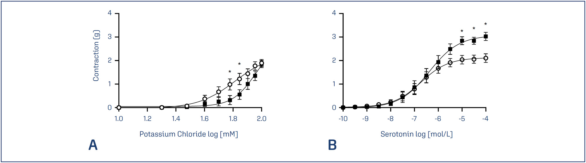
Potassium channels have an important role in the vascular adaptation during pregnancy and a reduction in the expression of adenosine triphosphate-sensitive potassium channels (Katp) has been linked to preeclampsia. Activation of Katp induces vasodilation; however, no previous study has been conducted to evaluate the effects of the inhibition of these channels in the contractility of preeclamptic arteries. Glibenclamide is an oral antihyperglycemic agent that inhibits Katp and has been widely used in vascular studies.
To investigate the effects of the inhibition of Katp, umbilical arteries of preeclamptic women and women with healthy pregnancies were assessed by vascular contractility experiments, in the presence or absence of glibenclamide. The umbilical arteries were challenged with cumulative concentrations of potassium chloride (KCl) and serotonin.
There were no differences between the groups concerning the maternal age and gestational age of the patients. The percentage of smokers, caucasians and primiparae per group was also similar. On the other hand, blood pressure parameters were elevated in the preeclamptic group. In addition, the preeclamptic group presented a significantly higher body mass index. The newborns of both groups presented similar APGAR scores and weights.
In the presence of glibenclamide, there was an increase in the KCl-induced contractions only in vessels from the PE group, showing a possible involvement of these channels in the disorder.
Summary
Rev Bras Ginecol Obstet. 2024;46:e-rbgo2
Potassium channels have an important role in the vascular adaptation during pregnancy and a reduction in the expression of adenosine triphosphate-sensitive potassium channels (Katp) has been linked to preeclampsia. Activation of Katp induces vasodilation; however, no previous study has been conducted to evaluate the effects of the inhibition of these channels in the contractility of preeclamptic arteries. Glibenclamide is an oral antihyperglycemic agent that inhibits Katp and has been widely used in vascular studies.
To investigate the effects of the inhibition of Katp, umbilical arteries of preeclamptic women and women with healthy pregnancies were assessed by vascular contractility experiments, in the presence or absence of glibenclamide. The umbilical arteries were challenged with cumulative concentrations of potassium chloride (KCl) and serotonin.
There were no differences between the groups concerning the maternal age and gestational age of the patients. The percentage of smokers, caucasians and primiparae per group was also similar. On the other hand, blood pressure parameters were elevated in the preeclamptic group. In addition, the preeclamptic group presented a significantly higher body mass index. The newborns of both groups presented similar APGAR scores and weights.
In the presence of glibenclamide, there was an increase in the KCl-induced contractions only in vessels from the PE group, showing a possible involvement of these channels in the disorder.
Summary
Rev Bras Ginecol Obstet. 2013;35(7):309-316
DOI 10.1590/S0100-72032013000700005
PURPOSE: To determine perinatal outcomes and factors associated with fetal brain sparing effect diagnosed by Doppler flow velocimetry in patients with arterial hypertension. METHODS: We performed a cross-sectional retrospective study including 129 pregnant women with arterial hypertension and submitted to Doppler flow velocimetry, within fifteen days before delivery. Women with multiple pregnancies, fetal malformations, genital bleeding, placenta praevia, premature rupture of membranes, smoking, illicit drug use and chronic diseases were excluded. We analyzed the biological, socio-demographic and obstetric characteristics, as well the perinatal outcomes. To determine the association between variables, we used the χ² test, Fisher's exact test and Student's t-test. Multiple logistic regression analysis was performed to determine the factors associated with fetal centralization. RESULTS: Pre-eclampsia was the most frequent hypertensive disorder (53.5%) and fetal brain sparing effect was observed in 24.0% of fetuses. The prenatal factors associated with fetal brain sparing were the persistence of bilateral protodiastolic notches in uterine arteries (OR 3.6; 95%CI 1.4 - 9.4; p=0.009) and intrauterine growth restriction (IUGR) (OR 3.3; 95%CI 1.2 - 9.3; p=0.02). The perinatal outcomes associated with fetal brain sparing were gestational age <32 weeks, small for gestational age (SGA) infants, birth weight <2,500 g and perinatal death. There was no association with other maternal or neonatal variables. CONCLUSIONS: The main factors associated with fetal brain sparing were persistence of uterine arteries notches, IUGR, and increased frequency of adverse perinatal outcomes.
Summary
Rev Bras Ginecol Obstet. 2013;35(2):71-77
DOI 10.1590/S0100-72032013000200006
PURPOSE: To evaluate the anthropometric characteristics of morbidity and mortality of premature newborns (NB) of hypertensive mothers according to the presence or absence of flow (DZ) or reverse (DR) diastolic flow in the dopplervelocimetry of the umbilical artery. METHODS: A prospective study was conducted on preterm newborns of pregnant women with hypertension between 25 and 33 weeks of gestational age, submitted to umbilical artery Doppler study during the five days before delivery. Delivery occurred at Hospital Regional da Asa Sul, Brasília - Federal District, between November 1st, 2009 and October 31st, 2010. The infants were stratified into two groups according to the results of Doppler velocimetry: Gdz/dr=absent end-diastolic velocity waveform or reversed end-diastolic velocity waveform, and Gn=normal Doppler velocimetry. Anthropometric measurements at birth, neonatal morbidity, and mortality were compared between the two groups. RESULTS: We studied 92 infants, as follows: Gdz/dr=52 infants and Gn=40 infants. In Gdz/dr, the incidence of infants small for gestational age was significantly greater, with a relative risk of 2.5 (95%CI 1.7 - 3.7). In Gdz/dr, infants remained on mechanical ventilation for a longer time: median 2 (0‒28) and Gn median 0.5 (0‒25) p=0.03. The need for oxygen at 28 days was higher in G dz/dr comparing to Gn (33 versus 10%; p=0.01). Neonatal mortality was higher in Gdz/dr compared to Gn (36 versus 10%; p=0.03; relative risk of 1.6; 95%CI 1.2‒2.2). Logistic regression showed that, with each 100 grams lower birth weight, the chance of death increased 6.7 times in G dz/dr (95%CI 2.0 - 11.3; p<0.01). CONCLUSION: In preterm infants of mothers with hypertensive changes in Doppler velocimetry of the umbilical artery, intrauterine growth restriction and neonatal prognosis are often worse, with a high risk of death related to birth weight.
Summary
Rev Bras Ginecol Obstet. 2011;33(4):157-163
DOI 10.1590/S0100-72032011000400002
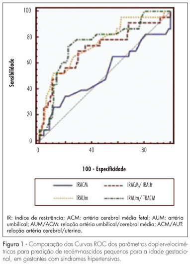
PURPOSE: to determine the best Doppler flow velocimetry index to predict small infants for gestational age (SGAI), in pregnant women with hypertensive syndromes. METHODS: a cross-sectional study was conducted enrolling 129 women with high blood pressure, submitted to dopplervelocimetry up to 15 days before delivery. Women with multiple fetuses, fetal malformations, genital bleeding, placental abruption, premature rupture of fetal membranes, smoking, use of illicit drugs, and chronic diseases were excluded. A receiver operating characteristic (ROC) curve for each Doppler variable was constructed to diagnose SGAI and the sensitivity (Se), specificity (Sp), positive (PLR) and negative (NLR) likelihood ratio were calculated. RESULTS: the area under the ROC curve for the middle cerebral artery resistance index was 52% (p=0.79) with Se, Sp, PLR, and NLR of 25.0, 89.1, 2.3 and 0.84% for a resistance index lower than 0.70, respectively. While the area under the ROC curve for the resistance index of the umbilical artery was 74% (p=0.0001), with Se=50.0%, Sp=90.0%, PLR=5.0 and NLR=0.56, for a resistance index higher or equal to 0.70. The area under the ROC curve for the resistance index umbilical artery/middle cerebral artery ratio was 75% (p=0.0001). When it was higher than 0.86, the Se, Sp, PLR and NLR were 70.8, 80.0, 3.4 and 0.36%, respectively. For the resistance index of the middle cerebral artery/uterine artery ratio, the area under the ROC curve was 71% (p=0.0001). We found a Se=52.2%, Sp=85.9%, PLR=3.7 and NLR=0.56, when the ratio was lower than 1.05. When we compared the area under the ROC curve of the four dopplervelocimetry indexes, we observed that only the resistance index umbilical artery/middle cerebral artery, resistance index middle cerebral artery/uterine artery and resistance index umbilical artery ratios seem to be useful for the prediction of SGA. CONCLUSION: in patients with high blood pressure during pregnancy, all dopplervelocimetry parameters, except the middle cerebral artery resistance index, can be used to predict SGAI. The umbilical artery/middle cerebral artery ratio seems to be the most recommended one.
Summary
Rev Bras Ginecol Obstet. 2011;33(4):157-163
DOI 10.1590/S0100-72032011000400002
PURPOSE: to determine the best Doppler flow velocimetry index to predict small infants for gestational age (SGAI), in pregnant women with hypertensive syndromes. METHODS: a cross-sectional study was conducted enrolling 129 women with high blood pressure, submitted to dopplervelocimetry up to 15 days before delivery. Women with multiple fetuses, fetal malformations, genital bleeding, placental abruption, premature rupture of fetal membranes, smoking, use of illicit drugs, and chronic diseases were excluded. A receiver operating characteristic (ROC) curve for each Doppler variable was constructed to diagnose SGAI and the sensitivity (Se), specificity (Sp), positive (PLR) and negative (NLR) likelihood ratio were calculated. RESULTS: the area under the ROC curve for the middle cerebral artery resistance index was 52% (p=0.79) with Se, Sp, PLR, and NLR of 25.0, 89.1, 2.3 and 0.84% for a resistance index lower than 0.70, respectively. While the area under the ROC curve for the resistance index of the umbilical artery was 74% (p=0.0001), with Se=50.0%, Sp=90.0%, PLR=5.0 and NLR=0.56, for a resistance index higher or equal to 0.70. The area under the ROC curve for the resistance index umbilical artery/middle cerebral artery ratio was 75% (p=0.0001). When it was higher than 0.86, the Se, Sp, PLR and NLR were 70.8, 80.0, 3.4 and 0.36%, respectively. For the resistance index of the middle cerebral artery/uterine artery ratio, the area under the ROC curve was 71% (p=0.0001). We found a Se=52.2%, Sp=85.9%, PLR=3.7 and NLR=0.56, when the ratio was lower than 1.05. When we compared the area under the ROC curve of the four dopplervelocimetry indexes, we observed that only the resistance index umbilical artery/middle cerebral artery, resistance index middle cerebral artery/uterine artery and resistance index umbilical artery ratios seem to be useful for the prediction of SGA. CONCLUSION: in patients with high blood pressure during pregnancy, all dopplervelocimetry parameters, except the middle cerebral artery resistance index, can be used to predict SGAI. The umbilical artery/middle cerebral artery ratio seems to be the most recommended one.
Summary
Rev Bras Ginecol Obstet. 2009;31(2):82-88
DOI 10.1590/S0100-72032009000200006
PURPOSE: to evaluate the effect of magnesium sulphate on the pulsatility index (PI) of the uterine, umbilical and fetal middle cerebral arteries, according to the persistency or not of the bilateral protodiastolic notch of the uterine arteries in severe pre-eclampsia. METHODS: a cohort study including 40 pregnant women with severe pre-eclampsia, 23 of them presenting bilateral protodiastolic notch, and 17, unilateral/absent notch. The patients were submitted to Doppler velocimetry before and 20 minutes after the intravenous administration of 6 g of magnesium sulphate. The examination was carried out with the patient in semi-Fowler position, the sonograms being obtained during fetal inactivity, in apnea and absent uterine contraction periods. All the exams were performed by two researchers, the average being considered as the final result. Wilcoxon's test was used to compare the PI, before and after magnesium sulphate in both groups. The difference between the two measurements (before and after magnesium sulphate) was compared between the groups (bilateral incision and unilateral/absent incision) using the Mann-Whitney test. RESULTS: there was a significant increase in the maternal heart rate (MHR) and decrease in the maternal blood pressure, and in the PI medians of the two uterine arteries and in the fetal middle cerebral artery, after magnesium sulphate in both groups. There was a significant decrease in the PI of the left uterine artery and in the umbilical artery, only in the protodiastolic unilateral/absent notch group. Nevertheless, it was not found any significant difference regarding the PI of the right uterine artery, or the cerebral/umbilical relationship, before and after magnesium sulphate in each group. No difference between the groups was found, before and after magnesium sulphate, for any of the studied outcomes. CONCLUSIONS: after the intravenous administration of 6 g of magnesium sulphate to patients with severe pre-eclampsia, a decrease in blood pressure and in the PI of the uterine, umbilical and fetal middle cerebral arteries occurs, besides the increase in the MHR, not influenced by the presence of bilateral protodiastolic notch in the uterine arteries.
Summary
Rev Bras Ginecol Obstet. 2006;28(8):453-459
DOI 10.1590/S0100-72032006000800003
PURPOSE: to analyze the fetal heart rate (FHR) and umbilical artery Dopplervelocimetry between 18th and 20th weeks of gestation in pregnant women complicated by pregestational diabetes mellitus. METHODS: twenty-eight pregnancies with pregestational diabetes and 27 normal pregnant women were analyzed prospectively, in a cross-sectional and case-control study. The inclusion criteria were the following: singleton pregnancy between 18 and 29 weeks, no other associated maternal diseases and no fetal abnormality. Ultrasonography was performed and FHR was calculated by the interval between the beginnings of two consecutive cardiac cycles, in the three umbilical artery Doppler sonograms, obtained in the umbilical cord near to the placental insertion, using color Doppler. Five consecutive FHR cycles from each sonogram were measured, to analyze mean FHR and its variation. The following Doppler indices were studied: systolic/diastolic ratio, pulsatility index (PI) and resistance index (RI). Student's t test and Mann-Whitney Utest were applied to comparative study. p values were considered significant when p<0.05. Results: no significant difference was observed in mean FHR between the studied groups (diabetic group: 149.2 bpm, control group: 147.2 bpm; p = 0.12). FHR variation revealed similar results between the groups (diabetic group: 5.3 bpm; control group: 5.3 bpm; p=0.50). No significant difference was found in the Doppler indices S/D (p=0.79), PI (p=0.25) and RI (p=0.71) between the groups. CONCLUSIONS: the absence of differences in FHR characteristics between the 18th and 20th gestational weeks indicates similar neurological maturation of FHR regulatory systems in this period, between fetuses of diabetic mothers and controls. Abnormalities in the uteroplacental resistance were not identified in the studied period, in pregnancies complicated by pregestational diabetes.
Summary
Rev Bras Ginecol Obstet. 2000;22(6):353-363
DOI 10.1590/S0100-72032000000600006
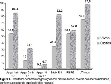
Purpose: to study the prognostic parameters for perinatal death in pregnancies with absent or reversed end-diastolic flow velocity on umbilical artery dopplervelocimetry. Methods: two hundred and four pregnancies were retrospectively reviewed. The methods used were cardiotocography, fetal biophysical profile, amniotic fluid index and dopplervelocimetry of ductus venosus, fetal aorta, middle cerebral artery, umbilical arteries and uterine artery. The logistic regression model was applied to one hundred and seventy cases in order to determine the most accurate variable for predicting perinatal death. Results: the mortality rates were: 28 cases of intrauterine fetal death (13.7%) and 45 neonatal deaths (22.1%). A statistically significant correlation was found between death and the studied variables. The perinatal death rate in the group with birth weight below 1,000 g was 74.7%, and in the group with gestational age at delivery below 31 weeks it was 66.3%. By logistic regression, birth weight was the most accurate variable for predicting perinatal death, and a probability curve for death according to this variable was obtained. Conclusions: absent or reversed end-diastolic flow velocity in the umbilical arteries is a severe fetal condition, where the risk of perinatal death is mainly related to birth weight and a gestational age at delivery below 31 weeks.
Summary
Rev Bras Ginecol Obstet. 2000;22(6):353-363
DOI 10.1590/S0100-72032000000600006
Purpose: to study the prognostic parameters for perinatal death in pregnancies with absent or reversed end-diastolic flow velocity on umbilical artery dopplervelocimetry. Methods: two hundred and four pregnancies were retrospectively reviewed. The methods used were cardiotocography, fetal biophysical profile, amniotic fluid index and dopplervelocimetry of ductus venosus, fetal aorta, middle cerebral artery, umbilical arteries and uterine artery. The logistic regression model was applied to one hundred and seventy cases in order to determine the most accurate variable for predicting perinatal death. Results: the mortality rates were: 28 cases of intrauterine fetal death (13.7%) and 45 neonatal deaths (22.1%). A statistically significant correlation was found between death and the studied variables. The perinatal death rate in the group with birth weight below 1,000 g was 74.7%, and in the group with gestational age at delivery below 31 weeks it was 66.3%. By logistic regression, birth weight was the most accurate variable for predicting perinatal death, and a probability curve for death according to this variable was obtained. Conclusions: absent or reversed end-diastolic flow velocity in the umbilical arteries is a severe fetal condition, where the risk of perinatal death is mainly related to birth weight and a gestational age at delivery below 31 weeks.
Search
Search in:
breast (42) breast cancer (42) breast neoplasms (95) Cesarean section (72) endometriosis (66) infertility (56) Maternal mortality (43) menopause (82) obesity (58) postpartum period (40) pregnancy (225) Pregnancy complications (99) Prenatal care (68) prenatal diagnosis (50) Prevalence (41) Quality of life (51) risk factors (94) ultrasonography (79) urinary incontinence (40) women's health (48)