-
Original Article04-30-2025
Effects of domestic violence on menopausal symptoms, sexual function, and quality of life: a cross-sectional study
Revista Brasileira de Ginecologia e Obstetrícia. 2025;47:e-rbgo16
Abstract
Original ArticleEffects of domestic violence on menopausal symptoms, sexual function, and quality of life: a cross-sectional study
Revista Brasileira de Ginecologia e Obstetrícia. 2025;47:e-rbgo16
Views99Abstract
Objective:
To investigate the association between lifetime experience of domestic violence and climacteric symptoms, sexual function, and quality of life in climacteric women in Rio Grande do Sul, Brazil.
Methods:
A cross-sectional study was conducted with 700 pre-, peri-, and postmenopausal women, recruited online via an anonymous questionnaire (REDCap platform). Women aged 40 to 65 years, residing in Rio Grande do Sul, and classified by the STRAW+10 criteria were included. Climacteric symptoms and sexual function were assessed using the 10-item Cervantes Scale (CS-10) and the 6-item Female Sexual Function Index (FSFI-6). Data were analyzed using SPSS version 18.0; quantitative data as median [IQR], qualitative as frequencies. Group comparisons used Kruskal-Wallis, Chi-Square, and Spearman's correlation between violence against women (VAW) and/or climacteric groups on CS-10 or FSFI-6. Significance was set at 5%.
Results:
The median [IQR] age of pre- (46 [43 - 50] years), peri- (50 [47 – 52] years), and postmenopausal (55 [51 – 58] years) were different among groups. Prevalence rates of psychological (38.8%), sexual (34.9%), and physical (21.3%) violence were observed. Postmenopausal women showed the poorest outcomes. Premenopausal women experiencing violence had severe anxiety, while postmenopausal women reported feeling worthless. Various sexual dysfunctions were associated with violence, including low desire, lubrication issues, and sexual pain.
Conclusions:
Domestic violence was linked to worse climacteric symptoms, sexual function, and quality of life, particularly in postmenopausal women. These findings underscore the need for improved care and public policies to enhance safety and well-being among women of all ages.
Key-words AnxietyClimactericDomestic violenceMenopausePostmenopauseQuality of lifeSexualitysurveys and questionnairesViolence against womenSee moreViews99

This is an Open Access article distributed under the terms of the Creative Commons Attribution License, which permits unrestricted use, distribution, and reproduction in any medium, provided the original work is properly cited. Abstract
Original ArticleEffects of domestic violence on menopausal symptoms, sexual function, and quality of life: a cross-sectional study
Revista Brasileira de Ginecologia e Obstetrícia. 2025;47:e-rbgo16
Views99Abstract
Objective:
To investigate the association between lifetime experience of domestic violence and climacteric symptoms, sexual function, and quality of life in climacteric women in Rio Grande do Sul, Brazil.
Methods:
A cross-sectional study was conducted with 700 pre-, peri-, and postmenopausal women, recruited online via an anonymous questionnaire (REDCap platform). Women aged 40 to 65 years, residing in Rio Grande do Sul, and classified by the STRAW+10 criteria were included. Climacteric symptoms and sexual function were assessed using the 10-item Cervantes Scale (CS-10) and the 6-item Female Sexual Function Index (FSFI-6). Data were analyzed using SPSS version 18.0; quantitative data as median [IQR], qualitative as frequencies. Group comparisons used Kruskal-Wallis, Chi-Square, and Spearman's correlation between violence against women (VAW) and/or climacteric groups on CS-10 or FSFI-6. Significance was set at 5%.
Results:
The median [IQR] age of pre- (46 [43 - 50] years), peri- (50 [47 – 52] years), and postmenopausal (55 [51 – 58] years) were different among groups. Prevalence rates of psychological (38.8%), sexual (34.9%), and physical (21.3%) violence were observed. Postmenopausal women showed the poorest outcomes. Premenopausal women experiencing violence had severe anxiety, while postmenopausal women reported feeling worthless. Various sexual dysfunctions were associated with violence, including low desire, lubrication issues, and sexual pain.
Conclusions:
Domestic violence was linked to worse climacteric symptoms, sexual function, and quality of life, particularly in postmenopausal women. These findings underscore the need for improved care and public policies to enhance safety and well-being among women of all ages.
Key-words AnxietyClimactericDomestic violenceMenopausePostmenopauseQuality of lifeSexualitysurveys and questionnairesViolence against womenSee moreThis is an Open Access article distributed under the terms of the Creative Commons Attribution License, which permits unrestricted use, distribution, and reproduction in any medium, provided the original work is properly cited. 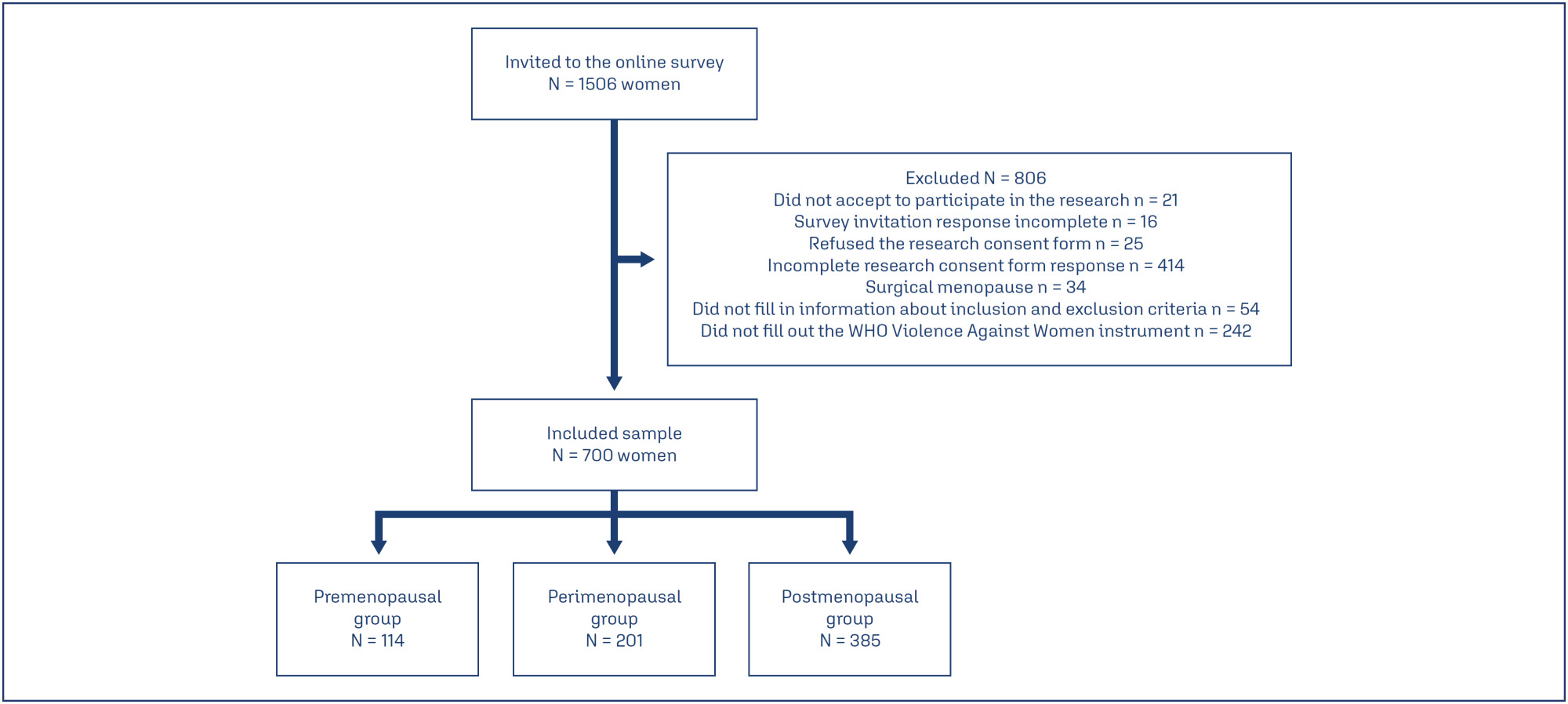
-
Original Article00-00-2024
Skeletal muscle mass obtained by anthropometric equation and presence of sarcopenia in postmenopausal women
- Thaís Loureiro Felipe
 ,
, - Patrícia Paula da Fonseca Grili ,
- Camila Vilarinho Vidigal ,
- Ben-Hur Albergaria ,
- Geise Ferreira da Cruz , [ ... ],
- Valdete Regina Guandalini
Abstract
Original ArticleSkeletal muscle mass obtained by anthropometric equation and presence of sarcopenia in postmenopausal women
Revista Brasileira de Ginecologia e Obstetrícia. 2024;46:e-rbgo9
- Thaís Loureiro Felipe ,
- Patrícia Paula da Fonseca Grili ,
- Camila Vilarinho Vidigal ,
- Ben-Hur Albergaria ,
- Geise Ferreira da Cruz ,
- José Luiz Marques-Rocha ,
- Valdete Regina Guandalini
Views436Abstract
Objective:
To analyze the amount of muscle and the presence of sarcopenia in postmenopausal women using different methods, verifying the agreement between them as to skeletal muscle mass (SMM).
Methods:
This cross-sectional observational study was conducted with postmenopausal women aged ≥ 50 years. SMM was obtained from a predictive equation, Bioelectrical Impedance (BIA), and Dual Energy X-Ray Absorptiometry (DXA). The skeletal muscle mass index (SMI) and the appendicular skeletal muscle mass index (ASMI) were calculated. The cut-off point of SMI was determined for the population itself. The agreement between the SMI obtained using the different methods was verified. Sarcopenia was diagnosed according to the criteria proposed by the European Working Group on Sarcopenia in Older People 2 (EWGSOP2). The significance level adopted for all tests was 5.0%.
Results:
A total of 112 women were evaluated, with an average age of 66.1 ± 5.65 years. Among them, 51.8% were sufficiently active and 43.8% were overweight and obese. The SMI cut-offs were 6.46 kg/m2 for the predictive equation and 7.66 kg/m2 for BIA, with high sensitivity and specificity. There was an excellent agreement in the identification of SMM by the predictive equation (0.89 [0.824-0.917], p < 0.001) and BIA (0.92 [0.883-0.945], p < 0.001), in reference to DXA. The prevalence of sarcopenia was 0.9%, 1.8%, and 2.7% according to BIA, DXA, and the predictive equation, respectively.
Conclusion:
The predictive equation showed the expected agreement in estimating skeletal muscle mass in postmenopausal women, offering a viable and accurate alternative.
Key-words AnthropometryBioelectrical impedanceBody compositionMuscle massObesityOverweightPostmenopausesarcopeniaSkeletal MuscleSee moreViews436This is an Open Access article distributed under the terms of the Creative Commons Attribution License, which permits unrestricted use, distribution, and reproduction in any medium, provided the original work is properly cited. Abstract
Original ArticleSkeletal muscle mass obtained by anthropometric equation and presence of sarcopenia in postmenopausal women
Revista Brasileira de Ginecologia e Obstetrícia. 2024;46:e-rbgo9
- Thaís Loureiro Felipe ,
- Patrícia Paula da Fonseca Grili ,
- Camila Vilarinho Vidigal ,
- Ben-Hur Albergaria ,
- Geise Ferreira da Cruz ,
- José Luiz Marques-Rocha ,
- Valdete Regina Guandalini
Views436Abstract
Objective:
To analyze the amount of muscle and the presence of sarcopenia in postmenopausal women using different methods, verifying the agreement between them as to skeletal muscle mass (SMM).
Methods:
This cross-sectional observational study was conducted with postmenopausal women aged ≥ 50 years. SMM was obtained from a predictive equation, Bioelectrical Impedance (BIA), and Dual Energy X-Ray Absorptiometry (DXA). The skeletal muscle mass index (SMI) and the appendicular skeletal muscle mass index (ASMI) were calculated. The cut-off point of SMI was determined for the population itself. The agreement between the SMI obtained using the different methods was verified. Sarcopenia was diagnosed according to the criteria proposed by the European Working Group on Sarcopenia in Older People 2 (EWGSOP2). The significance level adopted for all tests was 5.0%.
Results:
A total of 112 women were evaluated, with an average age of 66.1 ± 5.65 years. Among them, 51.8% were sufficiently active and 43.8% were overweight and obese. The SMI cut-offs were 6.46 kg/m2 for the predictive equation and 7.66 kg/m2 for BIA, with high sensitivity and specificity. There was an excellent agreement in the identification of SMM by the predictive equation (0.89 [0.824-0.917], p < 0.001) and BIA (0.92 [0.883-0.945], p < 0.001), in reference to DXA. The prevalence of sarcopenia was 0.9%, 1.8%, and 2.7% according to BIA, DXA, and the predictive equation, respectively.
Conclusion:
The predictive equation showed the expected agreement in estimating skeletal muscle mass in postmenopausal women, offering a viable and accurate alternative.
Key-words AnthropometryBioelectrical impedanceBody compositionMuscle massObesityOverweightPostmenopausesarcopeniaSkeletal MuscleSee moreThis is an Open Access article distributed under the terms of the Creative Commons Attribution License, which permits unrestricted use, distribution, and reproduction in any medium, provided the original work is properly cited. - Thaís Loureiro Felipe
-
Original Article10-18-2021
Sexual Function and Associated Factors in Postmenopausal Women
Revista Brasileira de Ginecologia e Obstetrícia. 2021;43(7):522-529
Abstract
Original ArticleSexual Function and Associated Factors in Postmenopausal Women
Revista Brasileira de Ginecologia e Obstetrícia. 2021;43(7):522-529
Views216See moreAbstract
Objective
To assess the sexual function and associated factors in postmenopausal women.
Methods
This a descriptive, cross-sectional study with 380 women aged 40 to 65 years, users of public health services in 2019. Questionnaires were applied on demographic characteristics, on climacteric symptoms (menopause rating scale) and on sexual function (sexual quotient, female version). Bivariate andmultiple analyses by logistic regression were performed, with adjusted odds ratios (ORad) and 95% confidence intervals (95%CIs).
Results
More than half (243/64%) of the participating women were at risk of sexual dysfunction, with lower scores in the domains of sexual desire and interest, comfort, orgasm, and satisfaction. Women with a partner (ORad 2.07; 95%CI 1.03-4.17) and those who reported sleep problems (ORad 2.72; 95%CI 1.77-4.19), depressed mood (ORad 2.03; 95%CI 1.32-3.10), sexual complaints (ORad 8.16; 95%CI 5.06-13.15), and vaginal dryness (ORad 3.44; 95%CI 2.22-5.32) showed greater chance of sexual dysfunction.
Conclusion
There was a high prevalence of sexual dysfunction, with the influence of conjugality and climacteric symptoms on sexual function.
Views216This is an Open Access article distributed under the terms of the Creative Commons Attribution License, which permits unrestricted use, distribution, and reproduction in any medium, provided the original work is properly cited. Abstract
Original ArticleSexual Function and Associated Factors in Postmenopausal Women
Revista Brasileira de Ginecologia e Obstetrícia. 2021;43(7):522-529
Views216See moreAbstract
Objective
To assess the sexual function and associated factors in postmenopausal women.
Methods
This a descriptive, cross-sectional study with 380 women aged 40 to 65 years, users of public health services in 2019. Questionnaires were applied on demographic characteristics, on climacteric symptoms (menopause rating scale) and on sexual function (sexual quotient, female version). Bivariate andmultiple analyses by logistic regression were performed, with adjusted odds ratios (ORad) and 95% confidence intervals (95%CIs).
Results
More than half (243/64%) of the participating women were at risk of sexual dysfunction, with lower scores in the domains of sexual desire and interest, comfort, orgasm, and satisfaction. Women with a partner (ORad 2.07; 95%CI 1.03-4.17) and those who reported sleep problems (ORad 2.72; 95%CI 1.77-4.19), depressed mood (ORad 2.03; 95%CI 1.32-3.10), sexual complaints (ORad 8.16; 95%CI 5.06-13.15), and vaginal dryness (ORad 3.44; 95%CI 2.22-5.32) showed greater chance of sexual dysfunction.
Conclusion
There was a high prevalence of sexual dysfunction, with the influence of conjugality and climacteric symptoms on sexual function.
This is an Open Access article distributed under the terms of the Creative Commons Attribution License, which permits unrestricted use, distribution, and reproduction in any medium, provided the original work is properly cited. -
Original Article07-30-2021
Gynecological/Obstetric Background and Rheumatoid Arthritis: A Cross-sectional Study in Brazilian Patients
Revista Brasileira de Ginecologia e Obstetrícia. 2021;43(5):357-361
Abstract
Original ArticleGynecological/Obstetric Background and Rheumatoid Arthritis: A Cross-sectional Study in Brazilian Patients
Revista Brasileira de Ginecologia e Obstetrícia. 2021;43(5):357-361
Views139See moreAbstract
Objective
To study a sample of rheumatoid arthritis (RA) patients for their gynecological/obstetric history and compare them to controls to determine their influences on number of pregnancies, menarche, menopause and reproductive years following RA onset.
Methods
This is a cross-sectional study of 122 RA patients and 126 controls. Patients and controls were questioned about age of menarche, age of menopause, number of pregnancies and abortions. Reproductive years were calculated as the difference between age at menopause and age at menarche. For comparison, we used the Mann-Whitney, unpaired t, chi-squared, and Spearman tests. The adopted significance was 5%.
Results
In the RA patients with disease beginning in the postmenopausal years, the period of reproductive years (age at menopause - age of menarche) showed a positive correlation with age at disease onset (rho=0.46; 95% confidence interval [CI]=0.20- 0.55 with p=0.0008). The number of pregnancies was higher in patients with postmenopausal disease onset when compared with those with premenopausal disease onset (median of 3 with interquartile range [IQR]=2-4 versus median of 2 with IQR=1-3; p=0.009), and RA patients had more pregnancies than controls (p=0.0002).
Conclusion
The present study shows that, in our population, the duration of reproductive years and the number of pregnancies are linked to the onset of RA.
Views139This is an Open Access article distributed under the terms of the Creative Commons Attribution License, which permits unrestricted use, distribution, and reproduction in any medium, provided the original work is properly cited. Abstract
Original ArticleGynecological/Obstetric Background and Rheumatoid Arthritis: A Cross-sectional Study in Brazilian Patients
Revista Brasileira de Ginecologia e Obstetrícia. 2021;43(5):357-361
Views139See moreAbstract
Objective
To study a sample of rheumatoid arthritis (RA) patients for their gynecological/obstetric history and compare them to controls to determine their influences on number of pregnancies, menarche, menopause and reproductive years following RA onset.
Methods
This is a cross-sectional study of 122 RA patients and 126 controls. Patients and controls were questioned about age of menarche, age of menopause, number of pregnancies and abortions. Reproductive years were calculated as the difference between age at menopause and age at menarche. For comparison, we used the Mann-Whitney, unpaired t, chi-squared, and Spearman tests. The adopted significance was 5%.
Results
In the RA patients with disease beginning in the postmenopausal years, the period of reproductive years (age at menopause - age of menarche) showed a positive correlation with age at disease onset (rho=0.46; 95% confidence interval [CI]=0.20- 0.55 with p=0.0008). The number of pregnancies was higher in patients with postmenopausal disease onset when compared with those with premenopausal disease onset (median of 3 with interquartile range [IQR]=2-4 versus median of 2 with IQR=1-3; p=0.009), and RA patients had more pregnancies than controls (p=0.0002).
Conclusion
The present study shows that, in our population, the duration of reproductive years and the number of pregnancies are linked to the onset of RA.
This is an Open Access article distributed under the terms of the Creative Commons Attribution License, which permits unrestricted use, distribution, and reproduction in any medium, provided the original work is properly cited. -
Original Article03-27-2020
The Effect of Testosterone Replacement on Intramedullary, Inguinal and Visceral Fat in Ovariectomized Rats
- Lorena Doretto da Silva ,
- Juliana Mora Veridiano ,
- Jussara Celi Conceição Oliveira ,
- Anna Carolina Haddad Sayeg ,
- Ana Maria Amaral Antonio Mader , [ ... ],
- Marcelo Luis Steiner
Abstract
Original ArticleThe Effect of Testosterone Replacement on Intramedullary, Inguinal and Visceral Fat in Ovariectomized Rats
Revista Brasileira de Ginecologia e Obstetrícia. 2020;42(1):43-50
- Lorena Doretto da Silva ,
- Juliana Mora Veridiano ,
- Jussara Celi Conceição Oliveira ,
- Anna Carolina Haddad Sayeg ,
- Ana Maria Amaral Antonio Mader ,
- Giuliana Petri ,
- Bianca Bianco ,
- César Eduardo Fernandes ,
- Olga Maria Szymanski de Toledo ,
- Luciano de Melo Pompei ,
- Marcelo Luis Steiner
Views225See moreAbstract
Objective
The present article aims to evaluate the impact of testosterone treatment on the expansion of visceral, subcutaneous and intramedullary adipose tissue of ovariectomized rats and the visceral and subcutaneous fat expression of peroxisome proliferator-activated receptors (PPARs) gamma.
Methods
In total 48 female Wistar rats were castrated and randomly divided into 6 treatment groups: group E2 was submitted to estradiol 5 μg/day; group T, to testosterone 5 μg/day; group E2+ T, to estradiol 5 μg/day + testosterone 5 μg/day; group TT, to testosterone 30 μg/day; group E2+ TT, to estradiol 5 μg/day+ testosterone 30 μg/day; and placebo was administered to group P. After 5 weeks, the rats were euthanized, the inguinal and visceral adipose tissues were harvested, weighted, and had their PPAR gamma expression evaluated by reverse transcription quantitative polymerase chain reaction (RTqPCR). The right femurs were harvested and histologically prepared to performthe number count of the intramedullary adipocytes.
Results
The expansion of visceral fat tissue was much higher in the TT group when compared with other treated groups (p < 0.001). The TT group also showed a higher expansion of inguinal fat (p < 0.01), and groups E2 +T and E2+ TT presented lower growth compared to the P group (p < 0.01). The number of femur intramedullary adipocytes only showed significant differences between groups TT and E2 + TT (p < 0.05). The expression of PPAR gamma showed no differences among the groups.
Conclusion
The use of testosterone in high doses leads to an important expansion in both visceral and inguinal adipose tissues. Association with estradiol exerts an expansion-repressive effect on the visceral and inguinal adipose tissues.
Views225This is an Open Access article distributed under the terms of the Creative Commons Attribution License, which permits unrestricted use, distribution, and reproduction in any medium, provided the original work is properly cited. Abstract
Original ArticleThe Effect of Testosterone Replacement on Intramedullary, Inguinal and Visceral Fat in Ovariectomized Rats
Revista Brasileira de Ginecologia e Obstetrícia. 2020;42(1):43-50
- Lorena Doretto da Silva ,
- Juliana Mora Veridiano ,
- Jussara Celi Conceição Oliveira ,
- Anna Carolina Haddad Sayeg ,
- Ana Maria Amaral Antonio Mader ,
- Giuliana Petri ,
- Bianca Bianco ,
- César Eduardo Fernandes ,
- Olga Maria Szymanski de Toledo ,
- Luciano de Melo Pompei ,
- Marcelo Luis Steiner
Views225See moreAbstract
Objective
The present article aims to evaluate the impact of testosterone treatment on the expansion of visceral, subcutaneous and intramedullary adipose tissue of ovariectomized rats and the visceral and subcutaneous fat expression of peroxisome proliferator-activated receptors (PPARs) gamma.
Methods
In total 48 female Wistar rats were castrated and randomly divided into 6 treatment groups: group E2 was submitted to estradiol 5 μg/day; group T, to testosterone 5 μg/day; group E2+ T, to estradiol 5 μg/day + testosterone 5 μg/day; group TT, to testosterone 30 μg/day; group E2+ TT, to estradiol 5 μg/day+ testosterone 30 μg/day; and placebo was administered to group P. After 5 weeks, the rats were euthanized, the inguinal and visceral adipose tissues were harvested, weighted, and had their PPAR gamma expression evaluated by reverse transcription quantitative polymerase chain reaction (RTqPCR). The right femurs were harvested and histologically prepared to performthe number count of the intramedullary adipocytes.
Results
The expansion of visceral fat tissue was much higher in the TT group when compared with other treated groups (p < 0.001). The TT group also showed a higher expansion of inguinal fat (p < 0.01), and groups E2 +T and E2+ TT presented lower growth compared to the P group (p < 0.01). The number of femur intramedullary adipocytes only showed significant differences between groups TT and E2 + TT (p < 0.05). The expression of PPAR gamma showed no differences among the groups.
Conclusion
The use of testosterone in high doses leads to an important expansion in both visceral and inguinal adipose tissues. Association with estradiol exerts an expansion-repressive effect on the visceral and inguinal adipose tissues.
This is an Open Access article distributed under the terms of the Creative Commons Attribution License, which permits unrestricted use, distribution, and reproduction in any medium, provided the original work is properly cited. - Lorena Doretto da Silva
-
Original Article06-01-2015
Assessment of functional fitness through the set of AAHPERD tests in women after menopause: Is there a decline between the fifth and sixth decades of life?
Revista Brasileira de Ginecologia e Obstetrícia. 2015;37(6):278-282
Abstract
Original ArticleAssessment of functional fitness through the set of AAHPERD tests in women after menopause: Is there a decline between the fifth and sixth decades of life?
Revista Brasileira de Ginecologia e Obstetrícia. 2015;37(6):278-282
DOI 10.1590/SO100-720320150005326
Views138See morePURPOSE:
to analize the level of functional fitness of a group of postmenopausal women in
the city of Presidente Prudente using the set of functional fitness tests of the
American Alliance for Health, Physical Education, Recreation and Dance and to
check whether there are differences between groups of women in the fifth and sixth
decade of life.METHODS:
This was a cross-sectional study conducted on 175 postmenopausal women (follicle
stimulating hormone level>26.72 mIU/L) in the city of Presidente Prudente in
2013. The inclusion criteria were not being part of any type of systematic motor
intervention for at least six months before the collection of research data;
absence of motor or cognitive impairment that would prevent the evaluation
protocols, and absence of chronic or degenerative disease, musculoskeletal injury
or comorbidity that could prevent or limit the evaluations. The women were
evaluated by the same trained examiners. The 50 to 59 year group showed a mean age
of 55.3±4.5 years, mean FSH values of 53.5±21.1 mIU/mL, mean coordination of
11.4±2.2 seconds, mean strength of 20.1±3.9 repetitions, mean flexibility of
51.7±11.8 cm, mean 23.2±2.8 seconds agility and mean aerobic resistance of
500±43/2 . The 60 to 69 year group had a mean age of 65.1±4.1 years with FSH
54.9±15.9, 11.6±2.6 seconds coordination, strength 20.3±4.7 repetitions, 54.6±11.2
cm flexibility, agility 24.7±4.3 seconds, and aerobic resistance of 508±51
seconds.CONCLUSION:
It was possible to analyze the functional fitness of postmenopausal women through
the set of the American Alliance testing for Health, Physical Education,
Recreation and Dance with no significant differences between groups for the
variables strength, flexibility, aerobic capacity and coordination, and with only
the speed variable showing significant differences. We recommend further studies
seeking to formulate normative values for the population in question.Views138This is an Open Access article distributed under the terms of the Creative Commons Attribution License, which permits unrestricted use, distribution, and reproduction in any medium, provided the original work is properly cited. Abstract
Original ArticleAssessment of functional fitness through the set of AAHPERD tests in women after menopause: Is there a decline between the fifth and sixth decades of life?
Revista Brasileira de Ginecologia e Obstetrícia. 2015;37(6):278-282
DOI 10.1590/SO100-720320150005326
Views138See morePURPOSE:
to analize the level of functional fitness of a group of postmenopausal women in
the city of Presidente Prudente using the set of functional fitness tests of the
American Alliance for Health, Physical Education, Recreation and Dance and to
check whether there are differences between groups of women in the fifth and sixth
decade of life.METHODS:
This was a cross-sectional study conducted on 175 postmenopausal women (follicle
stimulating hormone level>26.72 mIU/L) in the city of Presidente Prudente in
2013. The inclusion criteria were not being part of any type of systematic motor
intervention for at least six months before the collection of research data;
absence of motor or cognitive impairment that would prevent the evaluation
protocols, and absence of chronic or degenerative disease, musculoskeletal injury
or comorbidity that could prevent or limit the evaluations. The women were
evaluated by the same trained examiners. The 50 to 59 year group showed a mean age
of 55.3±4.5 years, mean FSH values of 53.5±21.1 mIU/mL, mean coordination of
11.4±2.2 seconds, mean strength of 20.1±3.9 repetitions, mean flexibility of
51.7±11.8 cm, mean 23.2±2.8 seconds agility and mean aerobic resistance of
500±43/2 . The 60 to 69 year group had a mean age of 65.1±4.1 years with FSH
54.9±15.9, 11.6±2.6 seconds coordination, strength 20.3±4.7 repetitions, 54.6±11.2
cm flexibility, agility 24.7±4.3 seconds, and aerobic resistance of 508±51
seconds.CONCLUSION:
It was possible to analyze the functional fitness of postmenopausal women through
the set of the American Alliance testing for Health, Physical Education,
Recreation and Dance with no significant differences between groups for the
variables strength, flexibility, aerobic capacity and coordination, and with only
the speed variable showing significant differences. We recommend further studies
seeking to formulate normative values for the population in question.This is an Open Access article distributed under the terms of the Creative Commons Attribution License, which permits unrestricted use, distribution, and reproduction in any medium, provided the original work is properly cited. -
Original Article02-01-2014
Frequency of sleep disturbances in overweight/obese postmenopausal women
Revista Brasileira de Ginecologia e Obstetrícia. 2014;36(2):90-96
Abstract
Original ArticleFrequency of sleep disturbances in overweight/obese postmenopausal women
Revista Brasileira de Ginecologia e Obstetrícia. 2014;36(2):90-96
DOI 10.1590/S0100-72032014000200008
Views98PURPOSE:
To evaluate the frequency of sleep disorders, such as obstructive sleep apnea,
restless leg syndrome and insomnia in overweight/obese postmenopausal women seen
in a climacteric sleep disorders clinic.METHODS:
Thirty-four postmenopausal women were selected using the following inclusion
criteria: age between 50 and 70 years; at least 12 months of amenorrhea; body mass
index (BMI) greater than or equal to 25 kg/m2; and sleep-related
complaints with at least one previous polysomnography. Patients provided responses
to 6 questionnaires related to sleep characteristics and menopausal symptoms.
Weight and height were measured using standardized scales, and abdomen and hip
circumferences were also measured. The statistical analyses were performed using
the χ2 test for qualitative variables and using Student's t-test for
quantitative variables.RESULTS:
Patients' characteristics were as follows: mean age of 60.35 years; mean BMI of
31.62; an average of 11.61 postmenopausal years and an average Kupperman Index of
19. A total of 85.2% of the patients had a waist/hip ratio of less than 0.8. The
Epworth Scale score was greater than or equal to 9 in 50% of patients; 68% had
sleep disturbances according to the Pittsburgh Index, and 68% were classified as
high-risk for sleep apnea by the Berlin Questionnaire. On polysomnography, 70.58%
of the patients had a sleep efficiency lower than 85%; 79.41% had a sleep latency
of less than 30 min; 58.82% had a REM sleep latency of less than 90 min, and
44.11% had mild apnea. When the groups were compared, a linear association was
identified between BMI and the AHI average, and a relationship between high BMI
and use of drugs for thyroid treatment was found.CONCLUSION:
There was a high prevalence of sleep-disordered breathing, initial insomnia,
fragmented sleep, and thyroid disorders in the group with higher BMI.Key-words ClimactericInsomniaObesityPostmenopauseRestless leg syndromeSleep apnea syndromeSleep disordersSee moreViews98This is an Open Access article distributed under the terms of the Creative Commons Attribution License, which permits unrestricted use, distribution, and reproduction in any medium, provided the original work is properly cited. Abstract
Original ArticleFrequency of sleep disturbances in overweight/obese postmenopausal women
Revista Brasileira de Ginecologia e Obstetrícia. 2014;36(2):90-96
DOI 10.1590/S0100-72032014000200008
Views98PURPOSE:
To evaluate the frequency of sleep disorders, such as obstructive sleep apnea,
restless leg syndrome and insomnia in overweight/obese postmenopausal women seen
in a climacteric sleep disorders clinic.METHODS:
Thirty-four postmenopausal women were selected using the following inclusion
criteria: age between 50 and 70 years; at least 12 months of amenorrhea; body mass
index (BMI) greater than or equal to 25 kg/m2; and sleep-related
complaints with at least one previous polysomnography. Patients provided responses
to 6 questionnaires related to sleep characteristics and menopausal symptoms.
Weight and height were measured using standardized scales, and abdomen and hip
circumferences were also measured. The statistical analyses were performed using
the χ2 test for qualitative variables and using Student's t-test for
quantitative variables.RESULTS:
Patients' characteristics were as follows: mean age of 60.35 years; mean BMI of
31.62; an average of 11.61 postmenopausal years and an average Kupperman Index of
19. A total of 85.2% of the patients had a waist/hip ratio of less than 0.8. The
Epworth Scale score was greater than or equal to 9 in 50% of patients; 68% had
sleep disturbances according to the Pittsburgh Index, and 68% were classified as
high-risk for sleep apnea by the Berlin Questionnaire. On polysomnography, 70.58%
of the patients had a sleep efficiency lower than 85%; 79.41% had a sleep latency
of less than 30 min; 58.82% had a REM sleep latency of less than 90 min, and
44.11% had mild apnea. When the groups were compared, a linear association was
identified between BMI and the AHI average, and a relationship between high BMI
and use of drugs for thyroid treatment was found.CONCLUSION:
There was a high prevalence of sleep-disordered breathing, initial insomnia,
fragmented sleep, and thyroid disorders in the group with higher BMI.Key-words ClimactericInsomniaObesityPostmenopauseRestless leg syndromeSleep apnea syndromeSleep disordersSee moreThis is an Open Access article distributed under the terms of the Creative Commons Attribution License, which permits unrestricted use, distribution, and reproduction in any medium, provided the original work is properly cited. -
Original Article01-11-2012
Periodontal disease in women in post-menopause and its relationship with osteoporosis
Revista Brasileira de Ginecologia e Obstetrícia. 2012;34(12):563-567
Abstract
Original ArticlePeriodontal disease in women in post-menopause and its relationship with osteoporosis
Revista Brasileira de Ginecologia e Obstetrícia. 2012;34(12):563-567
DOI 10.1590/S0100-72032012001200006
Views92See morePURPOSE: To investigate the relationship between periodontitis and osteoporosis, using a case-control study about periodontal status of postmenopausal women. METHODS: A total of 99 postmenopausal women were divided into three groups: normal bone (Gn, n=45), osteopenia (Gpenia, n=31) and osteoporosis (Gporosis, n=23). The categorization of bone mass was measured by dual energy absorptiometry with X-rays in the lumbar spine (L2 - L4), by assessing bone mineral density. Clinical attachment level (CAL), gingival bleeding index (GI), plaque index (PI), and probing depth (PD) were determined in all participants by a single examiner. The data were submitted to BioEstat 2.0 software through parametric analysis of variance (ANOVA) and the Bonferroni test, with the level of significance set at 5%. RESULTS: Women with osteoporosis presented the highest percentage of periodontal disease, with higher average CAL (2.6±0.4 mm) and PD (2.8±0.6 mm), GI (72.8±25.9 mm) and PI (72.9±24.2 mm). Statistical analysis revealed a significant difference in periodontal situation between Gn and Gporosis (p=0,01) and between Gpenia and Gporosis (p=0,03). CONCLUSION: Osteoporosis may have an influence on periodontal condition, based on the relation between periodontitis and osteoporosis in postmenopausal women.
Views92This is an Open Access article distributed under the terms of the Creative Commons Attribution License, which permits unrestricted use, distribution, and reproduction in any medium, provided the original work is properly cited. Abstract
Original ArticlePeriodontal disease in women in post-menopause and its relationship with osteoporosis
Revista Brasileira de Ginecologia e Obstetrícia. 2012;34(12):563-567
DOI 10.1590/S0100-72032012001200006
Views92See morePURPOSE: To investigate the relationship between periodontitis and osteoporosis, using a case-control study about periodontal status of postmenopausal women. METHODS: A total of 99 postmenopausal women were divided into three groups: normal bone (Gn, n=45), osteopenia (Gpenia, n=31) and osteoporosis (Gporosis, n=23). The categorization of bone mass was measured by dual energy absorptiometry with X-rays in the lumbar spine (L2 - L4), by assessing bone mineral density. Clinical attachment level (CAL), gingival bleeding index (GI), plaque index (PI), and probing depth (PD) were determined in all participants by a single examiner. The data were submitted to BioEstat 2.0 software through parametric analysis of variance (ANOVA) and the Bonferroni test, with the level of significance set at 5%. RESULTS: Women with osteoporosis presented the highest percentage of periodontal disease, with higher average CAL (2.6±0.4 mm) and PD (2.8±0.6 mm), GI (72.8±25.9 mm) and PI (72.9±24.2 mm). Statistical analysis revealed a significant difference in periodontal situation between Gn and Gporosis (p=0,01) and between Gpenia and Gporosis (p=0,03). CONCLUSION: Osteoporosis may have an influence on periodontal condition, based on the relation between periodontitis and osteoporosis in postmenopausal women.
This is an Open Access article distributed under the terms of the Creative Commons Attribution License, which permits unrestricted use, distribution, and reproduction in any medium, provided the original work is properly cited.