Summary
Revista Brasileira de Ginecologia e Obstetrícia. 2024;46:e-rbgo55
Our study evaluated the effectiveness of the Botucatu Abbreviated Protocol in breast magnetic resonance imaging (MRI) within Brazil’s public healthcare system, focusing on its impact on patient access to MRI exams.
This retrospective study involved 197 breast MRI exams of female patients over 18 years with histological breast carcinoma diagnosis, conducted at Hospital das Clínicas de Botucatu - UNESP between 2014 and 2018. Two experienced examiners prospectively and blindly analyzed the exams using an Integrated Picture Archiving and Communication System (PACS). They first evaluated the Botucatu Abbreviated Protocol, created from sequences of the complete protocol (PC), and after an average interval of 30 days, they reassessed the same 197 exams with the complete protocol. Dynamic and morphological characteristics of lesions were assessed according to BI-RADS 5th edition criteria. The study also analyzed the average number of monthly exams before and after the implementation of Botucatu Abbreviated Protocol.
The Botucatu Abbreviated Protocol showed high sensitivity (99% and 96%) and specificity (90.9% and 96%). There was a significant increase in the average monthly MRI exams from 6.62 to 23.8 post-implementation.
The Botucatu Abbreviated Protocol proved effective in maintaining diagnostic accuracy and improving accessibility to breast MRI exams, particularly in the public healthcare setting.
Summary
Revista Brasileira de Ginecologia e Obstetrícia. 2023;45(8):480-488
To present the update of the recommendations of the Brazilian College of Radiology and Diagnostic Imaging, the Brazilian Society of Mastology and the Brazilian Federation of Associations of Gynecology and Obstetrics for breast cancer screening in Brazil.
Scientific evidence published in Medline, EMBASE, Cochrane Library, EBSCO, CINAHL and Lilacs databases between January 2012 and July 2022 was searched. Recommendations were based on this evidence by consensus of the expert committee of the three entities.
Annual mammography screening is recommended for women at usual risk aged 40–74 years. Above 75 years, it should be reserved for those with a life expectancy greater than seven years. Women at higher than usual risk, including those with dense breasts, with a personal history of atypical lobular hyperplasia, classic lobular carcinoma in situ, atypical ductal hyperplasia, treatment for breast cancer or chest irradiation before age 30, or even, carriers of a genetic mutation or with a strong family history, benefit from complementary screening, and should be considered individually. Tomosynthesis is a form of mammography and should be considered in screening whenever accessible and available.
Summary
Revista Brasileira de Ginecologia e Obstetrícia. 2022;44(4):435-441
Antenatal recognition of severe cases of congenital diaphragmatic hernia (CDH) by ultrasound (US) and magnetic resonance imaging (MRI) may aid decisions regarding the indication of fetal endoscopic tracheal occlusion.
An integrative review was performed. Searches in MEDLINE and EMBASE used terms related to CDH, diagnosis, MRI, and US. The inclusion criteria were reviews and guidelines approaching US and MRI markers of severity of CDH published in English in the past 10 years.
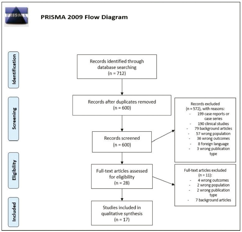
The search retrieved 712 studies, out of which 17 publications were included. The US parameters were stomach and liver positions, lung-to-head ratio (LHR), observed/expected LHR (o/e LHR), and quantitative lung index. The MRI parameters were total fetal lung volume (TFLV), observed/expected TFLV, relative fetal or percent predicted lung volumes, liver intrathoracic ratio, and modified McGoon index. None of the parameters was reported to be superior to the others.
The most mentioned parameters were o/e LHR, LHR, liver position, o/e TFLV, and TFLV.
Summary
Revista Brasileira de Ginecologia e Obstetrícia. 2021;43(12):985-987
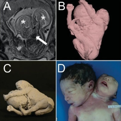
Conjoined twins (CTs) are a rare complication from monochorionic and monoamniotic twin pregnancies. We describe the use of 3D technologies, including 3D virtual and 3D physical models on prenatal evaluation of one parapagus CT. A 16-year-old G1P0 woman was referred for fetal magnetic resonance imaging (MRI) anatomical evaluation of a CT at 28 weeks of gestation. 3D images of the fetal surface were generated by the software during the examination for spatial comprehension of the relationship between the fetal parts. The pair of CTs died at the 32nd week of gestation, a few hours after cesarean section. 3D technologies are an important tool for parental counseling and preparation of the multidisciplinary care team for delivery and neonatal assistance and possible surgical planning for postnatal separation in CTs cases.
Summary
Revista Brasileira de Ginecologia e Obstetrícia. 2021;43(4):317-322
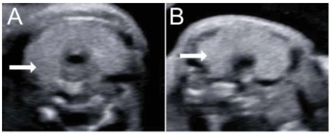
Fetal thyroid complications in pregnancy are uncommon, and are commonly related to the passage of substances through the placenta. The excessive iodine intake during the pregnancy is a well-known mechanism of fetal thyroid enlargement or goiter, and invasive procedures have been proposed for the treatment of fetal thyroid pathologies. In the present report, we demonstrate two cases from different centers of prenatal diagnosis of fetal thyroid enlargement and/or goiter in three fetuses (one pair of twins, wherein both fetuses were affected, and one singleton pregnancy). The anamnesis revealed the ingestion of iodine by the patients, prescribed from inadequate vitamin supplementation. In both cases, the cessation of iodine supplement intake resulted in a marked reduction of the volume of the fetal thyroid glands, demonstrating that conservative treatmentmay be an option in those cases. Also, clinicians must be aware that patients may be exposed to harmful dosages or substances during pregnancy.
Summary
Revista Brasileira de Ginecologia e Obstetrícia. 2021;43(1):46-53
Magnetic resonance imaging (MRI) has been considered another tool for use during the pre- and postoperative periods of the management of pelvic-organ prolapse (POP). However, there is little consensus regarding its practical use for POP and the association betweenMRI lines of reference and physical examination.We aimedto evaluate the mid- to long-term results of two surgical techniques for apical prolapse.
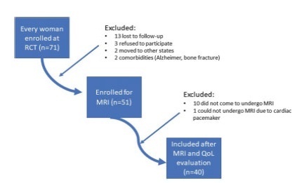
In total, 40 women with apical POP randomized from 2014 to 2016 underwent abdominal sacrocolpopexy (ASC group; n = 20) or bilateral vaginal sacrospinous fixation with an anterior mesh (VSF-AM group; n = 20). A physical examination using the POP Quantification System (POP-Q) for staging (objective cure) and the International Consultation on Incontinence Questionnaire-Vaginal Symptoms (ICIQ-VS: subjective cure), were applied and analyzed before and one year after surgery respectively. All MRI variables (pubococcigeous line [PCL], bladder base [BB], anorectal junction [ARJ], and the estimated levator ani subtended volume [eLASV]) were investigated one year after surgery. Significance was established at p < 0.05.
After a mean 27-month follow-up, according to the MRI criteria, 60% of the women were cured in the VSF-AM group versus 45% in ASC group (p= 0.52). The POP-Q and objective cure rates by MRI were correlated in the anterior vaginal wall (p= 0.007), but no correlationwas foundwith the subjective cure. The eLASVwas largeramongthe patients with surgical failure, and a cutoff of ≥ 33.5mm3 was associated with postoperative failure (area under the receiver operating characteristic curve [ROC]: 0.813; p= 0.002).
Both surgeries for prolapse were similar regarding theobjective variables (POP-Q measurements and MRI cure rates). Larger eLASV areas were associated with surgical failure.
Summary
Revista Brasileira de Ginecologia e Obstetrícia. 2019;41(1):17-23
To assess and compare the sensitivity and specificity of ultrasonography and magnetic resonance imaging in the diagnosis of placenta accreta in patients with placenta previa.
This retrospective cohort study included 37 women, and was conducted between January 2013 and October 2015; 16 out of the 37 women suffered from placenta accreta. Histopathology was considered the gold standard for the diagnosis of placenta accreta; in its absence, a description of the intraoperative findings was used. The associations among the variables were investigated using the Pearson chi-squared test and the Mann-Whitney U-test.
The mean age of the patients was 31.8 ± 7.3 years, the mean number of pregnancies was 2.8 ± 1.1, the mean number of births was 1.4 ± 0.7, and the mean number of previous cesarean sections was 1.2 ± 0.8. Patients with placenta accreta had a higher frequency of history of cesarean section than those without it (63.6% versus 36.4% respectively; p < 0.001). The mean gestational age at birth among women diagnosed with placenta previa accreta was 35.4 ± 1.1 weeks. The mean birth weight was 2,635.9 ± 374.1 g. The sensitivity of the ultrasound was 87.5%, with a positive predictive value (PPV) of 65.1%, and a negative predictive value (NPV) of 75.0%. The sensitivity of the magnetic resonance imaging was 92.9%, with a PPV of
The ultrasound and the magnetic resonance imaging showed similar sensitivity and specificity for the diagnosis of placenta accreta.
Summary
Revista Brasileira de Ginecologia e Obstetrícia. 2016;38(9):436-442
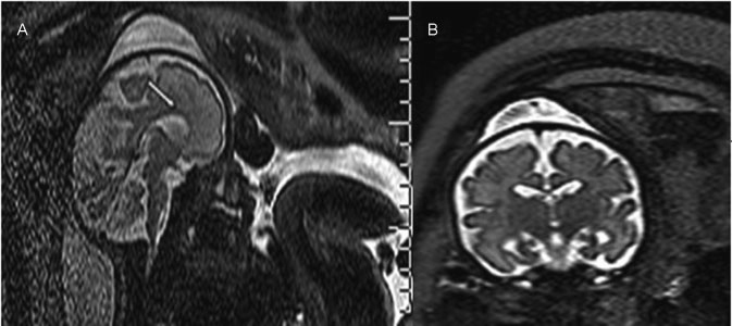
Ventriculomegaly (VM) is one the most frequent anomalies detected on prenatal ultrasound. Magnetic resonance imaging (MRI) may enhance diagnostic accuracy and prediction of developmental outcome in newborns.
The aim of this study was to assess the correlation between ultrasound and MRI in fetuses with isolated mild and moderate VM. The secondary aim was to report the neurodevelopmental outcome at 4 years of age.
Fetuses with a prenatal ultrasound (brain scan) diagnosis of VM were identified over a 4-year period. Ventriculomegaly was defined as an atrial width of 10- 15 mm that was further divided as mild (10.1-12.0 mm) and moderate (12.1-15.0 mm). Fetuses with VM underwent antenatal as well as postnatal follow-ups by brain scan and MRI. Neurodevelopmental outcome was performed using the Griffiths Mental Development Scales and conducted, where indicated, until 4 years into the postnatal period.
Sixty-two fetuses were identified. Ventriculomegaly was bilateral in 58% of cases. A stable dilatation was seen in 45% of cases, progression was seen in 13%, and regression of VM was seen in 4.5% respectively. Fetal MRI was performed in 54 fetuses and was concordant with brain scan findings in 85% of cases. Abnormal neurodevelopmental outcomes were seen in 9.6% of cases.
Fetuses in whom a progression of VM is seen are at a higher risk of developing an abnormal neurodevelopmental outcome. Although brain scan and MRI are substantially in agreement in defining the grade of ventricular dilatation, a low correlation was seen in the evaluation of VM associated with central nervous system (CNS) or non-CNS abnormalities.
