Summary
Revista Brasileira de Ginecologia e Obstetrícia. 2022;44(5):519-531
04-11-2022
To provide a survey of relevant literature on umbilical artery Doppler ultrasound use in clinical practice, technical considerations and limitations, and future perspectives.
Literature searches were conducted in PubMed and Medline, restricted to articles written in English. Additionally, the references of all analyzed studies were searched to obtain necessary information.
The use of this technique as a routine surveillance method is only recommended for high-risk pregnancies with impaired placentation. Meta-analyses of randomized trials have established that obstetric management guided by umbilical artery Doppler findings can improve perinatal mortality and morbidity. The values of the indices of Umbilical artery Doppler decrease with advancing gestational age; however, a lack of consensus on reference ranges prevails.
Important clinical decisions are based on the information obtained with umbilical artery Doppler ultrasound. Future efforts in research are imperative to overcome the current limitations of the technique.
Summary
Revista Brasileira de Ginecologia e Obstetrícia. 2022;44(2):118-124
04-08-2022
To assess the degree of correlation/agreement of maternal-fetal Doppler parameters between normal and growth-restricted fetuses (fetal growth restriction [FGR]).
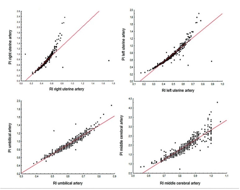
The present observational and retrospective study included 274 singleton pregnancies. The following maternal-fetal Doppler parameters were assessed: uterine artery (UAt), umbilical artery (UA), middle cerebral artery (MCA), cerebroplacental ratio (CPR), and umbilical-cerebral ratio (U/C). The assessment of FGR was based on the Figueiras and Gratacós9 criteria. Spearman correlation coefficients were estimated to assess the correlation between resistance (RI) and pulsatility (PI) indices of Doppler parameters. The agreement between two Doppler parameters was assessed by the Kappa coefficient.
In total, 502 Doppler examinations were included, and FGR was observed in 19 out of 274 fetuses. A strong correlation was observed between RI and PI of UAt, UA, and MCA in all of the samples (p<0.001). Of the 502 Doppler examinations, there was agreement between U/C and CPR percentiles for 480 (95.6%) and disagreement for 22 (4.4%), with Kappa coefficient of 0.26, thereby corresponding to weak agreement. Of the 68 cases with estimated fetal weight ≤ 9th percentile (small for gestational age [SGA]), there was agreement between U/C>1.0 and CPR<5th percentile in 61 (88.4%) and disagreement in 7 (5.8%) with Kappa coefficient of 0.49, thereby corresponding to moderate agreement.
Strong correlation was observed among RI and PI UAt, UA, and MCA Doppler examinations in the present study; however, weak agreement was observed between U/C and CPR in the normal and FGR fetuses. In SGA, U/C and CPR demonstrated moderate agreement.
Summary
Revista Brasileira de Ginecologia e Obstetrícia. 2022;44(2):118-124
04-08-2022
To assess the degree of correlation/agreement of maternal-fetal Doppler parameters between normal and growth-restricted fetuses (fetal growth restriction [FGR]).
The present observational and retrospective study included 274 singleton pregnancies. The following maternal-fetal Doppler parameters were assessed: uterine artery (UAt), umbilical artery (UA), middle cerebral artery (MCA), cerebroplacental ratio (CPR), and umbilical-cerebral ratio (U/C). The assessment of FGR was based on the Figueiras and Gratacós9 criteria. Spearman correlation coefficients were estimated to assess the correlation between resistance (RI) and pulsatility (PI) indices of Doppler parameters. The agreement between two Doppler parameters was assessed by the Kappa coefficient.
In total, 502 Doppler examinations were included, and FGR was observed in 19 out of 274 fetuses. A strong correlation was observed between RI and PI of UAt, UA, and MCA in all of the samples (p<0.001). Of the 502 Doppler examinations, there was agreement between U/C and CPR percentiles for 480 (95.6%) and disagreement for 22 (4.4%), with Kappa coefficient of 0.26, thereby corresponding to weak agreement. Of the 68 cases with estimated fetal weight ≤ 9th percentile (small for gestational age [SGA]), there was agreement between U/C>1.0 and CPR<5th percentile in 61 (88.4%) and disagreement in 7 (5.8%) with Kappa coefficient of 0.49, thereby corresponding to moderate agreement.
Strong correlation was observed among RI and PI UAt, UA, and MCA Doppler examinations in the present study; however, weak agreement was observed between U/C and CPR in the normal and FGR fetuses. In SGA, U/C and CPR demonstrated moderate agreement.
Summary
Revista Brasileira de Ginecologia e Obstetrícia. 2022;44(3):231-237
02-09-2022
To analyze whether acetylsalicylic (ASA) intake modifies the mean uterine arteries pulsatility index (UtA-PI) at the 2nd or 3rd trimester in a cohort of pregnant women with abnormal mean UtA-PI at between 11 and 14 weeks of gestation.
This is a retrospective cohort study. Singleton pregnancies with abnormal mean UtA-PI at between 11 and 14 weeks of gestation were studied. The participants were divided into 3 groups: 1) If the participant did not take ASA during pregnancy; 2) If the participant took ASA before 14 weeks of gestation; and 3) If the participant took ASA after 14 weeks of gestation. The mean UtA-PI was evaluated at the 2nd and 3rd trimesters, and it was considered to improve when it decreased below the 95th percentile. The prevalence ratio (PR) and the number needed to treat (NNT) werecalculated.
A total of 72 participants with a mean UtA-PI>95th percentile at the 1st trimester of gestation were evaluated. Out of the 18 participants who took ASA, 8 participants started it before 14 weeks of gestation and 10 after. A total of 33.3% of these participants had improved the mean UtA-PI at the 2nd and 3rd trimesters of gestation, although it was not statistically significant (p=0.154). The prevalence ratio was 0.95 (95% confidence interval [CI]: 0.31-1.89), but between the 1st and 2nd trimesters of gestation, the PR was 0.92 (95%CI: 0.21-0.99) and it was statistically significant.
The present work demonstrates a modification of the mean UtA-PI in participants who took ASA compared with those who did not. It is important to check if ASA can modify the normal limits of uterine arteries because this could have an impact on surveillance.
Summary
Revista Brasileira de Ginecologia e Obstetrícia. 2020;42(5):289-296
06-22-2020
Intrauterine growth restriction (IUGR) is associated with poor perinatal prognosis and a higher risk of stillbirth, neonatal death, and cerebral palsy. Its detection and the evaluation of its severity by new Doppler velocimetric parameters, such as aortic isthmus (AoI), are of great relevance for obstetrical practice. The AoI is a vascular segment that represents a point of communication between the right and left fetal circulations. It is considered to be a functional arterial shunt that reflects the relationship between the systemic and cerebral impedances, and has recently been proposed as a tool to detect the status of hemodynamic balance and prognosis of IUGR in fetuses. In the present review, we noticed that in healthy fetuses, the AoI net flow is always antegrade, but in fetuses with IUGR the deterioration of placental function leads to progressive reduction in its flow until it becomes mostly retrograde; this point is associated with a drastic reduction in oxygen delivery to the brain. The more impaired the AoI flow is, the greater is the risk of impairment in the Doppler velocimetry of other vessels; and the alterations of the AoI Doppler seem to precede other indicators of severe hypoxemia. Although there seems to be an association between the presence of retrograde flow in the AoI and the risk of long-term neurologic disability, its role in the prediction of perinatal morbi-mortality remains unclear. The AoI Doppler seems to be a promising tool in the management of fetuses with IUGR, but more studies are needed to investigate its employment in clinical practice.
Summary
Revista Brasileira de Ginecologia e Obstetrícia. 2018;40(5):287-293
05-01-2018
To perform a comprehensive review of the current evidence on the role of uterine artery Doppler, isolated or in combination with other markers, in screening for preeclampsia (PE) and fetal growth restriction (FGR) in the general population. The review included recently published large cohort studies and randomized trials.
A search of the literature was conducted usingMedline, PubMed, MeSH and ScienceDirect. Combinations of the search terms “preeclampsia,” “screening,” “prediction,” “Doppler,” “Doppler velocimetry,” “fetal growth restriction,” “small for gestational age” and “uterine artery” were used. Articles in English (excluding reviews) reporting the use of uterine artery Doppler in screening for PE and FGR were included.
Thirty articles were included. As a single predictor, uterine artery Doppler detects less than 50% of the cases of PE and no more than 40% of the pregnancies affected by FGR. Logistic regression-based models that allow calculation of individual risk based on the combination of multiple markers, in turn, is able to detect ~ 75% of the cases of preterm PE and 55% of the pregnancies resulting in small for gestational age infants.
The use of uterine artery Doppler as a single predictive test for PE and FGR has poor accuracy. However, its combined use in predictive models is promising, being more accurate in detecting preterm PE than FGR.
Summary
Revista Brasileira de Ginecologia e Obstetrícia. 2013;35(7):309-316
09-27-2013
DOI 10.1590/S0100-72032013000700005
PURPOSE: To determine perinatal outcomes and factors associated with fetal brain sparing effect diagnosed by Doppler flow velocimetry in patients with arterial hypertension. METHODS: We performed a cross-sectional retrospective study including 129 pregnant women with arterial hypertension and submitted to Doppler flow velocimetry, within fifteen days before delivery. Women with multiple pregnancies, fetal malformations, genital bleeding, placenta praevia, premature rupture of membranes, smoking, illicit drug use and chronic diseases were excluded. We analyzed the biological, socio-demographic and obstetric characteristics, as well the perinatal outcomes. To determine the association between variables, we used the χ² test, Fisher's exact test and Student's t-test. Multiple logistic regression analysis was performed to determine the factors associated with fetal centralization. RESULTS: Pre-eclampsia was the most frequent hypertensive disorder (53.5%) and fetal brain sparing effect was observed in 24.0% of fetuses. The prenatal factors associated with fetal brain sparing were the persistence of bilateral protodiastolic notches in uterine arteries (OR 3.6; 95%CI 1.4 - 9.4; p=0.009) and intrauterine growth restriction (IUGR) (OR 3.3; 95%CI 1.2 - 9.3; p=0.02). The perinatal outcomes associated with fetal brain sparing were gestational age <32 weeks, small for gestational age (SGA) infants, birth weight <2,500 g and perinatal death. There was no association with other maternal or neonatal variables. CONCLUSIONS: The main factors associated with fetal brain sparing were persistence of uterine arteries notches, IUGR, and increased frequency of adverse perinatal outcomes.
Summary
Revista Brasileira de Ginecologia e Obstetrícia. 2013;35(2):71-77
02-07-2013
DOI 10.1590/S0100-72032013000200006
PURPOSE: To evaluate the anthropometric characteristics of morbidity and mortality of premature newborns (NB) of hypertensive mothers according to the presence or absence of flow (DZ) or reverse (DR) diastolic flow in the dopplervelocimetry of the umbilical artery. METHODS: A prospective study was conducted on preterm newborns of pregnant women with hypertension between 25 and 33 weeks of gestational age, submitted to umbilical artery Doppler study during the five days before delivery. Delivery occurred at Hospital Regional da Asa Sul, Brasília - Federal District, between November 1st, 2009 and October 31st, 2010. The infants were stratified into two groups according to the results of Doppler velocimetry: Gdz/dr=absent end-diastolic velocity waveform or reversed end-diastolic velocity waveform, and Gn=normal Doppler velocimetry. Anthropometric measurements at birth, neonatal morbidity, and mortality were compared between the two groups. RESULTS: We studied 92 infants, as follows: Gdz/dr=52 infants and Gn=40 infants. In Gdz/dr, the incidence of infants small for gestational age was significantly greater, with a relative risk of 2.5 (95%CI 1.7 - 3.7). In Gdz/dr, infants remained on mechanical ventilation for a longer time: median 2 (0‒28) and Gn median 0.5 (0‒25) p=0.03. The need for oxygen at 28 days was higher in G dz/dr comparing to Gn (33 versus 10%; p=0.01). Neonatal mortality was higher in Gdz/dr compared to Gn (36 versus 10%; p=0.03; relative risk of 1.6; 95%CI 1.2‒2.2). Logistic regression showed that, with each 100 grams lower birth weight, the chance of death increased 6.7 times in G dz/dr (95%CI 2.0 - 11.3; p<0.01). CONCLUSION: In preterm infants of mothers with hypertensive changes in Doppler velocimetry of the umbilical artery, intrauterine growth restriction and neonatal prognosis are often worse, with a high risk of death related to birth weight.
Summary
Revista Brasileira de Ginecologia e Obstetrícia. 2013;35(1):33-38
01-11-2013
DOI 10.1590/S0100-72032013000100007
PURPOSE: To create longitudinal reference intervals for pulsatility index (PI) of the umbilical (UA), middle cerebral (MCA), uterine (UtA) arteries and ductus venosus (DV) in a Brazilian cohort. METHODS: A longitudinal observational study performed from February 2010 to May 2012. Low risk pregnancies were scanned fortnightly from 18 to 40 weeks for the measurements of PI of the UA, MCA, DV and UtA. Linear mixed models were used for the elaboration of longitudinal reference intervals (5th, 50th and 95th percentiles) of these measurements. PI obtained for the placental and abdominal portions of the umbilical artery were compared by the t-test for independent samples. Two-sided p values of less than 0.05 were considered statistically significant. RESULTS: A total of 164 patients underwent 1,242 scans. There was significant decrease in PI values of all vessels studied with gestational age (GA). From the 18th to the 40th week of pregnancy, the median PI values of UA (abdominal and placental ends of the cord), MCA, DV and the mean PI of the UtA ranged from 1.19 to 0.74, 1.33 to 0.78, 1.56 to 1.39, 0.58 to 0.41, and 0.98 to 0.66, respectively. The following equations were obtained for the prediction of the medians: PI-UA=1.5602786 - (0.020623 x GA); Logarithm of the PI-MCA=0.8149111 - (0.004168 x GA) - [0.02543 x (GA - 28.7756)²]; Logarithm of the PI-DV=-0.26691- (0.015414 x GA); PI-UtA = 1.2362403 - (0.014392 x GA). There was a significant difference between the PI-UA obtained at the abdominal and placental ends of the umbilical cord (p<0.001). CONCLUSIONS: Longitudinal reference intervals for the main gestational Doppler parameters were obtained in a Brazilian cohort. These intervals could be more adequate for the follow-up of maternal-fetal hemodynamic modifications in normal and abnormal pregnancies, a fact that still requires further validation.