You searched for:"Cleisson Fábio Andrioli Peralta"
We found (12) results for your search.Summary
Rev Bras Ginecol Obstet. 2012;34(10):466-472
DOI 10.1590/S0100-72032012001000006
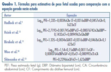
PURPOSES: To elaborate models for the estimation of fetal weight and longitudinal reference intervals of estimated fetal weight (EFW) using a sample of the Brazilian population. METHODS: Prospective observational study. Two groups of patients were evaluated: Group EFW (estimation of fetal weight): to elaborate (EFW-El) and validate (EFW-Val) a model for the prediction of fetal weight; Group LRI (longitudinal reference intervals): To elaborate (LRI-El) and validate (LRF-Val) conditional (longitudinal) percentiles of EFW. Polynomial regression analysis was applied to the data from subgroup EFW-El to elaborate a model for the estimation of fetal weight. The performance of this model was compared to those of previously published formulas. Linear mixed models were used for the elaboration of longitudinal reference intervals of EFW using data from subgroup LRI-El. Data obtained from subgroup LRI-Val were used to validate these intervals. RESULTS: Group EFW consisted of 458 patients (EFW-El: 367; EFW-Val: 91) and Group LRI consisted of 315 patients (LRI-El: 265; LRI-Val: 50). The model obtained for EFW was: EFW=-8.277+2.146xBPDxACxFL-2.449xFLxBPD². The performances of other models were significantly worse than those obtained with our formula. Equations for the prediction of conditional percentiles of EFW were derived from the longitudinal observation of patients of subgroup LRI-El and validated with data from subgroup LRI-Val. CONCLUSIONS: We described a method for customization of longitudinal reference intervals of EFW obtained using formulas generated from a sample of the Brazilian population.
Summary
Rev Bras Ginecol Obstet. 2012;34(10):466-472
DOI 10.1590/S0100-72032012001000006
PURPOSES: To elaborate models for the estimation of fetal weight and longitudinal reference intervals of estimated fetal weight (EFW) using a sample of the Brazilian population. METHODS: Prospective observational study. Two groups of patients were evaluated: Group EFW (estimation of fetal weight): to elaborate (EFW-El) and validate (EFW-Val) a model for the prediction of fetal weight; Group LRI (longitudinal reference intervals): To elaborate (LRI-El) and validate (LRF-Val) conditional (longitudinal) percentiles of EFW. Polynomial regression analysis was applied to the data from subgroup EFW-El to elaborate a model for the estimation of fetal weight. The performance of this model was compared to those of previously published formulas. Linear mixed models were used for the elaboration of longitudinal reference intervals of EFW using data from subgroup LRI-El. Data obtained from subgroup LRI-Val were used to validate these intervals. RESULTS: Group EFW consisted of 458 patients (EFW-El: 367; EFW-Val: 91) and Group LRI consisted of 315 patients (LRI-El: 265; LRI-Val: 50). The model obtained for EFW was: EFW=-8.277+2.146xBPDxACxFL-2.449xFLxBPD². The performances of other models were significantly worse than those obtained with our formula. Equations for the prediction of conditional percentiles of EFW were derived from the longitudinal observation of patients of subgroup LRI-El and validated with data from subgroup LRI-Val. CONCLUSIONS: We described a method for customization of longitudinal reference intervals of EFW obtained using formulas generated from a sample of the Brazilian population.
Summary
Rev Bras Ginecol Obstet. 2011;33(1):49-57
DOI 10.1590/S0100-72032011000100008
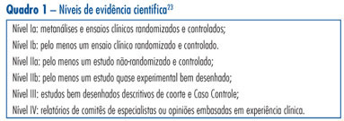
This is a traditional (narrative) review with the objective of highlighting the contribution of obstetric ultrasonography (US) between the 11th and 14th week of pregnancy, commonly called first trimester anomaly scan. In addition to being used for the screening of chromosomal anomalies, US can be employed during this period to confirm or determine gestational age, evaluate fetal anatomy, diagnose malformations, screen major structural abnormalities and genetic syndromes, define the prognosis of pregnancy, diagnose and characterize multiple pregnancies, and screen preeclampsia and intrauterine growth restriction. The most important studies about this subject published between 1990 and 2010 in the Cochrane and PubMed libraries were included. The selected studies can be classified with scientific levels I to III.
Summary
Rev Bras Ginecol Obstet. 2011;33(1):49-57
DOI 10.1590/S0100-72032011000100008
This is a traditional (narrative) review with the objective of highlighting the contribution of obstetric ultrasonography (US) between the 11th and 14th week of pregnancy, commonly called first trimester anomaly scan. In addition to being used for the screening of chromosomal anomalies, US can be employed during this period to confirm or determine gestational age, evaluate fetal anatomy, diagnose malformations, screen major structural abnormalities and genetic syndromes, define the prognosis of pregnancy, diagnose and characterize multiple pregnancies, and screen preeclampsia and intrauterine growth restriction. The most important studies about this subject published between 1990 and 2010 in the Cochrane and PubMed libraries were included. The selected studies can be classified with scientific levels I to III.
Summary
Rev Bras Ginecol Obstet. 2009;31(11):540-546
DOI 10.1590/S0100-72032009001100003
PURPOSE: to verify the association between ultrasonographic signs during gestation and post-delivery evolution in fetuses with bilateral obstructive uropathies, followed up in an expectant way. METHODS: fetuses with bilateral obstructive uropathies presenting severe oligoamnios and narrow thorax have been compared with fetuses with bilateral obstructive uropathies without those alterations, concerning the presence or absence of cysts in both kidneys, and the presence or absence of parenchymal hyperechogenicity in both kidneys. Cases of neonatal death were compared with cases of neonatal discharge from the nursery, regarding the same renal echographic aspects mentioned above, the presence of severe oligoamnios and narrow thorax. The sensitivity, specificity, positive and negative predictive value of the presence of bilateral renal cysts, bilateral renal hyperechogenicity, severe oligoamnios and narrow fetal thorax for the neonatal death were calculated. RESULTS: severe oligoamnios and narrow thorax were more frequent (p=0.03; p<0.001) in fetuses with bilateral renal cysts, as compared to those with echographically normal renal parenchyma. Neonatal death was more frequent among cases with severe oligoamnios (p<0.001), narrow thorax (p<0.001) and bilateral renal cysts (p<0.002), when respectively compared with cases without those alterations. The best values of sensitivity, specificity, positive and negative predictive value for the death of neonatal/breastfeeding infants were obtained using the echographic aspect of narrow thorax, and were 81.8, 100, 100 and 79.3%, respectively. CONCLUSIONS: in cases of fetuses with bilateral obstructive uropathies followed up in an expectant way, the ultrasonographic signs more associated to bad prognosis are severe oligoamnios, narrow fetal thorax and presence of bilateral renal cysts.
Summary
Rev Bras Ginecol Obstet. 2002;24(9):601-608
DOI 10.1590/S0100-72032002000900006
Purpose: to appraise the value of ultrasonographic parameters for the diagnosis of fetal Down syndrome (T21), in order to permit its use in routine clinical practice. Methods: this is a prospective cohort study using various ultrasonographic parameters for the prediction of T21. A total of 1662 scans were evaluated in the cohort study and 289 examinations were analyzed as a differential sample to test the normality curve from October 1993 to November 2000. The statistical analysis was based on the calculation of intra- and interobserver variations, the construction of normality curves for the studied parameters, as well as their validity tests, and the calculation of sensitivity, specificity, relative risk, likelyhood ratio and posttest predictive values. Results: among 1662 cases, 22 fetuses (1.32%) with T21 were identified. The normality curves were built for nucal fold thickness, femur/foot ratio and nasal bone length. Renal pelvis had a semiquantitative distribution and the proposed cutoff level was 4.0 mm. Sensitivity, specificity, false positive rate, relative risk and likelyhood ratio for nucal fold measurements above the 95th percentile were 54.5%, 95.2%, 4.9%, 20.2 and 11, respectively. For nasal bone measurements below the 5th percentile, 59.0%, 90.1%, 9.0%, 13.4 and 6.5. For femur/foot ratio below the 5th percentile, 45.5%, 84.4%, 15.6%, 3.7 and 2,6. For renal pelvis greater than 4.0 mm, 36.4%, 89.2%, 10.9%, 4.5 and 3.4. For absent fifth finger middle phalanx, 22.7%, 98.1%, 1.9%, 13.2 and 11.9. For the presence of major malformations, 31.8%, 98.7%, 1.3%, 27.2 and 24,8. After calculating the probability rates and the incidence of T21 in different maternal ages, a table for posttest risk using ultrasonographic parameters was set up. Conclusions: normality curves and indices for the assessment of risk for fetal Down syndrome on a population basis were established by the utilization of different maternal ages and by multiplying factors proposed by the authors. It was not possible to establish a normality curve for renal pelvis measurements, because of their semiquantitative distribution.