Summary
Revista Brasileira de Ginecologia e Obstetrícia. 2007;29(6):285-290
DOI 10.1590/S0100-72032007000600002
PURPOSE: to evaluate the spreading of endometrial cells to the peritoneal cavity during diagnostic hysteroscopy. METHODS: a prospective, descriptive study involving 76 patients divided in two groups: one with 61 patients without malignant endometrial cancer, and the other with 15 patients with endometrial cancer. Two samples of peritoneal fluid were collected, one before (PF-1) and the other immediately after (PF-2) the diagnostic hysteroscopy. Spread to the peritoneal cavity was defined by the presence of endometrial cells in PF-2, with the absence of such cells in PF-1. The 5 mm diameter Storz’s hysteroscopy was used. Distention was obtained by CO2 with electronically controlled flow pressure of 80 mmHg. The PF was fixated in absolute alcohol (ratio1:1). The PF samples were centrifuged and aliquots were smeared and stained using the Papanicolaou method. Analyses were performed by the same observer. RESULTS: during the study, four patients (5.26%) were excluded for presenting endometrial cells in PF-1. In the remaining 72 patients, there was no spread of cells to the peritoneal cavity. In the non-endometrial cancer group, 88.1% (52/59) presented secretory endometrial phase, with correlation of 80% between the hysteroscopy and the biopsy. In the group with endometrial cancer, most of the patients were in stage I (92.3%). There was a 100% correlation between the hysteroscopy/biopsy and histopathology of the surgical sample. CONCLUSIONS: the diagnostic hysteroscopy with CO2 at flow pressure of 80 mmHg did not cause spread of endometrial cells to the peritoneal cavity in both groups, thus suggesting that the diagnostic hysteroscopy is safe for patients at high risk for endometrial cancer.
Summary
Revista Brasileira de Ginecologia e Obstetrícia. 2011;33(10):286-291
DOI 10.1590/S0100-72032011001000003
PURPOSES: To determine the rate of low birth weight and some of the risk factors associated with this event among adolescents. METHODS: A cross-sectional study conducted between October 1994 and December 2009 at a maternity in Campinas, in Brazil, using information generated from the computerized obstetric form. After selection of adolescents who delivered at this hospital, two groups were created, with and without low birth weight, respectively. Relative risk and 95% confidence interval for all independent variables (risk factors) and the Χ2 test for some perinatal results were performed. The level of significance was set at 5%. RESULTS: During the study period, 24,000 births occurred at CAISM. Of these, 2,404 occurred among 2,357 teenagers (10.02%) and the frequency of low birth weight was 15.1%. Adolescent pregnancy recurred in 294 (8.2%). Age less than 15 years-old, anemia, smoking, and hypertension were not significantly associated with low birth weight. Antecedent of miscarriage and association with systemic lupus erythematosus increased the risk of low birth weight. Cesarean section and an Apgar score below seven were more prevalent among adolescents with low birth weight, and 85% of all adolescents had less than six prenatal visits. CONCLUSIONS: The prevalence of low birth weight is higher among adolescents than among adult women, and there was a large number of adolescents with less than six prenatal visits . The antecedent of miscarriage and the presence of systemic lupus erythematosus were risk factors associated with the occurrence of low birth weight among adolescents.
Summary
Revista Brasileira de Ginecologia e Obstetrícia. 2010;32(6):286-292
DOI 10.1590/S0100-72032010000600006
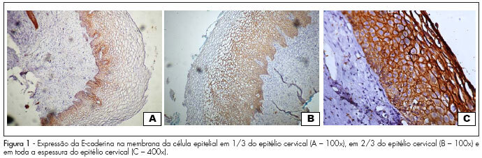
PURPOSE: to evaluate the expression of E-cadherin in cervical lesions of patients suffering from HIV infection. METHODS: we conducted a study with 77 patients with cervical HPV infection, 40 of them were HIV seropositive and 37 HIV seronegative who underwent colposcopy and a biopsy of the cervix. The material obtained by biopsy of the cervix was sent for histopathologic and immunohistochemical study. Sections were obtained and mounted on silanized slides and examined by an observer who was blind to patient serology. E-cadherin antibody, clone NHC-38 diluted 1:400 (DAKO) and the Novolink polymer system (Novocastra) were used. The expression of E-cadherin was determined on the epithelial cell membrane based on the extent of the stained area. The χ2 test with Yates correction or the Fisher's Exact test was used for comparison of the proportion in univariate analysis. All the variables with p<0.25 were included in the logistic regression model, called initial model. The analyses were carried out using the SPSS software, with the level of significance set at 5%. RESULTS: the expression of E-cadherin was observed in up to the internal 1/3 of the epithelium in 59.3% of cases and in up to 2/3 of the epithelium in 11.1% of cases, but in 29.6% of cases the expression was identified throughout the thickness of the epithelium in HIV-seronegative patients. In contrast, in HIV-seropositive patients, 45.9% showed expression up to 1/3 of the epithelium, 13.5% showed expression in up to 2/3 of the epithelium, and 40.5% showed expression throughout the thickness of the epithelium. E-cadherin expression did not differ between groups (p=0.5). However, the multivariate analysis identified a significant association between high-grade cervical injury and E-cadherin expression in 2/3 and 3/3 of the epithelium (p=0.001; χ2=36.9). CONCLUSIONS: the expression of E-cadherin in the epithelial cell membrane is not associated with infection by the human immunodeficiency virus, but with the degree of intraepithelial cervical injury.
Summary
Revista Brasileira de Ginecologia e Obstetrícia. 2010;32(6):286-292
DOI 10.1590/S0100-72032010000600006
PURPOSE: to evaluate the expression of E-cadherin in cervical lesions of patients suffering from HIV infection. METHODS: we conducted a study with 77 patients with cervical HPV infection, 40 of them were HIV seropositive and 37 HIV seronegative who underwent colposcopy and a biopsy of the cervix. The material obtained by biopsy of the cervix was sent for histopathologic and immunohistochemical study. Sections were obtained and mounted on silanized slides and examined by an observer who was blind to patient serology. E-cadherin antibody, clone NHC-38 diluted 1:400 (DAKO) and the Novolink polymer system (Novocastra) were used. The expression of E-cadherin was determined on the epithelial cell membrane based on the extent of the stained area. The χ2 test with Yates correction or the Fisher's Exact test was used for comparison of the proportion in univariate analysis. All the variables with p<0.25 were included in the logistic regression model, called initial model. The analyses were carried out using the SPSS software, with the level of significance set at 5%. RESULTS: the expression of E-cadherin was observed in up to the internal 1/3 of the epithelium in 59.3% of cases and in up to 2/3 of the epithelium in 11.1% of cases, but in 29.6% of cases the expression was identified throughout the thickness of the epithelium in HIV-seronegative patients. In contrast, in HIV-seropositive patients, 45.9% showed expression up to 1/3 of the epithelium, 13.5% showed expression in up to 2/3 of the epithelium, and 40.5% showed expression throughout the thickness of the epithelium. E-cadherin expression did not differ between groups (p=0.5). However, the multivariate analysis identified a significant association between high-grade cervical injury and E-cadherin expression in 2/3 and 3/3 of the epithelium (p=0.001; χ2=36.9). CONCLUSIONS: the expression of E-cadherin in the epithelial cell membrane is not associated with infection by the human immunodeficiency virus, but with the degree of intraepithelial cervical injury.
Summary
Revista Brasileira de Ginecologia e Obstetrícia. 2008;30(6):287-293
DOI 10.1590/S0100-72032008000600004
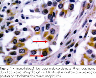
PURPOSE: to analyze the expression of matrix metalloproteinase-9 (MMP-9) and of vascular endothelial growth factor (EVGF) in a group of patients with primary breast cancer, and correlate them to one another and with other prognostic indicators. METHODS: transversal study that has analyzed the expression of MMP-9 and of VEGF in 88 consecutive cases of primary breast tumors. The samples were obtained from patients with primary breast cancer, submitted to surgical treatment in the Clinical Hospital of Porto Alegre of the Universidade Federal do Rio Grande do Sul, from January 2000 to December 2004. An immunohistochemical technique has been applied, using the avidin-biotin-peroxidase complex to evaluate the antigen immunoreactions in the tumors. The qualitative expression of proteins has been assessed through the observation of the brownish stain intensity of antibodies in the cytoplasm of malignant cells, when at least one of the tumoral cells presented clear and unequivocal staining with each of those markers. To determine the qualitative score (0=absent, 1=weak, 2=average and 3=strong), the stronger cytoplasmatic staining intensity on the glass slide has been taken into consideration, independently of the stained cells. The quantitative expression was determined by the average percentage of stained cells, observed in at least ten microscopic fields. The MMP-9 and VEGF final quantification expression has been done by the application of the HSCORE=Σ[(I+1)]xPC, where I and PC represent the staining intensity and the percentage of stained cells, respectively. RESULTS: MMP-9 and VEGF presented a significant correlation in the tumors studied. The final expression has shown a median score of 180 and 190, respectively. When MMP-9 and VEGF expression were compared with the variables "age", "tumoral diameter", "histological type", "histological grade", "axillary lymph node" and "vascular invasion", it was impossible to find any significant correlation. Compared to one another, MMP-0 and VEGF have presented a positive correlation (rho=0.23; p=0.03). The axillary lymph node positivity has presented a positive correlation with the larger tumoral diameter (2.7±1.1 cm; p<0.01) and with the presence of vascular invasion (84.1%; p<0.01). CONCLUSIONS: The present results do not show correlation between the MMP-9 and VEGF with the selected prognostic indicators, but shown a significant correlation between one another.
Summary
Revista Brasileira de Ginecologia e Obstetrícia. 2008;30(6):287-293
DOI 10.1590/S0100-72032008000600004
PURPOSE: to analyze the expression of matrix metalloproteinase-9 (MMP-9) and of vascular endothelial growth factor (EVGF) in a group of patients with primary breast cancer, and correlate them to one another and with other prognostic indicators. METHODS: transversal study that has analyzed the expression of MMP-9 and of VEGF in 88 consecutive cases of primary breast tumors. The samples were obtained from patients with primary breast cancer, submitted to surgical treatment in the Clinical Hospital of Porto Alegre of the Universidade Federal do Rio Grande do Sul, from January 2000 to December 2004. An immunohistochemical technique has been applied, using the avidin-biotin-peroxidase complex to evaluate the antigen immunoreactions in the tumors. The qualitative expression of proteins has been assessed through the observation of the brownish stain intensity of antibodies in the cytoplasm of malignant cells, when at least one of the tumoral cells presented clear and unequivocal staining with each of those markers. To determine the qualitative score (0=absent, 1=weak, 2=average and 3=strong), the stronger cytoplasmatic staining intensity on the glass slide has been taken into consideration, independently of the stained cells. The quantitative expression was determined by the average percentage of stained cells, observed in at least ten microscopic fields. The MMP-9 and VEGF final quantification expression has been done by the application of the HSCORE=Σ[(I+1)]xPC, where I and PC represent the staining intensity and the percentage of stained cells, respectively. RESULTS: MMP-9 and VEGF presented a significant correlation in the tumors studied. The final expression has shown a median score of 180 and 190, respectively. When MMP-9 and VEGF expression were compared with the variables "age", "tumoral diameter", "histological type", "histological grade", "axillary lymph node" and "vascular invasion", it was impossible to find any significant correlation. Compared to one another, MMP-0 and VEGF have presented a positive correlation (rho=0.23; p=0.03). The axillary lymph node positivity has presented a positive correlation with the larger tumoral diameter (2.7±1.1 cm; p<0.01) and with the presence of vascular invasion (84.1%; p<0.01). CONCLUSIONS: The present results do not show correlation between the MMP-9 and VEGF with the selected prognostic indicators, but shown a significant correlation between one another.
Summary
Revista Brasileira de Ginecologia e Obstetrícia. 2018;40(5):287-293
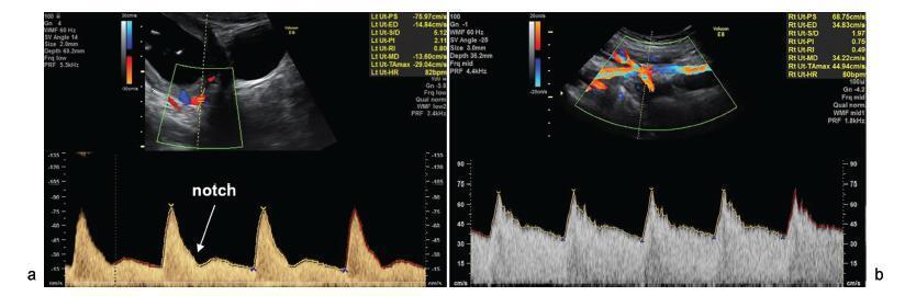
To perform a comprehensive review of the current evidence on the role of uterine artery Doppler, isolated or in combination with other markers, in screening for preeclampsia (PE) and fetal growth restriction (FGR) in the general population. The review included recently published large cohort studies and randomized trials.
A search of the literature was conducted usingMedline, PubMed, MeSH and ScienceDirect. Combinations of the search terms “preeclampsia,” “screening,” “prediction,” “Doppler,” “Doppler velocimetry,” “fetal growth restriction,” “small for gestational age” and “uterine artery” were used. Articles in English (excluding reviews) reporting the use of uterine artery Doppler in screening for PE and FGR were included.
Thirty articles were included. As a single predictor, uterine artery Doppler detects less than 50% of the cases of PE and no more than 40% of the pregnancies affected by FGR. Logistic regression-based models that allow calculation of individual risk based on the combination of multiple markers, in turn, is able to detect ~ 75% of the cases of preterm PE and 55% of the pregnancies resulting in small for gestational age infants.
The use of uterine artery Doppler as a single predictive test for PE and FGR has poor accuracy. However, its combined use in predictive models is promising, being more accurate in detecting preterm PE than FGR.
Summary
Revista Brasileira de Ginecologia e Obstetrícia. 2018;40(5):287-293
To perform a comprehensive review of the current evidence on the role of uterine artery Doppler, isolated or in combination with other markers, in screening for preeclampsia (PE) and fetal growth restriction (FGR) in the general population. The review included recently published large cohort studies and randomized trials.
A search of the literature was conducted usingMedline, PubMed, MeSH and ScienceDirect. Combinations of the search terms “preeclampsia,” “screening,” “prediction,” “Doppler,” “Doppler velocimetry,” “fetal growth restriction,” “small for gestational age” and “uterine artery” were used. Articles in English (excluding reviews) reporting the use of uterine artery Doppler in screening for PE and FGR were included.
Thirty articles were included. As a single predictor, uterine artery Doppler detects less than 50% of the cases of PE and no more than 40% of the pregnancies affected by FGR. Logistic regression-based models that allow calculation of individual risk based on the combination of multiple markers, in turn, is able to detect ~ 75% of the cases of preterm PE and 55% of the pregnancies resulting in small for gestational age infants.
The use of uterine artery Doppler as a single predictive test for PE and FGR has poor accuracy. However, its combined use in predictive models is promising, being more accurate in detecting preterm PE than FGR.
Summary
Revista Brasileira de Ginecologia e Obstetrícia. 2022;44(3):287-294
To evaluate the association between polycystic ovary syndrome (PCOS) and metabolic syndrome (MetS), adding liver assessment through elastography and ultrasound, for correlation with non-alcoholic fatty liver disease (NAFLD). Metabolic syndrome occurs in~43% of women with PCOS, and NAFLD is the hepatic expression of MetS.
One hundred women, 50 with PCOS and 50 controls, matched by age (18- 35 years) and body mass index (BMI) were included, restricted to patients with overweight and obesity grade 1, at the Assis Chateaubrian Maternity School, Universidade Federal do Ceará, Brazil. For the diagnosis of PCOS, we adopted the Rotterdam criteria, and for the diagnosis of MetS, the criteria of the National Cholesterol Education Program (NCEP/ATP III). Hepatic elastography and ultrasound were performed to assess liver stiffness and echotexture, respectively.
The average ages were 29.1 (±5.3) and 30.54 (±4.39) years, for the PCOS and the control group, respectively. Patients with PCOS had a risk 4 times higher of having MetS, odds ratio (95% confidence interval)=4.14, than those in the control group. Women with PCOS had higher average of abdominal circumference (100.9±9.08 cm vs 94.96±6.99 cm) and triglycerides (162±54.63 mg/dL vs 137.54±36.91mg/dL) and lower average of HDL cholesterol (45.66±6.88 mg/dL vs 49.78±7.05 mg/dL), with statistically significant difference. Hepatic steatosis was observed on ultrasound in women with PCOS; however, with no statistically significant difference. There was no change to NAFLD at elastography in any group.
Women with PCOS had 4-fold higher frequency of MetS andmore hepatic steatosis, with no statistically significant difference. There was no change in liver stiffness between the groups at elastography. The results can be extended only to populations of overweight and obesity grade 1, with PCOS or not. They cannot be generalized to other untested groups.
Summary
Revista Brasileira de Ginecologia e Obstetrícia. 2016;38(6):287-292
betatrophin has been reported to boost β cell expansion in insulin resistant states. Pregnancy is a well-recognized physiological state of insulin resistance. Betatrophin levels in pregnant women and their relationships with metabolic variables remain to be elucidated.
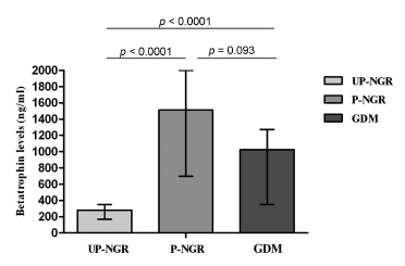
A total of 49 pregnant women and 31 age-matched unpregnant women with normal glucose regulation (UP-NGR) were included. Among these subjects, according to results from 75 g oral glucose tolerance test (OGTT), 22 women were diagnosed as having gestational diabetes mellitus ( GDM ).
Our study found that pregnant women, regardless of their glucose regulation status, had remarkably higher triglycerides (TG), total cholesterol (TC), fasting insulin (FINS), homeostasis model assessment of insulin resistance (HOMA-IR) and homeostasis model assessment of β-cell function (HOMA-β). However, GDM patients had much lower HOMA-β compared with those of pregnant women with normal glucose regulation (P-NGR). Participants of the P-NGR group had almost 4 times higher levels of betatrophin than those of the UP-NGR group. Although betatrophin levels were lower in the GDM group than those of the P-NGR group, the difference did not reach statistical significance. Spearman correlation analysis showed that betatrophin levels were positively and significantly associated with total cholesterol, triglycerides, highdensity lipoprotein cholesterol (HDL-c), FINS and HOMA-β. However, adjustments of TC, TG and HDL-c eliminated the association between HOMA-β and betatrophin.
Pregnant women have significantly higher betatrophin levels in comparison to unpregnant women. Betatrophin levels are positively and significantly associated with β cell function and lipid levels. Furthermore, lipids may contribute to
Summary
Revista Brasileira de Ginecologia e Obstetrícia. 2016;38(6):287-292
betatrophin has been reported to boost β cell expansion in insulin resistant states. Pregnancy is a well-recognized physiological state of insulin resistance. Betatrophin levels in pregnant women and their relationships with metabolic variables remain to be elucidated.
A total of 49 pregnant women and 31 age-matched unpregnant women with normal glucose regulation (UP-NGR) were included. Among these subjects, according to results from 75 g oral glucose tolerance test (OGTT), 22 women were diagnosed as having gestational diabetes mellitus ( GDM ).
Our study found that pregnant women, regardless of their glucose regulation status, had remarkably higher triglycerides (TG), total cholesterol (TC), fasting insulin (FINS), homeostasis model assessment of insulin resistance (HOMA-IR) and homeostasis model assessment of β-cell function (HOMA-β). However, GDM patients had much lower HOMA-β compared with those of pregnant women with normal glucose regulation (P-NGR). Participants of the P-NGR group had almost 4 times higher levels of betatrophin than those of the UP-NGR group. Although betatrophin levels were lower in the GDM group than those of the P-NGR group, the difference did not reach statistical significance. Spearman correlation analysis showed that betatrophin levels were positively and significantly associated with total cholesterol, triglycerides, highdensity lipoprotein cholesterol (HDL-c), FINS and HOMA-β. However, adjustments of TC, TG and HDL-c eliminated the association between HOMA-β and betatrophin.
Pregnant women have significantly higher betatrophin levels in comparison to unpregnant women. Betatrophin levels are positively and significantly associated with β cell function and lipid levels. Furthermore, lipids may contribute to