Summary
Revista Brasileira de Ginecologia e Obstetrícia. 2000;22(4):243-243
DOI 10.1590/S0100-72032000000400010
Summary
Revista Brasileira de Ginecologia e Obstetrícia. 2000;22(4):243-243
DOI 10.1590/S0100-72032000000400010
Summary
Revista Brasileira de Ginecologia e Obstetrícia. 2013;35(6):243-248
DOI 10.1590/S0100-72032013000600002
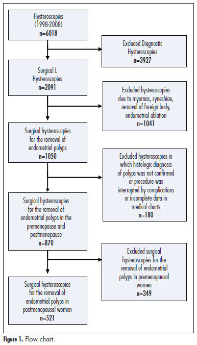
PURPOSE: To evaluate the accuracy of sonographic endometrial thickness and hysteroscopic characteristics in predicting malignancy in postmenopausal women undergoing surgical resection of endometrial polyps. METHODS: Five hundred twenty-one (521) postmenopausal women undergoing hysteroscopic resection of endometrial polyps between January 1998 and December 2008 were studied. For each value of sonographic endometrial thickness and polyp size on hysteroscopy, the sensitivity, specificity, positive predictive value (PPV) and negative predictive value (NPV) were calculated in relation to the histologic diagnosis of malignancy. The best values of sensitivity and specificity for the diagnosis of malignancy were determined by the Receiver Operating Characteristic (ROC) curve. RESULTS: Histologic diagnosis identified the presence of premalignancy or malignancy in 4.1% of cases. Sonographic measurement revealed a greater endometrial thickness in cases of malignant polyps when compared to benign and premalignant polyps. On surgical hysteroscopy, malignant endometrial polyps were also larger. An endometrial thickness of 13 mm showed a sensitivity of 69.6%, specificity of 68.5%, PPV of 9.3%, and NPV of 98% in predicting malignancy in endometrial polyps. Polyp measurement by hysteroscopy showed that for polyps 30 mm in size, the sensitivity was 47.8%, specificity was 66.1%, PPV was 6.1%, and NPV was 96.5% for predicting cancer. CONCLUSIONS: Sonographic endometrial thickness showed a higher level of accuracy than hysteroscopic measurement in predicting malignancy in endometrial polyps. Despite this, both techniques showed low accuracy for predicting malignancy in endometrial polyps in postmenopausal women. In suspected cases, histologic evaluation is necessary to exclude malignancy.
Summary
Revista Brasileira de Ginecologia e Obstetrícia. 2013;35(6):243-248
DOI 10.1590/S0100-72032013000600002
PURPOSE: To evaluate the accuracy of sonographic endometrial thickness and hysteroscopic characteristics in predicting malignancy in postmenopausal women undergoing surgical resection of endometrial polyps. METHODS: Five hundred twenty-one (521) postmenopausal women undergoing hysteroscopic resection of endometrial polyps between January 1998 and December 2008 were studied. For each value of sonographic endometrial thickness and polyp size on hysteroscopy, the sensitivity, specificity, positive predictive value (PPV) and negative predictive value (NPV) were calculated in relation to the histologic diagnosis of malignancy. The best values of sensitivity and specificity for the diagnosis of malignancy were determined by the Receiver Operating Characteristic (ROC) curve. RESULTS: Histologic diagnosis identified the presence of premalignancy or malignancy in 4.1% of cases. Sonographic measurement revealed a greater endometrial thickness in cases of malignant polyps when compared to benign and premalignant polyps. On surgical hysteroscopy, malignant endometrial polyps were also larger. An endometrial thickness of 13 mm showed a sensitivity of 69.6%, specificity of 68.5%, PPV of 9.3%, and NPV of 98% in predicting malignancy in endometrial polyps. Polyp measurement by hysteroscopy showed that for polyps 30 mm in size, the sensitivity was 47.8%, specificity was 66.1%, PPV was 6.1%, and NPV was 96.5% for predicting cancer. CONCLUSIONS: Sonographic endometrial thickness showed a higher level of accuracy than hysteroscopic measurement in predicting malignancy in endometrial polyps. Despite this, both techniques showed low accuracy for predicting malignancy in endometrial polyps in postmenopausal women. In suspected cases, histologic evaluation is necessary to exclude malignancy.
Summary
Revista Brasileira de Ginecologia e Obstetrícia. 2005;27(5):243-247
DOI 10.1590/S0100-72032005000500003
PURPOSE: to evaluate the association between p53 and Ki-67 expression in the tumor and clinicopathological features in patients with carcinoma of the cervix. METHODS: samples were taken from the tumor of 36 patients with stage IB (FIGO) cervical carcinoma submitted to radical hysterectomy. Tissue samples were taken from the tumor, fixed in formalin and embedded in paraffin. The specimens were analyzed by histopathology (hematoxylin and eosin) and immunohistochemically evaluated using monoclonal antibodies for p53 and Ki-67. Data were analyzed statistically by the chi2 test to evaluate eventual differences between the groups. RESULTS: the age of the patients ranged from 27 to 73 years (48.7±10.4 years). Clinical stage (FIGO) was IB1 in 27 cases (75%) and IB2 in 9 cases (25%). A positive tumoral expression of the p53 protein was found in half of the cases. In relation to the Ki-67 expression, a high cell proliferation index was shown in 73.3% of the cases. There was no association between tumoral p53 and Ki-67 expression with age (p=0.091 and 0.900), clinical stage (p=0.054 and 0.667), histological classification (p=0.674 and 0.674), grade of differentiation (p=0.070 and 0.282), presence of lymphatic vascular space invasion (p=0.248 and 0.667), parametrial involvement (p=0.729 and 0.763) and pelvic lymph node metastasis (p=0.729 and 0.636, respectively). CONCLUSIONS: tumoral expression of p53 and Ki-67 was not associated with the clinicopathological features in patients with stage IB carcinoma of the cervix.
Summary
Revista Brasileira de Ginecologia e Obstetrícia. 2003;25(4):243-248
DOI 10.1590/S0100-72032003000400004
PURPOSE: to analyze the influence of seminal parameters on intrauterine insemination (IUI) outcomes in patients with male factor and to emphasize the predictive value of each parameter for the successful result. METHODS: two hundred and thirty-nine IUI cycles (155 couples) were analyzed for 15 months. Female patients were submitted to ovary hyperstimulation according to the "I Consenso Brasileiro de Indução de Ovulação". Seminal analysis based on the World Health Organization (WHO) for sperm concentration and motility was used and sperm morphology was evaluated according to Kruger's criterion. Samples to be used in IUI were prepared by colloidal discontinued gradient (ISolate®). After IUI two patient groups were formed: group G - positive for pregnancy and group NG - negative for pregnancy. RESULTS: there was no statistical difference in total sperm concentration per mL, total motility and progressive motility before and after the ISolate® procedure. When sperm morphology was compared between the two groups, a statistical difference was observed (group G=10.6% normal morphology; group NG=6.4% normal morphology; p<0.05). Better pregnancy results were obtained when the number of inseminated spermatozoa was more than 15 x 10(6)/mL. CONCLUSIONS: sperm morphology and the number of inseminated sperm seem to be positive parameters for pregnancy and should be emphasized during male infertility propaedeutics.
Summary
Revista Brasileira de Ginecologia e Obstetrícia. 2000;22(4):244-244
Summary
Revista Brasileira de Ginecologia e Obstetrícia. 2000;22(4):244-244
Summary
Revista Brasileira de Ginecologia e Obstetrícia. 2000;22(4):244-244
DOI 10.1590/S0100-72032000000400012
Summary
Revista Brasileira de Ginecologia e Obstetrícia. 2000;22(4):244-244
DOI 10.1590/S0100-72032000000400012
Summary
Revista Brasileira de Ginecologia e Obstetrícia. 2014;36(6):244-250
DOI 10.1590/S0100-720320140005004
This study investigated short-term changes in body composition, handgrip strength, and presence of lymphedema in women who underwent breast cancer surgery.
Ninety-five women participated in a cross-sectional study, divided into two groups: Control (n=46), with healthy women, and Experimental (n=49), with women six months after breast cancer surgery . The Experimental Group was subdivided into right total mastectomy (RTM, n=15), left total mastectomy (LTM, n=11), right quadrant (RQ, n=13), and left quadrant (LQ, n=10). It was also redistributed among women with presence (n=10) or absence (n=39) of lymphedema. Presence of lymphedema, handgrip strength, and body composition were assessed.
Trunk lean mass and handgrip strength were decreased in the Experimental Group. Total lean mass was increased in the LTM compared to RTM or LQ. Left handgrip strength in LTM was decreased compared to RTM and RQ and in LQ compared to RTM and RQ. Finally, total lean mass, trunk fat mass, trunk lean mass, right and left arm lean mass were increased in women with lymphedema.
Breast cancer survivors have changes in their body composition and in handgrip strength six months after surgery; however, the interaction between the type of surgery and its impact is unclear. Furthermore, women who developed lymphedema in this period showed more significant changes in the body composition, but they were not enough to cause impairment in handgrip strength.
Summary
Revista Brasileira de Ginecologia e Obstetrícia. 2006;28(4):244-250
DOI 10.1590/S0100-72032006000400007
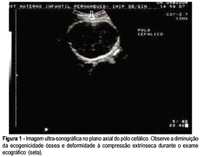
Osteogenesis imperfecta is a connective tissue disorder due to quantitative and qualitative anomalies in type 1 collagen, genetically transmitted by a dominant or recessive autosomal gene, leading to bone fragility. We report a case of a 19-year-old G1 PO patient referred to our institution following a screening ultrasound that demonstrated short limb fetal extremities. A level 3 scan was performed which evidenced an irregular cranial shape and compression of the cephalic pole with moderate transducer pressure. Limb shortening, decreased echoes and fractures of long bones were found on our scan evaluation. A vaginal delivery occurred at 35 weeks of gestation. The male newborn, weighing 1.990 grams had 6 and 8 in Apgar scores. The neonate was clearly abnormal, presenting irregular cranial shape, with poor ossification on X-ray, blue sclera, fractures and limb deformities. Postnatal evaluation was satisfactory and the neonate was discharged in good conditions. Prenatal diagnosis is important for an adequate pregnancy follow-up. Postnatal outcome was not related to vaginal delivery, as there were no recent fractures in the newborn.
Summary
Revista Brasileira de Ginecologia e Obstetrícia. 2006;28(4):244-250
DOI 10.1590/S0100-72032006000400007
Osteogenesis imperfecta is a connective tissue disorder due to quantitative and qualitative anomalies in type 1 collagen, genetically transmitted by a dominant or recessive autosomal gene, leading to bone fragility. We report a case of a 19-year-old G1 PO patient referred to our institution following a screening ultrasound that demonstrated short limb fetal extremities. A level 3 scan was performed which evidenced an irregular cranial shape and compression of the cephalic pole with moderate transducer pressure. Limb shortening, decreased echoes and fractures of long bones were found on our scan evaluation. A vaginal delivery occurred at 35 weeks of gestation. The male newborn, weighing 1.990 grams had 6 and 8 in Apgar scores. The neonate was clearly abnormal, presenting irregular cranial shape, with poor ossification on X-ray, blue sclera, fractures and limb deformities. Postnatal evaluation was satisfactory and the neonate was discharged in good conditions. Prenatal diagnosis is important for an adequate pregnancy follow-up. Postnatal outcome was not related to vaginal delivery, as there were no recent fractures in the newborn.