Summary
Revista Brasileira de Ginecologia e Obstetrícia. 2005;27(2):51-57
DOI 10.1590/S0100-72032005000200002
PURPOSE: to estimate the validity of visual inspection of cervical intraepithelial neoplasia (CIN) and HPV-induced lesion screening, after acetic acid application (VIA), and to compare its performance with that of colpocytology and colposcopy. METHODS: a diagnostic test validation study involving 893 women aged 18 to 65 years, simultaneously screened with colpocytology, VIA and colposcopy was carried out at a public health unit in Recife, PE. VIA was performed by applying 5% acetic acid onto the cervix and observing it with the help of a clinical spotlight. The finding of any aceto-white lesion on the cervix was considered positive. The gold standard was the histopathology of cervical biopsy, carried out whenever any of the three test results was abnormal. Validity indicators were estimated for each test, within 95% confidence intervals. The analysis of agreement between test results was done by the kappa coefficient. RESULTS: of 303 women submitted to biopsy, the histopathological study was abnormal in 24. Among this total, VIA was positive in 22, yielding an estimated 91.7% sensibility, 68.9% specificity, and 7.5% positive predictive value and 99.7% negative predictive value. Comparing 95% confidence intervals, VIA was more sensitive than colpocytology, despite a lower specificity and positive predictive value. There was poor agreement between VIA and colpocytology (k=0.02) and excellent agreement with colposcopy (k=0.93). CONCLUSION: VIA was much more sensitive than colpocytology in the screening of CIN and HPV-induced lesions and presented a performance similar to colposcopy. Its low specificity determined a high number of false-positive results.
Summary
Revista Brasileira de Ginecologia e Obstetrícia. 2005;27(2):58-63
DOI 10.1590/S0100-72032005000200003
PURPOSE: to evaluate the distribution of yeast species isolated from the vagina in two cities of the South of Brazil and compare the in vitro susceptibility profile of these yeasts against some antifungals, which are used in clinical routine. METHODS: all women attended from January to June 2004 for vaginal routine examinations, independent of being symptomatic or not were included in the study. Only those who presented immunodeficiency like AIDS or any other genital infection were excluded. Samples of vaginal discharge from the women (Jaraguá do Sul - SC (n=130) and Maringá - PR (n=97)) were cultivated. The yeasts were identified and submitted to the susceptibility test against the antifungals fluconazole, nystatin and amphotericin B. RESULTS: the frequency of positive cultures for yeasts was the same in both cities; C. albicans was the most prevalent species (about 24%), but its frequency was different: in SC it corresponded to 77.4% of the yeasts both in symptomatic and asymptomatic women and in PR it was 50.0% with predominance in symptomatic women. We observed high rates of susceptibility to fluconazole and amphotericin B, but 51.1% of the yeasts presented dose-dependent susceptibility (DDS) to nystatin. C. albicans showed a higher tendency to be nystatin resistant (52.8% DDS) than non-albicans species (44.4%). CONCLUSIONS: our data showed geographic differences among the species of yeasts isolated from the vagina and suggest that this fact has clinical relevance considering the differences in susceptibility, especially regarding nystatin, which could be important for the management of vulvovaginal candidiasis.
Summary
Revista Brasileira de Ginecologia e Obstetrícia. 2005;27(2):64-68
DOI 10.1590/S0100-72032005000200004
PURPOSE: to determine the frequency of Mycoplasma hominis and Ureaplasma urealyticum infection, and relate it to the associated clinical variables of infertile women. METHODS: transversal study involving 322 infertile women, submitted to collection of endocervix swab for research of Mycoplasma hominis and Ureaplasma urealyticum infecction, from October 2002 to May 2004. All patients were submitted to a basic infertility investigation protocol. As control, a historical series of 51 non-pregnant women previously investigated as for the studied infectious agents, was used. RESULTS: the frequency of Mycoplasma hominis and Ureaplasma urealyticum infection was 4.9% in the infertile women and 13.8% in the control group. Among the infertile patients, a relationship between the presence of the two agents and changes in the histerosalpingography result (OR: 3.20; IC 95%: 1.05-9.73), presence of dyspareunia (OR: 10.72; IC 95%: 3.21-35.77) and vaginal discharge (OR: 8.5; IC 95%: 2.83-26.02), besides endocervical culture positive for Escherichia coli (OR: 6.09; IC 95%: 4.95-52.25) was observed. CONCLUSION: Mycoplasma hominis and Ureaplasma urealyticum infection rate is low in infertile patients and is associated with reproductive sequels.
Summary
Revista Brasileira de Ginecologia e Obstetrícia. 2005;27(2):69-74
DOI 10.1590/S0100-72032005000200005
PURPOSE: to determine the prevalence of overweight, obesity, and associated factors among women who visited a general gynecologic clinic in a secondary hospital of reference. METHODS: the following variables were studied: age, race, educational level, family income, job (paid work done by the women), type of the women's job, current partner, menstrual cycle characteristics at the time of interview, and body mass index (BMI). The patients were divided into three groups, according to their BMI values: <25 kg/m² (normal), between 25-29 kg/m² (overweight) and >30 kg/m² (obesity). The odds ratio (OR) and respective 95% confidence interval (95% CI) were calculated in the overweight and obese groups. Subsequently, the OR was calculated and adjusted for other variables. RESULTS: among the 676 studied women, 89.8% had received up to 8 years of formal education, 83.0% had a partner, 77.6% were Caucasian, 61.4% earned less than 5 minimum wages, and 36.0% of these women were menopausal. The prevalence of overweight was 35.6% and of obesity 24.6%. Overweight was related to age ranging from 50 to 59 years (OR: 3.22; 95% CI: 1.67-6.20) and menopause (OR: 1.52; 95% CI: 1.03-2.26), and obesity was related to menopause (OR: 2.57; 95% CI: 1.66-4.00) and to age range above 40 years (OR: 2.95; 95% CI: 1.37-6.37). According to the multiple regression analysis, only obesity was associated with age range above 40 years (OR: 2,51; 95% CI: 1.05-6.00). CONCLUSION: the prevalence rates of overweight and obesity were high in our sample of low-income women and those with less education. Obesity was associated with women aged over 40. Attempts should be made to reduce the prevalence of overweight and obesity in women.
Summary
Revista Brasileira de Ginecologia e Obstetrícia. 2005;27(2):75-79
DOI 10.1590/S0100-72032005000200006
PURPOSE: to evaluate perinatal outcomes in cases of oligohydramnios without premature rupture of membranes. METHODS: a total of 51 consecutive cases of oligohydramnios (amniotic fluid index, AFI < 5 cm) born between March 1998 and September 2001 were studied retrospectively. Data were compared to 61 cases with intermediate and normal volume of amniotic fluid AFI >5). Maternal and neonatal variables, as well as fetal mortality, early neonatal, and perinatal mortality rates were analyzed. For statistical analysis the c² test with Yates correction and Student's t test were used with level of signicance set at 5%. RESULTS: there were no significant differences between groups when the presence of gestational hypertensive syndromes, meconium-stained amniotic fluid, 1- and 5-minute Apgar score, need of neonatal intensive center unit, and preterm birth were analyzed. Oligohydramnios was associated with the way of delivery (p<0.0002; RR=0.3), fetal distress (p<0.0004; RR=2.2) and fetal malformations (p<0.01; RR=5.4). Fetal malformation rates were 17.6 and 3.3% in oligohydramnios and normal groups, respectively. Fetal mortality (2.0 vs 1.6%), early neonatal (5.9 vs 1.6%) and perinatal mortality (7.8 vs 3.3%) rates in both groups did not show statistical significance. CONCLUSION: Oligohydramnios was related to increased risk factor for cesarean section, fetal distress and fetal malformations.
Summary
Revista Brasileira de Ginecologia e Obstetrícia. 2005;27(2):80-85
DOI 10.1590/S0100-72032005000200007
PURPOSE: to evaluate the evolution of pregnancy and the maternofetal prognosis in women with uterine leiomyomas. METHODS: a descriptive retrospective analysis of the medical records of 75 pregnant women with leiomyomas attended at the University Hospital, Faculty of Medicine of Ribeirão Preto, University of São Paulo, from January 1992 to January 2002. RESULTS: seventy-five pregnant women with leiomyomas were identified in a population of 34,467 pregnant women attended during this period (incidence of 0.2%). The diagnosis was made before pregnancy in 18 patients (24%), during the current pregnancy in 41 (54.6%), and during cesarean section in 16 (21.3%), of whom only six were not submitted to ultrasound scan during the prenatal period. Ten deliveries with preterm fetuses and five cases of premature rupture of the amniotic membranes were observed. Forty-seven patients (75.8%) were submitted to cesarean section, with the indication being directly related to the leiomyomas in 38.3% of them (anomalous presentation, obstruction of the birth canal, or uterine scar due to a previous myomectomy). Four cases of central necrosis, two cases of hyaline degeneration and one case of malignant potential of the leiomyoma were identified in patients submitted to postpartum myomectomy or hysterectomy. Sixty-one newborns (98.4%) had an Apgar score above 7 at the fifth minute of life, and surgery did not lead to a worse maternofetal prognosis when performed during pregnancy. CONCLUSIONS: the incidence of leiomyomas during pregnancy was 0.2% during the study period, with ultrasonography failing to diagnose 10 patients. Cesarean section was frequently indicated for this group of patients, but the presence of leiomyomas during pregnancy did not compromise the Apgar score of the newborns.
Summary
Revista Brasileira de Ginecologia e Obstetrícia. 2005;27(1):1-6
DOI 10.1590/S0100-72032005000100002
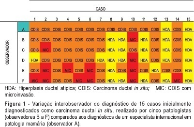
PURPOSE: to perform a critical evaluation of the histopathological diagnosis of ductal carcinoma in situ (DCIS) of the breast, through the analysis of interobserver variation related to diagnosis, architectural pattern, nuclear grade, and histological grade. METHODS: eighty-five cases with an initial diagnosis of DCIS were reviewed by the same pathologist, specialist in breast pathology, who selected 15 cases for interobserver analysis. The analysis was carried out by five pathologists and an international expert in breast pathology, who received the same slides and a protocol for classifying the lesions as atypical ductal hyperplasia (ADH), DCIS, or ductal carcinoma in situ with microinvasion (DCIS-MIC). If the diagnosis was DCIS, the pathologists should classify it according to the dominant architectural pattern, nuclear grade, and histological grade. The results were analyzed using percent concordance and the kappa test. RESULTS: there was a great interobserver diagnostic variation. In one case we had all diagnoses, from ADH, DCIS to DCIS-MIC. The kappa test for the comparison among the five observers' and the expert's diagnoses showed minimum interobservers' concordance (<0.40). Regarding DCIS classification related to the dominant architectural pattern and the histological grade, the kappa test values were considered poor among the pathologists. The best results were obtained for the nuclear grading, with a kappa index up to 0.80, considered as good concordance. CONCLUSION: the low index of interobserver concordance in diagnosis and classification of DCIS of the breast indicates the difficulty in using the most common diagnostic criteria of the literature and the need for specific training of non-specialist pathologists in breast pathology for the diagnosis of these lesions.

Summary
Revista Brasileira de Ginecologia e Obstetrícia. 2005;27(1):12-19
DOI 10.1590/S0100-72032005000100005
PURPOSE: to evaluate and compare results of female pelvic floor surface electromyography in different positions: lying, sitting and standing. METHODS: twenty-six women with the diagnosis of stress urinary incontinence treated with a protocol of exercises to strengthen the pelvic floor muscle were evaluated. Pelvic floor surface electromyography was performed with an intravaginal sensor connected to Myotrac 3G TM equipment, as follows: initial rest of 60 s, five phasic contractions, one 10-s tonic contraction and one 20-s tonic contraction. The amplitudes were obtained from the difference between the final contraction amplitude and the amplitude at rest (in µV). Wilcoxon test was applied for nonparametric data (p value <0.05). RESULTS: the amplitudes of contractions were higher in the lying position, decreasing in the sitting and standing positions. In the lying position, the median values of phasic and tonic contractions were 23.5 (5-73), 18.0 (3-58) and 17.0 (2-48), respectively. In the sitting position, they were 20.0 (2-69), 16.0 (0-58) and 15.5 (1-48). In the standing position they were 16.5 (3-67), 12.5 (2-54) and 13.5 (2-41). All amplitude values were significantly lower in the standing position compared to the lying position (p<0.001, p<0.001 and p=0.003). Similar results were also found in comparison to the sitting position. However, there was no significant difference between the lying and the sitting positions. CONCLUSION: all female pelvic floor contraction amplitudes were lower in the standing position, suggesting that the muscle strength should be intensified in that position.