Summary
Revista Brasileira de Ginecologia e Obstetrícia. 2009;31(6):305-310
DOI 10.1590/S0100-72032009000600007
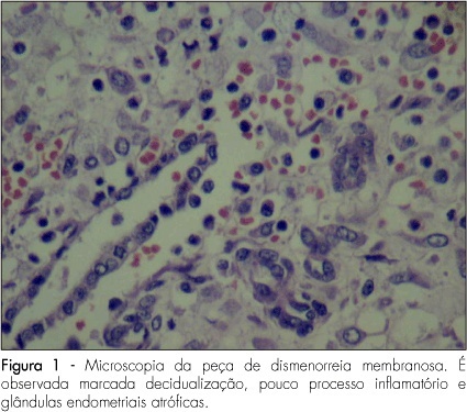
PURPOSE: to present a series of cases of membranous dysmenorrhea. METHODS: all the patients selected were under diagnostic suspicion, after being clinically attended in a private medical office due to the report of painful dysmenorrhea associated with spontaneous elimination of elastic material with uterine shape. Only relevant facts about the pain condition have been described, together with the present and previous medical history and life habits. The material eliminated was forwarded to the pathology laboratory, where the macro and microscopic analyses were done. Cases with no confirmation of membranous material elimination were not selected. After the diagnostic confirmation, literature up to 2008 was carried out using the MeSH method, with the words "membranous dysmenorrheal". RESULTS: three cases of dysmenorrhea were transcribed. Besides the characteristic picture of pain and vaginal elimination of elastic material, all the cases were associated with the use of hormonal contraceptive methods. CONCLUSIONS: despite the fact that there are only sporadic reports of cases of membranous dysmenorrhea in the scientific literature, this etiology must be considered in cases of pain associated with vaginal bleeding plus elimination of elastic or solid material. The final diagnosis depends on anatomopathological exam, which should not be dismissed. We highlight the need for more discussion about this pathology, to keep the professionals updated with the aim of exerting adequate diagnosis and therapeutics.

Summary
Revista Brasileira de Ginecologia e Obstetrícia. 2009;31(5):219-223
DOI 10.1590/S0100-72032009000500003
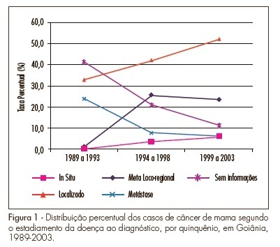
PURPOSE: To analyze the temporal changes of breast cancer staging at diagnosis among women living in Goiânia, Goiás, Brazil, between 1989 and 2003. METHODS: Retrospective and descriptive study in which the cases were identified from the Population-Based Cancer Registry of Goiânia for the period from 1989 to 2003. The variables studied were age, diagnostic method, topographic sublocation, morphology and breast cancer staging. Frequency analyses were carried out on the variables and means, and the medians for the age were determined. The SPSS® 15.0 software was used for statistical analyses. RESULTS: A total of 3,204 breast cancer cases were collected. The mean age was 56 years (sd±16 years). With regard to clinical staging, 45.6% of the cases were found to be localized in the breast, with an increased rate of 19.25% between the first and the third five-year period (p<0.001; CI 95%=0.14-0.23) and 10.2% of cases were with distant metastases. However, a reduction of 17.74% for metastatic cases in the same interval (p<0.001 e CI 95%=0.14-21) was observed. The in situ case rate was 0.2% in 1989-1993 and increased to 6.2% in 1999-2003 (p<0.001, IC95%=4.9-7.4). CONCLUSION: The diagnostic profile of breast cancer in the city of Goiânia is changing. Substantial increases in the number of early breast cancer cases are being found in relation to the number of advanced cases.

Summary
Revista Brasileira de Ginecologia e Obstetrícia. 2009;31(5):224-229
DOI 10.1590/S0100-72032009000500004
PURPOSE: to identify the pattern of myoelectrical activity of muscles from the scapular region, after axillary lymphadenectomy in breast cancer. METHODS: prospective cohort study including all the women submitted to axillary lymphadenectomy for surgical treatment of breast cancer, in a breast cancer reference center, from June to August 2006. The women were evaluated before, and after 3 and 12 months from the surgery, through physical and electromyographic examinations of the serratus anterior, upper trapezius and middle deltoid muscles. RESULTS: the patients' average age was 60.3 years old (DP±14.1), and the incidence of winged scapula at the physical examination was 64.9%. At the third-months evaluation, a reduction of 28.3 µV was observed in the myoelectrical activity of the serratus anterior muscle. At the twelveth-months evaluation and between the 3rd and the 12th month, there was an increment of 23.3 µV and 43.6 µV, respectively. For the upper trapezius, the increase was of 23.1 µV at the third-months evaluation, and 23.3 µV and 43.6 µV between the 3rd and the 12th months. As compared to before the surgery, the evaluation of the middle deltoid muscle did no present significant differences. CONCLUSIONS: considering muscle activity evaluated by surface electromyography, there was a decrease in the myoelectrical activity of the serratus anterior, due to lesion of the long thoracic nerve (neuropraxia), in the immediate postoperative evaluation. The increase of the mean square root of the electromyographic signal of the upper trapezius muscle, since the preoperative evaluation, suggests a muscular compensation related to the serratus anterior muscle's deficit.
Summary
Revista Brasileira de Ginecologia e Obstetrícia. 2009;31(5):230-234
DOI 10.1590/S0100-72032009000500005
PURPOSE: to evaluate the patient's age as an outcome predictor in an in vitro fertilization (IVF) program. METHODS: transversal study, which has included 302 women with ages varying from 24 to 46 years old, submitted to IVF, from May 2005 to July 2007. The patients were divided in three groups, according to their age: G<35 (n=161), G 36-39 (n=89) e G>40 (n=52). The number of collected oocytes, the fertilization rates, the number of transferred embryos, the embryonary quality and the pregnancy rate were evaluated. Statistical analysis was realized through Kruskal-Wallis variance analysis and χ2 test. RESULTS: in the G<35 group, an average of 8.8 oocytes by patient was obtained; in the G 36-49 group, 7.4; and in the G>40 group, 1.6. The number of oocytes obtained in G>40 group was significantly lower than in the other two groups (p<0.001).The fertilization rate was similar in the three groups, 61.4, 65.8 e 64.6% (p=0.2288), respectively. The percentage of good quality embryos was not statistically different among the three groups either, with rates of 57.4, 63.2 and 56.0% (p=0.2254), respectively. The average number of transferred embryos in each group was 3.1 (G<35), 2.8 (G 36-39) and 1.5 (G>40), respectively, with statistically significant decrease in the G>40 group (p<0.001). Concerning pregnancy rates, the G>40 group has presented a rate of 9.6%, a result which is significantly lower (p=0.0330) than the one presented by the G<35 and G 36-39 groups (26.1 e 27.0%, respectively), with no significant difference between themselves. CONCLUSIONS: though the embryonary quality is not different among women from different age groups, the number of collected oocytes, the number of transferred embryos and the pregnancy rate indicate that the women's age is an important predictive factor of success for the techniques of assisted reproduction and should be taken into consideration when this kind of treatment is proposed to women over 40.
Summary
Revista Brasileira de Ginecologia e Obstetrícia. 2009;31(5):235-240
DOI 10.1590/S0100-72032009000500006
PURPOSE: to study infection prevalence by Chlamydia trachomatis (CT) and Neisseria gonorrhoeae (NG), among adolescent and young women in a family planning outpatient clinic. METHODS: a total of 230 women up to 24 years old and history of up to four sexual partners have been followed-up for 48 months, with urine collection to search CT and NG, by the polymerase chain reaction method at the 1st, 12nd, 24th, 36th and 48th months. The variables studied were age group, schooling, marital status, number of gestations, abortions and children alive, age at the onset of sexual life, previous and present use of condom, previous use of intrauterine device, number of sexual partners in the previous six months and follow-up time. Bivariate analysis of variables according to positive tests for CT and NG, and multiple analyses by logistic regression were done. RESULTS: the ratio of infections by CT was 13.5% and by NG, 3%. Two women presented both tests as positive. The previous intrauterine device use was associated with positive tests for NG. CONCLUSIONS: the prevalence of infections by CT and NG was higher among the age group studied and the screening of young women must be taken into consideration in our services, to control the dissemination of sexually transmitted diseases and prevention of sequels.
Summary
Revista Brasileira de Ginecologia e Obstetrícia. 2009;31(5):241-248
DOI 10.1590/S0100-72032009000500007
OBJECTIVE: to determine the frequency of macrosomia in babies born alive at a reference obstetric service, and its association with maternal risk factors. METHODS: a transversal descriptive study, including 551 women at puerperium, hospitalized at Instituto de Saúde Elpídio de Almeida, in Campina Grande (PB), Brazil, from August to October, 2007. Women, whose deliveries had been assisted at the institution, with babies born alive from one single gestation and approached in the first postpartum day, were included in the study. The nutritional and sociodemographic maternal characteristics were analyzed, and the ratio of macrosomia (birth weight >4.000 g) and its association with maternal variables were determined. Macrosomia was classified as symmetric or asymmetric according to Rohrer's index. Statistical analysis has been done through Epi-Info 3.5 software; the prevalence ratio (PR) and the confidence interval at 95% (CI 95%) were calculated. The research protocol was approved by the local Ethics Committee and all the participants signed the informed consent. RESULTS: the mean maternal age was 24.7 years old, and the mean gestational age was 38.6 weeks. Excessive gestational weight gain was observed in 21.3% of the pregnant women, and 2.1% of the participants had a diagnosis of diabetes mellitus (gestational or clinic). A ratio of 5.4% of macrosomic newborns was found, 60 were asymmetric. There was no significant association between macrosomia, mother's age and parity. There was an association between macrosomia and overweight/obesity in the pre-gestational period (PR=2.9; CI 95%=1.0-7.8) and at the last medical appointment (PR=4.9; CI 95%=1.9-12.5), excessive weight gain (PR = 6.9; CI 95%:2.8-16.9), clinical or gestational diabetes (PR = 8.9; CI 95%:4.1-19.4) and hypertension (PR=2.9; CI 95%=1.1-7.9). The factors that persisted significantly associated with macrosomia in the multivariate analysis were the excessive weight gain during the gestation (RR=6.9; CI 95%=2.9-16.9) and the presence of diabetes mellitus (RR=8.9, CI 95%=4.1-19.4). CONCLUSIONS: considering that excessive gestational weight gain and diabetes mellitus were the factors more strongly associated with macrosomia, it is important that precocious detection measurements and adequate follow-up of such conditions be taken, aiming at preventing unfavorable perinatal outcomes.
Summary
Revista Brasileira de Ginecologia e Obstetrícia. 2009;31(5):249-253
DOI 10.1590/S0100-72032009000500008
PURPOSE: to compare the expression of tumor necrosis factor-alpha (TNF-α) in ovular membranes with premature rupture (MPR) and with opportune rupture; to verify the association between the expression of the TNF-α in ovular membranes and the degree of chorioamnionitis, correlating the expression of the TNF-α and the membranes' time of rupture. METHODS: ovular membranes from 31 parturients with MPR, with gestational ages over 34 weeks, and from parturients with opportune membranes' rupture, with gestational ages equal or over 37 weeks. Chorioamnionitis detection has been done by histopathological analysis. The evaluation of the TNF-α expression has been done by immune-histochemical technique, using the labile streptavidin-biotin-peroxidase (LSAB) method. RESULTS: the average rupture time was 16.6 hours. The ratio of the TNF-α expression in the Control and Study Groups did not show a significant difference (χ2=6.6; p=0.08). In the Study Group, there was no correlation between the degree of chorioamnionitis and the intensity of TNF-α expression (Spearman's coefficient (Rs)=0.4; p=0.02). CONCLUSIONS: there was no significant difference between the TNF-α expression in ovular membranes with premature or opportune rupture; in the Study Group, there was significant association between TNF-α expression and the degree of chorioamnionitis, and there was no association between rupture time and the intensity of TNF-α expression.
Summary
Revista Brasileira de Ginecologia e Obstetrícia. 2009;31(3)