Summary
Revista Brasileira de Ginecologia e Obstetrícia. 2005;27(10):613-618
DOI 10.1590/S0100-72032005001000008
PURPOSE: to evaluate the results of 14 cases of laparoscopic surgical treatment of patients with deep endometriosis of the rectovaginal septum in the Sector of Gynecological Endoscopy of the 'Hospital do Servidor Público Estadual "Francisco Morato de Oliveira"'. METHODS: a retrospective analysis was accomplished with data from the records, associated with postoperative evaluation of the patients operated between February 2002 and February 2004. The patients' age varied from 33 to 44 years, with a mean of 38.4. The parity ranged from 0 to 3, with a mean of 1.1. The main preoperative symptoms were: dysmenorrhea in 14 (100%), deep dyspareunia in 12 (85.7%), non-ciclic pelvic pain in 10 (71.4%), pain at defecation in two (14.3%), rectal bleeding in two (14.3%), and infertility in two (14.3%). The plasma level of CA-125 ranged from 3.6 to 100.3 U/mL, with a mean of 52.9 U/mL. RESULTS: the histological examination of the lesions of the rectovaginal septum was compatible with endometriosis in nine (64.3%) patients. Concerning painful symptoms, there was total regression in seven (50%) patients, partial regression (more than 80% relief) in two (14.3%), no improvement in four (28.6%), and worsening in one (7.1%). The incidence of complications was 14.3%: a ureter lesion associated with lesion of the sigmoid and a lesion of the rectum diagnosed on the 8th postoperative day. Conclusion: it can be concluded that endometriosis of the rectovaginal septum can be treated through laparoscopic surgery with low morbidity, leading to a complete or almost complete relief of the symptoms in most of the patients.
Summary
Revista Brasileira de Ginecologia e Obstetrícia. 2005;27(10):619-626
DOI 10.1590/S0100-72032005001000009
PURPOSE: to evaluate the prevalence of cytologic, colposcopic and histopathologic alterations observed in the uterine cervix of adolescents with suspected cervical neoplasia and to compare it with young adult women. METHODS: a cross-sectional, retrospective study that analyzed 366 medical records of females referred to clarify diagnosis of the suspected cervical neoplasia. The patients had been classified into two groups defined by age. The Adolescent group was composed of 129 females between 13 and 19 years and the Adult group was composed of 237 females between 20 and 24 years. Data were analyzed statistically by the prevalence ratio (PR), respective confidence intervals (CI) at 95% for each variable, chi2 test, or Fisher exact test used to compare proportion. RESULTS: the first sexual intercourse coitarche occurred on average at 15.0 years in the Adolescent group and 16.6 years in the Adult group. The possibility of diagnosis of cytological alterations in the first Papanicolaou smears (PR=2.61; CI 95%: 2.0-3,4), the condition of non-clarified cervical intraepithelial neoplasia (CIN) (PR=1.78; CI 95%: 1.26-2,52), and the colposcopic impressions of low grade (PR=1.42; CI 95%: 1.08-1.86) were statistically significant in the Adolescent group. The histopathologic analysis did not show differences at any grade of CIN. However, two cases of microinvasive carcinoma, one in each group, and three cases of clinical invasive carcinoma in the Adult group were identified. CONCLUSION: our study suggests that cervical cancer is rare among adolescents, but we verified that alterations associated with it occurred even in younger women. The evaluation of cervical intraepithelial neoplasia with the careful application of the same tools used for adult women was appropriate also in adolescence.
Summary
Revista Brasileira de Ginecologia e Obstetrícia. 2005;27(9):509-514
DOI 10.1590/S0100-72032005000900002
PURPOSE: to evaluate the luteal function in adolescents with regular menstrual cycles. METHODS: this prospective cohort study included 55 adolescents, aged 14-19 years, with menarche at 12.2 years. Ovulation was identified by ultrasound, starting on the second or fifth day of the cycle. The corpus luteum vascularization and the resistence index of the ovarian vessels were measured by Doppler on the tenth postovulatory day. Progesterone was measured by chemoluminescence on days 6, 9 and 12 of the luteal phase. The endometrial biopsy was performed 8 to 10 days after ovulation. The results were analyzed using the SPSS software and were considered significant when p<0.05. RESULTS: on average ovulation was on day 17. Progesterone levels were 11.4, 10.9 and 3.9 ng/mL on days 6, 9, and 12 after ovulation, respectively; the progesterone mean during the whole luteal phase was 10.3 ng/ml. Luteal vascularization was scarce in 34.6%, mild in 23.6% and exuberant in 41.8%. The resistance index was 0.441. On the tenth day post-ovulation the endometrium was normal in 85.5% and out-of-phase in 14.5%. There was no correlation between the ovulation day and endometrial dating (p=0.294), levels of progesterone and endometrial dating (p=0.454), progesterone and corpus luteum vascularization (p=0.994), or resistance index (p=0.237). There also was no association between endometrium development and degree of vascularization (p=0.611). CONCLUSION: abnormal luteal function in adolescents with regular menstrual cycles was found in 14.5%. Degree of vascularization, resistance index, and serum progesterone were not related to endometrium development.
Summary
Revista Brasileira de Ginecologia e Obstetrícia. 2005;27(9):515-523
DOI 10.1590/S0100-72032005000900003
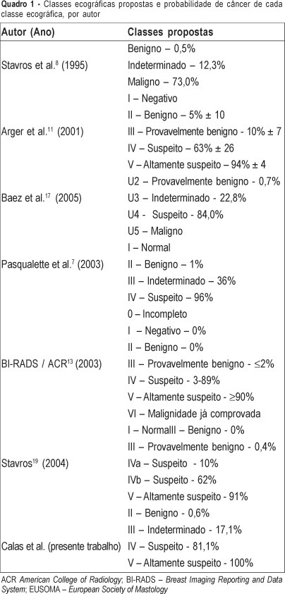
PURPOSE: the technological improvements in image quality have increased the importance of ultrasound as an imaging method in the study of breast pathologies. The need for a standardized method for lesion characterization, description and reporting in image analysis motivated the development of a breast sonographic report classification system. METHODS: the classification grouped the breast sonographic images in five classes: I - normal; II - benign; III - indeterminate, IV - suspect, and V - highly suspect. The used morphologic ultrasound features were shape, border, contour, echogenicity, echotexture, sound transmission, orientation, and secondary signals. The gold standard test, in the study of 450 lesions, considered sonographic follow-up of the lesions for a period from 6 to 24 months and the histopathology of surgical cases. RESULTS: breast sonographic classification for the diagnosis of breast cancer showed a sensitivity of 90.2% (CI: 82.8-94.9%), a specificity of 96.2% (CI: 94.0-97.6%), a positive predictive value of 84.1% (CI: 76.0-89.9%), and a negative predictive value of 97.8% (CI: 95.9-98.9%), obtaining an accuracy of 95.1%. CONCLUSIONS: the adoption of a sonographic classification system results in the standardization and optimization of the reports. It also aids the comparison with clinical findings, histopathological tests and breast images, avoiding unnecessary procedures and therefore leading to more adequate therapeutical management.

Summary
Revista Brasileira de Ginecologia e Obstetrícia. 2005;27(9):524-528
DOI 10.1590/S0100-72032005000900004
PURPOSE: to evaluate the morphological changes in murine lacrimal glands by metoclopramide-induced hyperprolactinemia during the proestrus phase or pregnancy. METHODS: forty adult mice were divided into two groups: CTR1 (control) and MET1 (treated with metoclopramide). After fifty days, half of the mice were sacrificed. The remaining animals were mated, and then labeled as pregnant controls (CTR2). Part of these animals were treated with metoclopramide and constituted the metoclopramide-treated pregnant (MET2) group. The CTR2 and MET2 groups were sacrificed on the 6th day of pregnancy. The blood was collected for determination of the hormonal levels of estradiol and progesterone by a chemoluminescent method. The lacrimal glands were then removed, fixed in 10% formaldehyde and stained with HE. The morphometric analysis was performed using the Axion Vision program (Carl Zeiss) to measure acinar nuclear and cellular volumes. RESULTS: the nuclear and cellular volumes of the lacrimal glands in the MET1-(152.2±8.7; 6.3±1.6 µm³) and MET2-(278.3±7.9; 27.5±0.9 µm³) treated groups were lower than those in CTR1 (204.2±7.4; 21.9±1.3 µm³) and CTR2 (329.4±2.2; 35.5±2.0 µm³), respectively. There was a significant hormonal level reduction in the animals that received metoclopramide compared to controls (CTR1: estradiol = 156.6±42.2 pg/ml; progesterone = 39.4±5.1 ng/ml; MET1: estradiol = 108.0±33.1 pg/ml; progesterone = 28.0±6.4 ng/ml; CTR2: estradiol = 354.0±56.0 pg/ml; progesterone = 251.0±56.0 ng/ml; MET2: estradiol = 293.0±43.0 pg/ml, progesterone = 184.0±33.0 ng/ml). CONCLUSION: metoclopramide-induced hyperprolactinemia produced morphological signs of reduction of cellular activity in lacrimal glands during the proestrus phase and pregnancy. It is hypothesized that this effect might be related to the hyperprolactinemia-induced decrease in the hormonal production of estrogen and progesterone.

Summary
Revista Brasileira de Ginecologia e Obstetrícia. 2005;27(9):529-533
DOI 10.1590/S0100-72032005000900005
PURPOSE: to study the histological modifications that occur in the endometrium of women before and six months after tubal ligation (TL) and to correlate these findings with progesterone (P4) levels. METHODS: the study was conducted on 16 women with normal menstrual cycles who were evaluated before and in the sixth cycle after TL. P4 levels were determined from the 8th day at 2-day intervals until ovulation and on the 8th, 10th and 12th day after ovulation or on the 24th day of the cycle. An endometrial biopsy was obtained between the 10th and 12th day after ovulation or on the 24th day of the cycle and a correlation with P4 was determined. Data were analyzed statistically by the nonparametric McNemar test for the evaluation of hormonal determination and by the exact Fisher test for the histological evaluation of the endometrium, with the level of significance set at p<0.05. RESULTS: mean age was 34.1±1.3 years. The intermenstrual interval was 27.1±2.6 days and the duration of bleeding was 3 to 5 days, with no difference between the studied periods. Before TL, 8/16 (50.0%) of the cases had a secretory endometrium according to the cycle, 3/16 (18.8%) had a secretory endometrium not according to the cycle and 3/16 (18.8%) had a dysfunctional endometrium, suggesting a defect in the luteal phase in 6/16 (37.5%). After TL, 7/16 (43.8%) had a secretory endometrium according to the cycle, 3/16 (18.8%) a secretory endometrium not according to the cycle and 4/16 (25.0%) had a dysfunctional endometrium, suggesting a defect in the luteal phase in 7/16 (43.8%). In 2/16 (12.5%) of the cases before TL and in 2/16 (12.5%) other cases after TL it was not possible to perform histological evaluation due to insufficient material or unspecfiic endometritis. In the luteal phase after TL, mean P4 levels were significantly lower on days +8, +10 and +12 than before TL, being 15.1, 18.0 and 20.7 ng/ml, respectively, before TL and 10.6, 8.0 and 5.4 ng/ml after TL (p<0.05). Before TL, 5/8 (62.5%) of the cases with a secretory endometrium according to the cycle had P4 >10 ng/ml and 3/8 (37.5%) had P4 <10 ng/ml. After TL, when the endometrium was secretory according to the cycle, P4 was >10 ng/ml in 4/7 (57.1%) and <10 ng/ml in 3/7 (42.9%). These differences were nonsignificant (p>0.05). CONCLUSION: six months after TL, the intermenstrual interval and the duration of bleeding were unchanged. P4 levels decreased during the luteal phase although this did not interfere in the endometrial response.
Summary
Revista Brasileira de Ginecologia e Obstetrícia. 2005;27(9):534-540
DOI 10.1590/S0100-72032005000900006
PURPOSE: to evaluate the role of morphological (12) and Doppler velocimetry (17) ultrasonographic features, in the detection of lymph node metastases in breast cancer patients. METHODS: 179 women (181 axillary cavities) were included in the study from January to December 2004. The ultrasonographic examinations were performed with a real-time linear probe (Toshiba-Power Vision-6000 (model SSA-370A)). The morphological parameters were studied with a frequency of 7.5-12 MHz. A frequency of 5 MHz was used for the Doppler velocimetry parameters. Subsequently, the women were submitted to level I, II and III axillary dissection (158), or to the sentinel lymph node technique (23). Sensitivity, specificity, and positive and negative predictive values were calculated for each parameter. The decision tree test was used for parameter association. The cutoff points were established by the ROC curve. RESULTS: at least one lymph node was detected in 173 (96%) of the women by the ultrasonographic examinations. Histological examination detected lymph node metastases in 87 women (48%). The best sensitivity among the morphological paramenters was found with the volume (62%), the antero-posterior diameter (62%) and the fatty hilum placement (56%). Though the specificity of the extracapsular invasion (100%), border regularity (92%) and cortex echogenicity (99%) were high, the sensitivity of these features was too low. None of the Doppler velocimetry parameters reached 50% sensitivity. The decision tree test selected the ultrasonographic parametners: fatty hilum placement, border regularity and cortex echogenicity, as the best parameter association. CONCLUSION: the detection of axillary cavity lymph node stage by a noninvasive method still remains an unfulfilled goal in the treatment of patients with breast cancer.

Summary
Revista Brasileira de Ginecologia e Obstetrícia. 2005;27(9):541-547
DOI 10.1590/S0100-72032005000900007
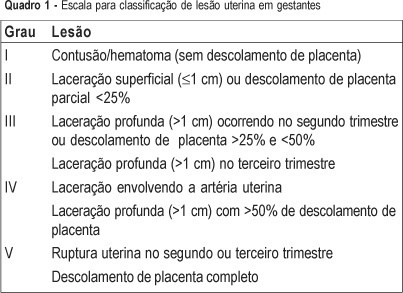
PURPOSE: to evaluate the predictors (clinical findings and physiological and anatomical scores) of the maternal and fetal outcomes among pregnant women victims of abdominal trauma who were submitted to laparotomy and to discuss particularities of assessment in this situation. METHODS: retrospective analysis of the medical records of 245 women with abdominal trauma and surgical treatment, from 1990 to 2002. Thirteen pregnant women with abdominal injury were identified. All cases were registered in the Epi-Info 6.04 protocol and data were analyzed statistically by the Fisher exact test, with confidence interval of 95%. RESULTS: ages ranged from 13 to 34 years (mean of 22.5). Six women (46.2%) were in the third trimester of pregnancy. Penetrating trauma accounted for 53.8% of injuries and in six of these patients the mechanism of trauma was gunshot wounds. Three patients had uterine injuries associated with fetal death. There were no maternal deaths and fetal mortality was 30.7%. The use of trauma scores was not associated with maternal and fetal mortality. Uterine injury was the only predictive risk factor for fetal loss (p=0.014). CONCLUSIONS: this is a retrospective study analyzing a small number of pregnant women victims of severe trauma. However, the results show that there are no predictive accuracy scores to evaluate maternal and fetal outcomes.
