Summary
Revista Brasileira de Ginecologia e Obstetrícia. 2005;27(11):650-655
DOI 10.1590/S0100-72032005001100003
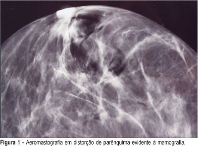
PURPOSE: to asses the efficiency of the radioguided localization and removal of occult breast lesions using radiopharmaceuticals injected directly into the lesions or close to them with posterior air injection as a radiological control. METHODS: twenty-nine consecutive patients with thirty-two occult breast lesions detected mammographically or by ultrasound, and categorized 3, 4 and 5 BI-RADS®, were included in this observational study with results expressed in percentages. The radiopharmaceutical used was human serum albumin labeled with 99mTc-HSA injected inside or close to the lesion using mammographic or ultrasonographic guidance. The injection of the radiopharmaceutical was followed immediately by air injection through the needle used for stereotaxis as a radiological control of the radiopharmaceutical placement. The excision biopsy was carried out with the aid of a hand-held gamma-detecting probe and the entire removal of the lesion was verified by X-ray of the surgical specimens or by intraoperative frozen section examination. RESULTS: breast cancer was found in 10.0% (1/10) of the 3 BI-RADS® lesions, in 31.5% (6/19) of the 4 BI-RADS® and in 66.6% (2/3) of the 5 BI-RADS®. The radiotracer was correctly positioned in 96.8% of the specimens (31/32) allowing the removal of also 96.8% of the studied non-palpable breast lesions. To show the entire removal, X-ray was used in 23 cases (71.8%), intraoperative frozen section study in 21.8% (7/32) and both methods in 6.2% (2/32). CONCLUSIONS: radioguided surgery showed to be an important tool in the removal of non-palpable breast lesions, as a simple, fast and feasible method that can be implemented in the clinical routine of these patients.
Summary
Revista Brasileira de Ginecologia e Obstetrícia. 2005;27(11):650-655
DOI 10.1590/S0100-72032005001100003
PURPOSE: to asses the efficiency of the radioguided localization and removal of occult breast lesions using radiopharmaceuticals injected directly into the lesions or close to them with posterior air injection as a radiological control. METHODS: twenty-nine consecutive patients with thirty-two occult breast lesions detected mammographically or by ultrasound, and categorized 3, 4 and 5 BI-RADS®, were included in this observational study with results expressed in percentages. The radiopharmaceutical used was human serum albumin labeled with 99mTc-HSA injected inside or close to the lesion using mammographic or ultrasonographic guidance. The injection of the radiopharmaceutical was followed immediately by air injection through the needle used for stereotaxis as a radiological control of the radiopharmaceutical placement. The excision biopsy was carried out with the aid of a hand-held gamma-detecting probe and the entire removal of the lesion was verified by X-ray of the surgical specimens or by intraoperative frozen section examination. RESULTS: breast cancer was found in 10.0% (1/10) of the 3 BI-RADS® lesions, in 31.5% (6/19) of the 4 BI-RADS® and in 66.6% (2/3) of the 5 BI-RADS®. The radiotracer was correctly positioned in 96.8% of the specimens (31/32) allowing the removal of also 96.8% of the studied non-palpable breast lesions. To show the entire removal, X-ray was used in 23 cases (71.8%), intraoperative frozen section study in 21.8% (7/32) and both methods in 6.2% (2/32). CONCLUSIONS: radioguided surgery showed to be an important tool in the removal of non-palpable breast lesions, as a simple, fast and feasible method that can be implemented in the clinical routine of these patients.
Summary
Revista Brasileira de Ginecologia e Obstetrícia. 2005;27(11):642-649
DOI 10.1590/S0100-72032005001100002
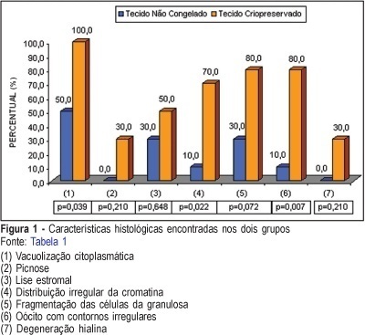
PURPOSE: to evaluate follicular preservation and histologic characteristics of the cryopreserved ovarian tissue and to compare with the fresh one, in rabbits. METHODS: ten adult female white rabbits were submitted to right oophorectomy. The dried ovary was dissected and the cortex was maintained with approximately 1.5 millimeter thickness. The tissue was fractionated into small sections, some reserved for control histologic study and others destined for cryopreservation. Six weeks later the ovarian tissue was thawed and evaluated histologically. After histologic processing, the control and the experimental samples were stained with hematoxylin and eosin and treated immunohistochemically by the PCNA technique for evaluation of DNA preservation. Histologic alterations present in the fresh and in the cryopreserved tissues were identified, and cryopreserved tissue viability was evaluated. RESULTS: in the cryopreserved tissue only primordial follicles persisted. Reversible alterations were identified: cytoplasmatic vacuolation (p=0,039), stromal lysis (p=0.648) and oocytes with irregular contours (p=0.007). Irreversible alterations: (hyalin degeneration and pyknosis) were found, but not at significant levels (p=0.210). The immunohistochemical analysis showed PCNA staining of follicles at different stages of development in the fresh tissue and primordial follicles in the cryopreserved tissue, indicating the presence of active DNA in both tissues. CONCLUSION: in the cryopreserved ovarian tissue the following were observed: survival of only primordial follicles; significant reversible histologic alterations (cytoplasmic vacuolation, stromal lysis and oocytes with irregular contours); irreversible alterations (hyalin degeneration and pyknosis), and PCNA staining of all follicles.
Summary
Revista Brasileira de Ginecologia e Obstetrícia. 2005;27(11):642-649
DOI 10.1590/S0100-72032005001100002
PURPOSE: to evaluate follicular preservation and histologic characteristics of the cryopreserved ovarian tissue and to compare with the fresh one, in rabbits. METHODS: ten adult female white rabbits were submitted to right oophorectomy. The dried ovary was dissected and the cortex was maintained with approximately 1.5 millimeter thickness. The tissue was fractionated into small sections, some reserved for control histologic study and others destined for cryopreservation. Six weeks later the ovarian tissue was thawed and evaluated histologically. After histologic processing, the control and the experimental samples were stained with hematoxylin and eosin and treated immunohistochemically by the PCNA technique for evaluation of DNA preservation. Histologic alterations present in the fresh and in the cryopreserved tissues were identified, and cryopreserved tissue viability was evaluated. RESULTS: in the cryopreserved tissue only primordial follicles persisted. Reversible alterations were identified: cytoplasmatic vacuolation (p=0,039), stromal lysis (p=0.648) and oocytes with irregular contours (p=0.007). Irreversible alterations: (hyalin degeneration and pyknosis) were found, but not at significant levels (p=0.210). The immunohistochemical analysis showed PCNA staining of follicles at different stages of development in the fresh tissue and primordial follicles in the cryopreserved tissue, indicating the presence of active DNA in both tissues. CONCLUSION: in the cryopreserved ovarian tissue the following were observed: survival of only primordial follicles; significant reversible histologic alterations (cytoplasmic vacuolation, stromal lysis and oocytes with irregular contours); irreversible alterations (hyalin degeneration and pyknosis), and PCNA staining of all follicles.
Summary
Revista Brasileira de Ginecologia e Obstetrícia. 2005;27(3):106-111
DOI 10.1590/S0100-72032005000300002
PURPOSE: to evaluate CD4+ T lymphocyte cell count and HIV viral load influence on the presence of cervical squamous intraepithelial lesions (SIL). METHODS: cross-sectional study of 134 HIV-infected women submitted to uterine cervical biopsy, HIV viral load quantification and CD4+ T lymphocyte cell count. Viral load and CD4+ T lymphocyte cell count were performed before biopsy timing. Three different levels of viral load (<400 copies/mL; 401 to 50,000 copies/mL; >50,000 copies/mL) and CD4+ T lymphocyte count (<200 cells/mm³; 200 to 350 cells/mm³; >350 cells/mm³) were defined. Data were statistically analyzed by the chi2 test, linear tendency chi2 test, Mantel-Haenszel test, and analysis of variance, with level of significance set at p<0.05 and 95% confidence interval. RESULTS: there was no risk tendency for HIV-infected women to show SIL with viral load level increase or CD4+ T lymphocyte reduction. Comparing viral load with the presence or absence of SIL, stratified by quantification timing, there was a significant difference for values over 400 copies/mL (p=0.048; OR: 3.17; 95% CI: 1,02-9.93). No association was found between CD4+ T lymphocyte cell count and SIL. CONCLUSION: patients with HIV viral load higher than 400 copies/mL, performed before uterine cervical biopsy, showed a 3.17 times greater chance to develop SIL. CD4+ T lymphocyte count had no influence on the development of SIL.
Summary
Revista Brasileira de Ginecologia e Obstetrícia. 2005;27(3):155-160
DOI 10.1590/S0100-72032005000300010
PURPOSE: to evaluate the outcome of fetuses with risk of chromosomal anomalies over 1:300, based on the nuchal translucency measurement, according to the Fetal Medicine Program. METHODS: in the pregnancies with risk of chromosomal anomalies over 1:300, variables like fetal karyotype, spontaneous or induced abortion, prematurity, stillbirth, neonatal death, malformations, and healthy newborn were considered. We used Fisher's exact test to compare differences in proportions between groups. RESULTS: we selected 193 (3.6%) single pregnancies with risk of chromosomal anomalies over 1:300. Only 165 cases fulfilled the inclusion criteria. Of these only 32.1% underwent fetal karyotyping and of which 8.5% had chromosomal anomalies (85.7% had trisomy 21). Regarding pregnancy outcomes, 4.2% were spontaneous miscarriages, 4.2% induced abortions, 4.8% were premature, 1.8% had neonatal death, 1.8% were stillborn, and 4.2% had structural malformation (85.7% congenital heart diseases). Almost 85.0% were healthy newborns. Patients with abnormal karyotyping had more induced abortions (p<0.001) and more structural malformations (p<0.001) than patients with normal karyotyping. None of the genetic diseases or miscarriages was associated with invasive procedures. Sixty-six percent of the pregnancies with prenatal diagnosis of abnormal karyotype were interrupted. CONCLUSION: nuchal translucency is an important screening tool for chromosomal diseases especially for low-risk pregnancies. However, counseling pregnancies with high risk of chromosomal anomalies should consider that, although these fetuses have a worse prognosis, most of the outcomes are favorable.
Summary
Revista Brasileira de Ginecologia e Obstetrícia. 2005;27(3):149-154
DOI 10.1590/S0100-72032005000300009
PURPOSE: to analyze the efficacy, safety and real advantage of vesicoamniotic shunt catheter in the intrauterine treatment of obstructive uropathy. METHODS: a retrospective and descriptive study, in which the evolution of 35 fetuses with obstructive uropathy, submitted to vesicoamniotic shunt from 1990 to 2004 in a Fetal Medical Center was evaluated. All these fetuses fitted the selection criteria defined by a protocol of this service, and had the parents' consent for the procedure. The Pediatric Nephrology Sector of the Hospital das Clínicas of UFMG assessed all of them after delivery to confirm the prenatal diagnosis and outcome. The dead neonates were studied by the Pathological Anatomy Sector of UFMG. Descriptive analysis of the following parameters was performed: prenatal diagnosis of the uropathy, gestational age at shunt insertion, time of catheter utilization, post-surgery complications, perinatal mortality and neonatal survival. RESULTS: posterior urethral valve was the most common uropathy (62.8%). The mean gestational age at the vesicoamniotic shunt placement was 26.1weeks and the mean time of its presence was 46 days (1-119 days). There were four intrauterine fetal deaths and 17 in the neonatal period (60% perinatal mortality). The main cause of death was pulmonary hypoplasia. Olygohidramnios was present in 33/35 fetuses (94.3%) and it was reversed in 23 of them (70%); fourteen fetuses survived the neonatal period. At present, there are 4 children followed up by the Pediatric Sector of Nephrology of Hospital das Clínicas. Two of them have been treated with peritoneal dialysis, awaiting renal transplantation. The other two have normal renal function. Their age varies from 2 months to 4 years. CONCLUSION: the vesicoamniotic shunt may be a viable intrauterine treatment for severe obstructive uropathy, with 40% of survival rate of fetuses that might have progressed to death. However, the procedure's success was directly related to the adequate selection, and to the early intervention in the uterus, performed before 32 weeks of gestation in fetuses with bilateral obstruction, without any associated malformation and with still preserved renal function. Olygohidramnios reversion did not guarantee a good prognosis. It remains controversial if the vesicoamniotic shunt can really ensure long-term renal function.
Summary
Revista Brasileira de Ginecologia e Obstetrícia. 2005;27(3):143-148
DOI 10.1590/S0100-72032005000300008
PURPOSE: to describe etiology, evolution and prevalence of hydrops fetalis in a cohort of pregnant women during a period of ten years (1992 to 2002) in a tertiary maternity. METHODS: a retrospective study was carried out in patients referred to the maternity of the Fernandes Figueira Institute, with diagnosis of hydrops fetalis, detected by ultrasonography, during the period from 1992 to 2002. The cases were selected according to etiology (immune or nonimmune) and evolution, performed invasive procedures and survival were compared between both groups. Analysis of variables was performed by Epi-Info 6.0 and a p value less than 0.05 was considered to be statistical significant. RESULTS: in ten years of follow-up, 80 patients with an initial diagnosis of hydrops were attended. The frequency of hydrops in this population was 1 in 157 live births. Rh immunization (immune group) was detected in 13 cases (16.2%), and for 67 cases (83.8%) nonimmune causes (nonimmune group) were considered. Major causes of nonimmune hydrops fetalis were idiopathic (40.2%), genetic (20.8%), infectious diseases (20.7%), and cardiopathy (7.4%). A difference was found in relation to maternal age in the immune group (mean = 32.8 years) when compared with the nonimmune group (mean = 28.7 years) (p=0.03), but gestational age at delivery was similar in both groups (mean = 33.6 weeks in the immune group and 33.1 weeks in the nonimmune group) (p=0.66). Amniocentesis and blood transfusion in utero were carried out more frequently in the immune group (p<0.001) and perinatal mortality was 53.8% in the immune group and 68.6% in the nonimmune group (p=0.47). Complementary research of IgG anti-parvovirus B19 antibodies was carried out in 41 of 67 cases of nonimmune hydrops, with 16 being positive for the presence of anti-B19 IgG antibodies. CONCLUSION: nonimmune etiology was the most common form of presentation of hydrops fetalis in our study. Perinatal mortality of this entity is still high and a substantial number of cases had no identified cause. Characterization of fetal karyotype and performance of specific parvovirus B19 serology could increase causal identification of nonimmune hydrops classified as idiopathic.
Summary
Revista Brasileira de Ginecologia e Obstetrícia. 2005;27(3):137-142
DOI 10.1590/S0100-72032005000300007
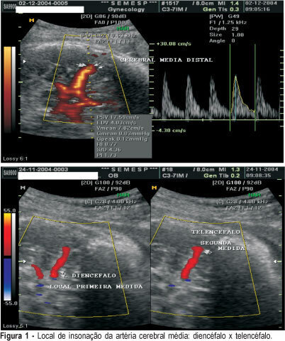
PURPOSE: to evaluate if there is any difference between Doppler indexes in the middle cerebral artery in two different sites of insonation in healthy patients and in patients with diseases. METHODS: a random prospective survey, in the period from June 2003 to March 2004 that analyzed the Doppler indexes of 100 patients: patient group (n = 50) included patients admitted to Clemente Farias University Hospital, which is part of UNIMONTES-MG, havinfg as inclusion criteria: to be in the 28th to 34th gestational week, diagnosis of chronic arterial hypertension, pre-eclampsia, intrauterine growth restriction. As control group, 50 healthy pregnant patients between the 28th and the 34th week, originary from SEMESP's clinic. The Doppler variables were the resistance index (RI), the pulsatility index (PI) and the relation systole/diastole (SD). All three Doppler indexes were assessed at two different sites of the cerebral artery: the first measurement in the diencephalons region, soon after the beginning of the middle cerebral artery and the second on a distal location in the telencephalon. The median Doppler indexes in the patient group in the first and second measurements were 1.55 and 1.69 for the PI, 0.77 and 0.79 for RI and 4.29 and 4.86 for SD, respectively. In the control group, the values were 1.73 and 1.86 for the PI; 0.83 and 0.79 for RI and 5.83 and 5.46 for SD. There were no differences between sites with a p value of 0.38, 0.29 and 0.39 for PI, RI and SD, respectively. In 15t fetuses with centralization (brain sparing effects), in the diencephalon the median indexes were 1.02 for PI, 0.63 for RI and 2.68 for SD. In the epencephalon the median indexes were 0.95 for IP, 0.62 for RI and 2.44 for SD. There were no differences between sites, with a p value of 0.53 for PI; 0.56 for IR and 0.31 for SD. The Doppler index site of assessment in the middle cerebral arteries does not interfere with the results.
Summary
Revista Brasileira de Ginecologia e Obstetrícia. 2005;27(3):137-142
DOI 10.1590/S0100-72032005000300007
PURPOSE: to evaluate if there is any difference between Doppler indexes in the middle cerebral artery in two different sites of insonation in healthy patients and in patients with diseases. METHODS: a random prospective survey, in the period from June 2003 to March 2004 that analyzed the Doppler indexes of 100 patients: patient group (n = 50) included patients admitted to Clemente Farias University Hospital, which is part of UNIMONTES-MG, havinfg as inclusion criteria: to be in the 28th to 34th gestational week, diagnosis of chronic arterial hypertension, pre-eclampsia, intrauterine growth restriction. As control group, 50 healthy pregnant patients between the 28th and the 34th week, originary from SEMESP's clinic. The Doppler variables were the resistance index (RI), the pulsatility index (PI) and the relation systole/diastole (SD). All three Doppler indexes were assessed at two different sites of the cerebral artery: the first measurement in the diencephalons region, soon after the beginning of the middle cerebral artery and the second on a distal location in the telencephalon. The median Doppler indexes in the patient group in the first and second measurements were 1.55 and 1.69 for the PI, 0.77 and 0.79 for RI and 4.29 and 4.86 for SD, respectively. In the control group, the values were 1.73 and 1.86 for the PI; 0.83 and 0.79 for RI and 5.83 and 5.46 for SD. There were no differences between sites with a p value of 0.38, 0.29 and 0.39 for PI, RI and SD, respectively. In 15t fetuses with centralization (brain sparing effects), in the diencephalon the median indexes were 1.02 for PI, 0.63 for RI and 2.68 for SD. In the epencephalon the median indexes were 0.95 for IP, 0.62 for RI and 2.44 for SD. There were no differences between sites, with a p value of 0.53 for PI; 0.56 for IR and 0.31 for SD. The Doppler index site of assessment in the middle cerebral arteries does not interfere with the results.
Summary
Revista Brasileira de Ginecologia e Obstetrícia. 2005;27(3):130-136
DOI 10.1590/S0100-72032005000300006
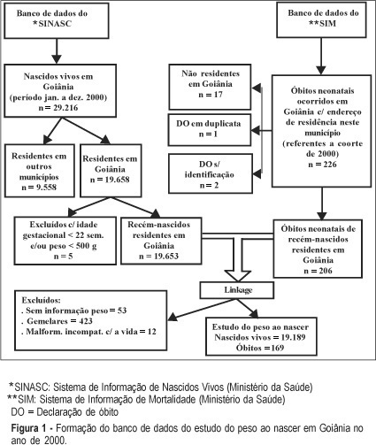
PURPOSE: to analyze birth weight in a cohort of newborns for the year 2000, in Goiânia, by determining the coefficient of mortality and neonatal survival probability, stratified by categories of birth weight, and also, through the identification of factors associated with low birth weight (LBW). METHODS: a retrospective cohort study, made possible by the linkage of data from the ISM (Information System on Mortality) and ISLB (Information System on Live Births) files. Coefficients of neonatal mortality were calculated for the categories of birth weight and a neonatal survival probability chart was constructed with the help of linear regression analysis. Risk factors for LBW were identified by univariate analysis (RR) and logistic regression analysis, and the level of significance was set at 5%. RESULTS: the incidence of LBW was 6.9% and 140 (66.8%) neonatal deaths took place in this group. Thirty percent of these deaths occurred in the 1,500-2,500 g weight bracket. The following risk factors were identified for LBW: preterm pregnancy, presence of congenital malformations, mothers at the extreme ages for reproduction, mothers living in the northwestern region of the city, insufficient prenatal appointments with the doctor, delivery in a public hospital, and female babies. CONCLUSION: Goiânia had an incidence of LBW which is comparable to that of developed countries and coefficients of neonatal mortality by category of weight were below those found for those countries. These results recommend that we pay attention to: prematurity, public hospitals, and the northwestern region of Goiânia.
Summary
Revista Brasileira de Ginecologia e Obstetrícia. 2005;27(3):130-136
DOI 10.1590/S0100-72032005000300006
PURPOSE: to analyze birth weight in a cohort of newborns for the year 2000, in Goiânia, by determining the coefficient of mortality and neonatal survival probability, stratified by categories of birth weight, and also, through the identification of factors associated with low birth weight (LBW). METHODS: a retrospective cohort study, made possible by the linkage of data from the ISM (Information System on Mortality) and ISLB (Information System on Live Births) files. Coefficients of neonatal mortality were calculated for the categories of birth weight and a neonatal survival probability chart was constructed with the help of linear regression analysis. Risk factors for LBW were identified by univariate analysis (RR) and logistic regression analysis, and the level of significance was set at 5%. RESULTS: the incidence of LBW was 6.9% and 140 (66.8%) neonatal deaths took place in this group. Thirty percent of these deaths occurred in the 1,500-2,500 g weight bracket. The following risk factors were identified for LBW: preterm pregnancy, presence of congenital malformations, mothers at the extreme ages for reproduction, mothers living in the northwestern region of the city, insufficient prenatal appointments with the doctor, delivery in a public hospital, and female babies. CONCLUSION: Goiânia had an incidence of LBW which is comparable to that of developed countries and coefficients of neonatal mortality by category of weight were below those found for those countries. These results recommend that we pay attention to: prematurity, public hospitals, and the northwestern region of Goiânia.