Summary
Revista Brasileira de Ginecologia e Obstetrícia. 2009;31(2):54-60
DOI 10.1590/S0100-72032009000200002
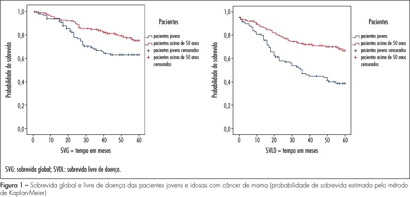
PURPOSE: the objective of this study was to evaluate the clinical, pathological and molecular characteristics in very young women and postmenopausal women with breast cancer. METHODS: we selected 106 cases of breast cancer of very young women (<35 years) and 130 cases of postmenopausal women. We evaluated clinical characteristics of patients (age at diagnosis, ethnic group, family history of breast cancer, staging, presence of distant metastases, overall and disease-free survival), pathological characteristics of tumors (tumor size, histological type and grade, axillary lymph nodes status) and expression of molecular markers (hormone receptors, HER2, p53, p63, cytokeratins 5 and 14, and EGFR), using immunohistochemistry and tissue microarray. RESULTS: when comparing clinicopathologic variables between the age groups, younger women demonstrated greater frequency of nulliparity (p=0.03), larger tumors (p<0.000), higher stage disease (p=0.01), lymph node positivity (p=0.001), and higher grade tumors (p=0.004). Most of the young patients received chemotherapy (90.8%) and radiotherapy (85.2%) and less tamoxifen therapy (31.5%) comparing with postmenopausal women. Lower estrogen receptor positivity 49.1% (p=0.01) and higher HER2 overexpression 28.7% (p=0.03) were observed in young women. In 32 young patients (29.6%) and in 20% of the posmenopausal women, the breast carcinomas were of the triple-negative phenotype (p=0.034). In 16 young women (50%) and in 10 postmenopausal women (7.7%), the tumors expressed positivity for cytokeratin 5 and/or 14, basal phenotype (p=0.064). Systemic metastases were detected in 55.3% of the young women and in 39.2% of the postmenopausal women. Breast cancer overall survival and disease-free survival in five years were, respectively, 63 and 39% for young women and 75 and 67% for postmenopausal women. CONCLUSIONS: breast cancer arising in very young women showed negative clinicobiological characteristics and more aggressive tumors.
Summary
Revista Brasileira de Ginecologia e Obstetrícia. 2009;31(2):54-60
DOI 10.1590/S0100-72032009000200002
PURPOSE: the objective of this study was to evaluate the clinical, pathological and molecular characteristics in very young women and postmenopausal women with breast cancer. METHODS: we selected 106 cases of breast cancer of very young women (<35 years) and 130 cases of postmenopausal women. We evaluated clinical characteristics of patients (age at diagnosis, ethnic group, family history of breast cancer, staging, presence of distant metastases, overall and disease-free survival), pathological characteristics of tumors (tumor size, histological type and grade, axillary lymph nodes status) and expression of molecular markers (hormone receptors, HER2, p53, p63, cytokeratins 5 and 14, and EGFR), using immunohistochemistry and tissue microarray. RESULTS: when comparing clinicopathologic variables between the age groups, younger women demonstrated greater frequency of nulliparity (p=0.03), larger tumors (p<0.000), higher stage disease (p=0.01), lymph node positivity (p=0.001), and higher grade tumors (p=0.004). Most of the young patients received chemotherapy (90.8%) and radiotherapy (85.2%) and less tamoxifen therapy (31.5%) comparing with postmenopausal women. Lower estrogen receptor positivity 49.1% (p=0.01) and higher HER2 overexpression 28.7% (p=0.03) were observed in young women. In 32 young patients (29.6%) and in 20% of the posmenopausal women, the breast carcinomas were of the triple-negative phenotype (p=0.034). In 16 young women (50%) and in 10 postmenopausal women (7.7%), the tumors expressed positivity for cytokeratin 5 and/or 14, basal phenotype (p=0.064). Systemic metastases were detected in 55.3% of the young women and in 39.2% of the postmenopausal women. Breast cancer overall survival and disease-free survival in five years were, respectively, 63 and 39% for young women and 75 and 67% for postmenopausal women. CONCLUSIONS: breast cancer arising in very young women showed negative clinicobiological characteristics and more aggressive tumors.
Summary
Revista Brasileira de Ginecologia e Obstetrícia. 2009;31(2):82-88
DOI 10.1590/S0100-72032009000200006
PURPOSE: to evaluate the effect of magnesium sulphate on the pulsatility index (PI) of the uterine, umbilical and fetal middle cerebral arteries, according to the persistency or not of the bilateral protodiastolic notch of the uterine arteries in severe pre-eclampsia. METHODS: a cohort study including 40 pregnant women with severe pre-eclampsia, 23 of them presenting bilateral protodiastolic notch, and 17, unilateral/absent notch. The patients were submitted to Doppler velocimetry before and 20 minutes after the intravenous administration of 6 g of magnesium sulphate. The examination was carried out with the patient in semi-Fowler position, the sonograms being obtained during fetal inactivity, in apnea and absent uterine contraction periods. All the exams were performed by two researchers, the average being considered as the final result. Wilcoxon's test was used to compare the PI, before and after magnesium sulphate in both groups. The difference between the two measurements (before and after magnesium sulphate) was compared between the groups (bilateral incision and unilateral/absent incision) using the Mann-Whitney test. RESULTS: there was a significant increase in the maternal heart rate (MHR) and decrease in the maternal blood pressure, and in the PI medians of the two uterine arteries and in the fetal middle cerebral artery, after magnesium sulphate in both groups. There was a significant decrease in the PI of the left uterine artery and in the umbilical artery, only in the protodiastolic unilateral/absent notch group. Nevertheless, it was not found any significant difference regarding the PI of the right uterine artery, or the cerebral/umbilical relationship, before and after magnesium sulphate in each group. No difference between the groups was found, before and after magnesium sulphate, for any of the studied outcomes. CONCLUSIONS: after the intravenous administration of 6 g of magnesium sulphate to patients with severe pre-eclampsia, a decrease in blood pressure and in the PI of the uterine, umbilical and fetal middle cerebral arteries occurs, besides the increase in the MHR, not influenced by the presence of bilateral protodiastolic notch in the uterine arteries.
Summary
Revista Brasileira de Ginecologia e Obstetrícia. 2009;31(2):75-81
DOI 10.1590/S0100-72032009000200005
PURPOSE: to evaluate the factors leading to delays in obtaining definitive diagnosis of suspicious lesions for breast cancer. METHODS: a cross-sectional, observational study was carried out with 104 women attending a cancer hospital with a diagnosis or suspected diagnosis of breast cancer. A semistructured questionnaire on the patients' demographic, clinical characteristics and the use of services was applied.Variables were compared using t-Student test, Mann-Whitney test, Pearson's χ2 test or Fisher's exact test, as appropriate. In order to identify the variables associated with delays in breast cancer diagnosis, the Odds Ratio (OR) were calculated together with their respective 95% confidence intervals (95%CI) and a logistic regression model was constructed. RESULTS: age of patients was 54±12.6 years (mean±standard deviation). Most of the women were white (48.1%), married (63.5%), living in the city of Rio de Janeiro (57.7%) and poorly educated (60.6%). The median time between the first sign or symptom of the disease and first consultation was one month and the mean time between first consultation and confirmation of diagnosis was 6.5 months. In 51% of the women, diagnosis was late (stages II-IV). Symptomatic presentation and longer delay between symptom onset and the first evaluation and between symptom onset and the diagnosis were found to be significant factors (p<0.05) for delays in obtaining definitive diagnosis of suspicious lesions. CONCLUSIONS: the results of this study suggest that efforts must be made to reduce the time needed to get an appointment with a doctor and to confirm a diagnosis of suspicious lesions, as well as to educate physicians and the women themselves regarding the importance of breast symptoms and the value of prompt evaluation, diagnosis, and treatment.
Summary
Revista Brasileira de Ginecologia e Obstetrícia. 2009;31(2):68-74
DOI 10.1590/S0100-72032009000200004
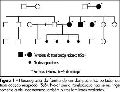
PURPOSE: to asses the prevalence and clinical characteristics of couples with history of recurrent spontaneous abortion and chromosome abnormality, attended at the present service. METHODS: all the couples referred to our service due to history of recurrent spontaneous abortion, from January 1975 to June 2008, were evaluated. Only the ones whose chromosome karyotype analysis by GTG bands has been successfully made were included in the study. Clinical data on their age, as well as on the number of abortions, stillbirth, multiple malformations, livebirth per couple, and the result of the karyotype exam were collected. Fisher's exact test (p<0.05) has been used to compare the incidence of chromosome alterations found in our study, with data in the literature. RESULTS: there were 108 couples in the sample. Their ages varied from 21 to 58 years old among the men (average of 31.4 years old), and from 19 to 43 among the women (average of 29.9 years old). In ten couples, one of the mates (9.3%) presented chromosome alterations, which corresponded respectively to three cases (30%) of reciprocal translocation [two of t(5;6) and one of t(2;13)], two (20%) of Robertsonian translocation [two of der(13;14) and one of der(13;15)], five(50%) of mosaicism (mos) [two cases of mos 45,X/46,XX, one of mos 46,XX/47,XXX, one of mos 46,XY/47,XXY and one of mos 46,XY/47,XYY] and one (10%) of chromosome inversion [inv(10)]. In one of the couples, the female presented two concomitant alterations: t(2;13) and der(13;14). Chromosome abnormalities were found in 5% of the couples with a history of two abortions, in 10.3% with three abortions, and in 14.3% with four or more abortions. CONCLUSIONS: the incidence of chromosome abnormalities seen in our study (9.3%) was similar to most of the studies carried out in the last 20 years, varying from 4.8 to 10.8%. Nevertheless the high percentage of patients with mosaicism in our sample, has called our attention. It is believed that this fact may be associated to the high number of metaphases ordinarily analyzed in the present service.
Summary
Revista Brasileira de Ginecologia e Obstetrícia. 2009;31(2):68-74
DOI 10.1590/S0100-72032009000200004
PURPOSE: to asses the prevalence and clinical characteristics of couples with history of recurrent spontaneous abortion and chromosome abnormality, attended at the present service. METHODS: all the couples referred to our service due to history of recurrent spontaneous abortion, from January 1975 to June 2008, were evaluated. Only the ones whose chromosome karyotype analysis by GTG bands has been successfully made were included in the study. Clinical data on their age, as well as on the number of abortions, stillbirth, multiple malformations, livebirth per couple, and the result of the karyotype exam were collected. Fisher's exact test (p<0.05) has been used to compare the incidence of chromosome alterations found in our study, with data in the literature. RESULTS: there were 108 couples in the sample. Their ages varied from 21 to 58 years old among the men (average of 31.4 years old), and from 19 to 43 among the women (average of 29.9 years old). In ten couples, one of the mates (9.3%) presented chromosome alterations, which corresponded respectively to three cases (30%) of reciprocal translocation [two of t(5;6) and one of t(2;13)], two (20%) of Robertsonian translocation [two of der(13;14) and one of der(13;15)], five(50%) of mosaicism (mos) [two cases of mos 45,X/46,XX, one of mos 46,XX/47,XXX, one of mos 46,XY/47,XXY and one of mos 46,XY/47,XYY] and one (10%) of chromosome inversion [inv(10)]. In one of the couples, the female presented two concomitant alterations: t(2;13) and der(13;14). Chromosome abnormalities were found in 5% of the couples with a history of two abortions, in 10.3% with three abortions, and in 14.3% with four or more abortions. CONCLUSIONS: the incidence of chromosome abnormalities seen in our study (9.3%) was similar to most of the studies carried out in the last 20 years, varying from 4.8 to 10.8%. Nevertheless the high percentage of patients with mosaicism in our sample, has called our attention. It is believed that this fact may be associated to the high number of metaphases ordinarily analyzed in the present service.
Summary
Revista Brasileira de Ginecologia e Obstetrícia. 2009;31(1):22-27
DOI 10.1590/S0100-72032009000100005
PURPOSE: to analyze complications, morbidity, mortality and survival rate in a group of patients with cervical cancer with central pelvic relapse after primary radiotherapy treatment. METHODS: retrospective study of a series of 16 cases of pelvic exenteration after primary radiotherapy treatment. Descriptive statistics, survival curve through Kaplan-Meier's method, and regression analysis to evaluate prognosis were performed. RESULTS: sixteen patients have undergone pelvic exenteration. Epidermoid carcinoma, IIb stage and undifferentiated grade were the most frequent conditions. Post-operatory tumor relapse occurred in half the cases. Eleven patients presented peri or post-surgical complications, the most frequent being pelvic infection, that of the surgical wound, and urinary fistulae. Global survival rate was 64.3%, with average follow-up of 11 months. Regression analysis did not detect any significant prognosis factor for the patient survival. CONCLUSIONS: the survival rate was 64.3%. No particular factor associated to poor prognosis has been found in the present series of cases.
Summary
Revista Brasileira de Ginecologia e Obstetrícia. 2009;31(1):17-21
DOI 10.1590/S0100-72032009000100004
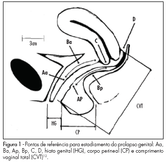
PURPOSE: to evaluate risk factors for the development of genital prolapse in the Brazilian population. METHODS: case-control study involving 316 patients submitted to prolapse staging, according to the pelvic organ prolapse quantification system. The patients were divided into two groups: in the Case Group there were 107 patients with prolapse at stage III or IV, and in the Control Group, 209 women at stage 0 or I. In the anamnesis, the selected women have been questioned about the presence of possible risk factors for genital prolapse, such as: age, menopause age, parturition, delivery type (vaginal, caesarean section or forceps), occurrence of fetal macrosomia, family history of genital dystopia in first degree relatives, chronic cough and intestinal constipation. RESULTS: The variables that were different between the groups were: age, body mass index, parturition, number of vaginal, caesarean section or forceps deliveries, newborn weight and positive family history for prolapse. Race, menopause age, chronic cough and intestinal constipation did not present differences between the groups. After logistic regression, only three variables have been shown to be independent risk factors: presence of at least one vaginal delivery, fetal macrosomia and positive family history for dystopia. Cesarean section was shown to be a protective factor. CONCLUSION: in the Brazilian population, the independent risk factor for genital prolapse were: personal antecedent of at least one vaginal delivery, fetal macrosomia and family history of dystopia.
Summary
Revista Brasileira de Ginecologia e Obstetrícia. 2009;31(1):17-21
DOI 10.1590/S0100-72032009000100004
PURPOSE: to evaluate risk factors for the development of genital prolapse in the Brazilian population. METHODS: case-control study involving 316 patients submitted to prolapse staging, according to the pelvic organ prolapse quantification system. The patients were divided into two groups: in the Case Group there were 107 patients with prolapse at stage III or IV, and in the Control Group, 209 women at stage 0 or I. In the anamnesis, the selected women have been questioned about the presence of possible risk factors for genital prolapse, such as: age, menopause age, parturition, delivery type (vaginal, caesarean section or forceps), occurrence of fetal macrosomia, family history of genital dystopia in first degree relatives, chronic cough and intestinal constipation. RESULTS: The variables that were different between the groups were: age, body mass index, parturition, number of vaginal, caesarean section or forceps deliveries, newborn weight and positive family history for prolapse. Race, menopause age, chronic cough and intestinal constipation did not present differences between the groups. After logistic regression, only three variables have been shown to be independent risk factors: presence of at least one vaginal delivery, fetal macrosomia and positive family history for dystopia. Cesarean section was shown to be a protective factor. CONCLUSION: in the Brazilian population, the independent risk factor for genital prolapse were: personal antecedent of at least one vaginal delivery, fetal macrosomia and family history of dystopia.
Summary
Revista Brasileira de Ginecologia e Obstetrícia. 2009;31(1):10-16
DOI 10.1590/S0100-72032009000100003
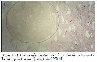
PURPOSE: to determine the prevalence of histopathological changes, in human placentas, related to hypertensive syndromes. METHODS: a transversal study that compares histopathological changes identified in 43 placentae from hypertensive pregnant women (HypPr), with the ones from 33 placentae from normotensive pregnant women (NorPr). The weight, volume and macroscopic and microscopic occurrence of infarctions, clots, hematomas, atherosis (partial obliteration, thickness of layers and presence of blood vessels hyalinization) and Tenney-Parker changes (absent, discreet and prominent), as well as the locating of infarctions and clots (central, peripheral or the association of both) have been analyzed. The χ2 and t Student tests have been used for the statistical analysis, as well as medians, standard deviations and ratios. It has been considered as significant, p<0.05. RESULTS: the macroscopic study of HypPr placentae have presented lower weight (461.1 versus 572.1 g) and volume (437.4 versus 542.0 cm³), higher infarction (51.2 versus 45.5%; p<0.05: OR=1.15) and clots (51.2 versus 15.1%; p<0.05; OR=5.4) ratios, as compared to the NorPr's. In the HypPr and NorPr, microscopic clots have occurred in 83.7 versus 45.5% (p<0.05; OR=4.3), respectively. Atherosis and Tenney-Parker changes have been statistically associated to the hypertensive syndromes (p<0.05). CONCLUSIONS: the obtained data allow us to associate lower placentary weight and volume, higher ratio of macro and microscopic infarction, clots, atherosis and Tenney-Parker changes to placentae of gestations occurring with hypertensive syndromes.
Summary
Revista Brasileira de Ginecologia e Obstetrícia. 2009;31(1):10-16
DOI 10.1590/S0100-72032009000100003
PURPOSE: to determine the prevalence of histopathological changes, in human placentas, related to hypertensive syndromes. METHODS: a transversal study that compares histopathological changes identified in 43 placentae from hypertensive pregnant women (HypPr), with the ones from 33 placentae from normotensive pregnant women (NorPr). The weight, volume and macroscopic and microscopic occurrence of infarctions, clots, hematomas, atherosis (partial obliteration, thickness of layers and presence of blood vessels hyalinization) and Tenney-Parker changes (absent, discreet and prominent), as well as the locating of infarctions and clots (central, peripheral or the association of both) have been analyzed. The χ2 and t Student tests have been used for the statistical analysis, as well as medians, standard deviations and ratios. It has been considered as significant, p<0.05. RESULTS: the macroscopic study of HypPr placentae have presented lower weight (461.1 versus 572.1 g) and volume (437.4 versus 542.0 cm³), higher infarction (51.2 versus 45.5%; p<0.05: OR=1.15) and clots (51.2 versus 15.1%; p<0.05; OR=5.4) ratios, as compared to the NorPr's. In the HypPr and NorPr, microscopic clots have occurred in 83.7 versus 45.5% (p<0.05; OR=4.3), respectively. Atherosis and Tenney-Parker changes have been statistically associated to the hypertensive syndromes (p<0.05). CONCLUSIONS: the obtained data allow us to associate lower placentary weight and volume, higher ratio of macro and microscopic infarction, clots, atherosis and Tenney-Parker changes to placentae of gestations occurring with hypertensive syndromes.
Summary
Revista Brasileira de Ginecologia e Obstetrícia. 2009;31(1):5-9
DOI 10.1590/S0100-72032009000100002
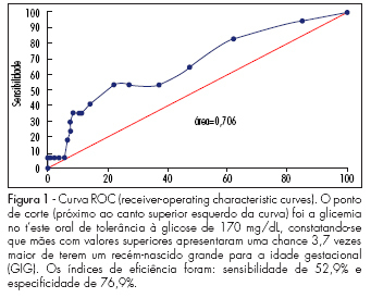
PURPOSE: to evaluate factors related to the presence of neonatal macrosomia in pregnant women with gestational diabetes mellitus. METHODS: 157 pregnant women presenting gestational diabetes mellitus in follow-up were retrospectively selected from January 2004 to July 2006. This group has been divided into two subgroups: one with newborns with weight in accordance with the gestational age (n=136) and another with macrosomic newborns (n=21). Maternal characteristics have been compared between the groups. The t-Student test was used for the analysis of equality hypothesis between the averages of the two groups, and chi-square test, to check the groups' homogeneity concerning ratios. RESULTS: the groups did not show any significant difference concerning the gestational age, body mass index, weight gain along the gestation, number of previous pregnancies, fast glycemia in the oral glucose tolerance test after the ingestion of 75 g (TOTG 75 g), gestational age at delivery, glycemic values during the treatment, and the type of treatment used (p>0.05). In the group with neonatal macrosomia, there was a higher two-hour-glycemia in the TOTG 75 g (p=0.02), higher gestational age at the treatment onset (p=0.02), and a lower number of appointments at the health service (p<0.01). When adjusted to a logistic regression model, the most important factor (p<0.01) found to predict neonatal macrosomia was the two-hour-glycemia in the TOTG 75 g. CONCLUSIONS: the factors more frequently related to neonatal macrosomia were late treatment onset and, consequently, lower number of appointments and chiefly, high two-hour-glycemia in the TOTG 75 g.
Summary
Revista Brasileira de Ginecologia e Obstetrícia. 2009;31(1):5-9
DOI 10.1590/S0100-72032009000100002
PURPOSE: to evaluate factors related to the presence of neonatal macrosomia in pregnant women with gestational diabetes mellitus. METHODS: 157 pregnant women presenting gestational diabetes mellitus in follow-up were retrospectively selected from January 2004 to July 2006. This group has been divided into two subgroups: one with newborns with weight in accordance with the gestational age (n=136) and another with macrosomic newborns (n=21). Maternal characteristics have been compared between the groups. The t-Student test was used for the analysis of equality hypothesis between the averages of the two groups, and chi-square test, to check the groups' homogeneity concerning ratios. RESULTS: the groups did not show any significant difference concerning the gestational age, body mass index, weight gain along the gestation, number of previous pregnancies, fast glycemia in the oral glucose tolerance test after the ingestion of 75 g (TOTG 75 g), gestational age at delivery, glycemic values during the treatment, and the type of treatment used (p>0.05). In the group with neonatal macrosomia, there was a higher two-hour-glycemia in the TOTG 75 g (p=0.02), higher gestational age at the treatment onset (p=0.02), and a lower number of appointments at the health service (p<0.01). When adjusted to a logistic regression model, the most important factor (p<0.01) found to predict neonatal macrosomia was the two-hour-glycemia in the TOTG 75 g. CONCLUSIONS: the factors more frequently related to neonatal macrosomia were late treatment onset and, consequently, lower number of appointments and chiefly, high two-hour-glycemia in the TOTG 75 g.