Summary
Revista Brasileira de Ginecologia e Obstetrícia. 2011;33(5):211-218
DOI 10.1590/S0100-72032011000500002
PURPOSE: the aim of this study was to analyze conjoined twins in terms of antenatal, delivery and postnatal aspects. METHODS: a retrospective descriptive analysis of prenatally diagnosed conjoined twins. Prenatal ultrasound and echocardiography, delivery details, postnatal follow-up, surgical separation and post mortem data were reviewed. The twins were classified according to the type of fusion between fetal structures. The following data were analyzed: ultrasound and echocardiographic findings, antenatal lethality and possibility of surgical separation, delivery details and survival rates. RESULTS: forty cases of conjoined twins were included in the study. There were 72.5% cases of thoracophagus, 12.5% of paraphagus, 7.5% of omphalo-ischiophagus, 5.0% of omphalophagus, and 2.5% of cephalophagus. Judicial termination of pregnancy was requested in 58.8% of the cases. Cesarean section was performed in all cases in which pregnancy was not terminated. The mean gestational age at delivery was 35 weeks; all twins were live births with a mean birth weight of 3,860 g and 88% died postnatally. Ten percent of the live borns were submitted to surgical separation with a 60% survival rate. The total survival rate was 7.5% and postnatal survival was 12%. Antenatal evaluation of lethality and possibility of surgical separation were precise. There were no maternal complications related to delivery. CONCLUSION: conjoined twins present a dismal prognosis mainly related to the complex cardiac fusion present in the majority of cases with thoracic sharing. At referring centers, prenatal ultrasound and echocardiographic evaluation accurately delineate fetal prognosis and the possibility of postnatal surgical separation.
Summary
Revista Brasileira de Ginecologia e Obstetrícia. 2011;33(4):188-195
DOI 10.1590/S0100-72032011000400007
PURPOSE: to evaluate the prevalence of urinary symptoms and association between pelvic floor muscle function and urinary symptoms in primiparous women 60 days after vaginal delivery with episiotomy and cesarean section after labor. METHODS: a cross-sectional analysis was conducted on women from an out patient clinic in São Paulo state, Brazil, 60 days after delivery. Pelvic floor muscle function was assessed by surface electromyography (basal tone, maximal voluntary contraction and mean sustained contraction) and by a manual muscle test (grades 0-5). In an interview, the urinary symptoms were identified and women with difficulty to understand, with motor/neurological impairment, pelvic surgery, diabetes, restriction for vaginal palpation and practicing exercises forpelvic floor muscles were excluded. The χ2 and Fisher Exact test were used to compare proportions and the Mann-Whitney test was used to analyze mean differences. RESULTS: 46 primiparous were assessed on average 63.7 days postpartum. The most prevalent symptoms were nocturia (19.6%), urgency (13%) and increased daytime urinary frequency (8.7%). Obese and overweight women had 4.6 times more of these symptoms (PR=4.6 [95%CI; 1.2-18.6; p value=0.0194]). Stress urinary incontinence was the most prevalent incontinence (6.5%). The mean values found for the basic tone, maximal voluntary contraction and sustained contraction were: 3 µV, 14.6 µV and 10.3 µV. Most of the women (56.5%) had grade 3 muscular strength. There was no association between urinary symptoms and pelvic floor muscle function. CONCLUSION: the prevalence of urinary symptoms was low 60 days postpartum and there was no association between pelvic floor muscle function and urinary symptoms.
Summary
Revista Brasileira de Ginecologia e Obstetrícia. 2011;33(4):182-187
DOI 10.1590/S0100-72032011000400006
PURPOSE: to translate into Portuguese, culturally adapt and validate the Incontinence Severity Index (ISI) questionnaire. METHODS: two Brazilian translators carried out the translation of the ISI into Portuguese and a version was generated by consensus. This version was back-translated by two other native English speaking translators. The differences between versions were resolved and the version was pre-tested in a pilot study. One week later, the ISI was reapplied to complete the retest. The final version of the ISI was applied together with the one-hour pad test to women with stress urinary incontinence. For the validation of the ISI, the reliability (internal consistency and test-retest) and the construct were evaluated. RESULTS: the reliability of the instrument was tested using the Cronbach α coefficient, with a general result of 0.93, demonstrating excellent reliability and consistency of the instrument. The intraclass correlation coefficient and the standard errors of measurement were 0.96 and 0.43, respectively. The Pearson correlation revealed a strong positive correlation (r=0.72, p<0.0001) between the results of the ISI questionnaire and the one-hour pad test. CONCLUSION: the culturally adapted version of the ISI translated into Brazilian Portuguese presented satisfactory reliability and survey validity and was considered to be valid for the evaluation of the severity of urinary incontinence.
Summary
Revista Brasileira de Ginecologia e Obstetrícia. 2011;33(4):176-181
DOI 10.1590/S0100-72032011000400005
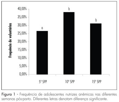
PURPOSE: to evaluate changes in the nutritional status of lactating adolescents in different postpartum weeks. METHOD: this is an analytical, observational, longitudinal study. Lactating adolescents were followed-up from the 5th to the 15th postpartum week (PPW). The nutritional status was evaluated in the 5th, 10th and 15th PPW by the Body Mass Index (BMI/age). A colorimetric method was used to determine hemoglobin level and microcentrifugation to define hematocrit. ANOVA with repeated measures was used to compare means, followed by the Tukey post-test. The level of significance was 5%. RESULTS: modification in nutritional status was observed from the pregestational period to the 15th PPW, with a reduction in the frequency of lactating adolescents with low weight (from 21% to 9%) and a rise in the frequency of overweight (21% to 27%) and eutrophic (58% to 64%) adolescents. Although mean hemoglobin (12.3±1.7 g/dL) and hematocrit (39.0±4.0%) levels were normal, a high frequency of anemia (30%) was observed throughout the study period. CONCLUSION: the present results show that the body weight of lactating adolescents rises during the lactation period and could lead to a higher frequency of obesity among adolescents. Anemia is still a nutritional problem, not only during pregnancy, but also during the postpartum period. It is necessary to prevent and treat probable subclinical nutritional deficiencies at this biological time.

Summary
Revista Brasileira de Ginecologia e Obstetrícia. 2011;33(4):164-169
DOI 10.1590/S0100-72032011000400003
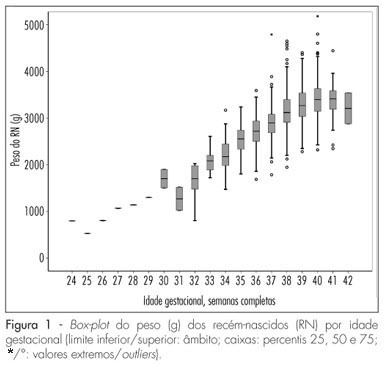
PURPOSE: to assess the validity of several fetal weight charts, commonly used in Portugal, to classify its population. METHODS: observational retrospective study. Singleton birth data was analyzed, from a two- year period (May 2008 to April 2010), from pregnancies with an ultrasound in the same institution, between the 8th and 14th gestational week. Upon data validation, percentiles for each completed gestational week were created, smoothed by a quadratic function, analyzed and compared to the tables more commonly utilized, in the institution and country, by using Z-scores, percentile comparison, sample 10th percentile detection sensibility and birthweight means comparison. RESULTS: a total of 5,378 newborns (NB) were born in the period; 2,195 (42%) NB were included, born from the 24th to 42nd gestational week, allowing statistical analysis from the 34th to the 41st week. There were differences in the mean birthweight for each gestational age, between references and with the sample, as well as between sexes. The 10th percentile from some references has shown differences ranging from -288g at 37 weeks (-11% in Lubchenco et al. data), with and +133g at 34 weeks (+7,6% with Carrascosa et al. data) compared to the values found with the sample. Differences were also found concerning the sensitivity of the identification of a sample birthweight below the 10th percentile, which was between 14.1 and 100%, depending on the reference used. DISCUSSION: the limitation of these kinds of reference values must be remembered and minimized, with the adoption of regionally or nationally produced references, contemplating other variables, such as sex, with precisely known gestation duration and with validation of the utilized references in loco.

Summary
Revista Brasileira de Ginecologia e Obstetrícia. 2011;33(4):157-163
DOI 10.1590/S0100-72032011000400002
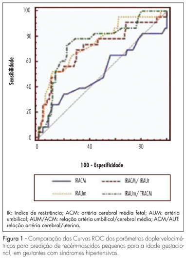
PURPOSE: to determine the best Doppler flow velocimetry index to predict small infants for gestational age (SGAI), in pregnant women with hypertensive syndromes. METHODS: a cross-sectional study was conducted enrolling 129 women with high blood pressure, submitted to dopplervelocimetry up to 15 days before delivery. Women with multiple fetuses, fetal malformations, genital bleeding, placental abruption, premature rupture of fetal membranes, smoking, use of illicit drugs, and chronic diseases were excluded. A receiver operating characteristic (ROC) curve for each Doppler variable was constructed to diagnose SGAI and the sensitivity (Se), specificity (Sp), positive (PLR) and negative (NLR) likelihood ratio were calculated. RESULTS: the area under the ROC curve for the middle cerebral artery resistance index was 52% (p=0.79) with Se, Sp, PLR, and NLR of 25.0, 89.1, 2.3 and 0.84% for a resistance index lower than 0.70, respectively. While the area under the ROC curve for the resistance index of the umbilical artery was 74% (p=0.0001), with Se=50.0%, Sp=90.0%, PLR=5.0 and NLR=0.56, for a resistance index higher or equal to 0.70. The area under the ROC curve for the resistance index umbilical artery/middle cerebral artery ratio was 75% (p=0.0001). When it was higher than 0.86, the Se, Sp, PLR and NLR were 70.8, 80.0, 3.4 and 0.36%, respectively. For the resistance index of the middle cerebral artery/uterine artery ratio, the area under the ROC curve was 71% (p=0.0001). We found a Se=52.2%, Sp=85.9%, PLR=3.7 and NLR=0.56, when the ratio was lower than 1.05. When we compared the area under the ROC curve of the four dopplervelocimetry indexes, we observed that only the resistance index umbilical artery/middle cerebral artery, resistance index middle cerebral artery/uterine artery and resistance index umbilical artery ratios seem to be useful for the prediction of SGA. CONCLUSION: in patients with high blood pressure during pregnancy, all dopplervelocimetry parameters, except the middle cerebral artery resistance index, can be used to predict SGAI. The umbilical artery/middle cerebral artery ratio seems to be the most recommended one.

Summary
Revista Brasileira de Ginecologia e Obstetrícia. 2011;33(3):123-127
DOI 10.1590/S0100-72032011000300004
PURPOSE: to evaluate the frequency and clinical picture of patients with incisional endometriosis. METHODS: retrospective descriptive study performed from the medical records of patients that underwent nodules resection in the surgical scar at Faculdade de Medicina do ABC, from November 1990 to September 2003. The age, parity, number of cesarean sections, symptoms, tumor location, initial diagnosis, treatment, and recurrences were surveyed and analyzed. The results were reported as percentage, mean, and standard deviation. RESULTS: we found 42 patients that were diagnosed with scar endometriosis. From these 42 cases, 37 were of endometriosis on cesarean section scar; 3 cases of episiotomies and 2 cases on bladder in scar of hysterography. The mean age of the patients was 32.4 years old, standard deviation of ±6.2 years. All of them had previous obstetric surgery, and the main complaint was nodulation with perimenstrual pain in 40% of the cases. In 57% of the patients, the clinical evaluation was confirmed by pelvic or transvaginal ultrasonography. Patients were treated with total resection, and recurrence occurred in only two cases. CONCLUSION: scar surgical endometriosis is uncommon; however, the clinical diagnosis is easy when the signs and symptoms are known. The effective treatment is surgical resection.