-
Original Article
Accurate evaluation of mode of delivery and labor progression with angle of progression: a prospective cross-sectional
Revista Brasileira de Ginecologia e Obstetrícia. 2025;47:e-rbgo5
03-18-2025
Summary
Original ArticleAccurate evaluation of mode of delivery and labor progression with angle of progression: a prospective cross-sectional
Revista Brasileira de Ginecologia e Obstetrícia. 2025;47:e-rbgo5
03-18-2025Views61See moreAbstract
Objective:
To determine the validity of the angle of progression (AoP) in predicting delivery mode among women in the second stage of labor.
Designs:
This prospective cohort study was conducted at the Obstetrics and Gynecology unit (OBGYN) of two hospitals in Vietnam. Transperineal ultrasound was performed for each woman to measure the progression angle in the second phase of labor.
Participants:
A total of 725 women with singleton pregnancies with cephalic presentation at term
Methods:
Transperineal ultrasound was used to measure the angle of progression in the second labor phase and to identify the delivery method.
Results:
The rate of vaginal birth in women with an AoP ≥ 120° on transperineal ultrasound was 70.2%. The optimal cutoff point of AOP ≥122° with sensitivity and specificity for vaginal birth were 87.8% and 80.7%, respectively the area under the ROC curve of 0.887 (p<0.0001). The study's sample size was restricted owing to deficiencies in resources and time.
Conclusion:
The likelihood of achieving spontaneous vaginal delivery can be predicted by the angle of progression measured with transperineal intrapartum ultrasonography during the second stage of labor in women.
Views61

This is an Open Access article distributed under the terms of the Creative Commons Attribution License, which permits unrestricted use, distribution, and reproduction in any medium, provided the original work is properly cited. Summary
Original ArticleAccurate evaluation of mode of delivery and labor progression with angle of progression: a prospective cross-sectional
Revista Brasileira de Ginecologia e Obstetrícia. 2025;47:e-rbgo5
03-18-2025Views61See moreAbstract
Objective:
To determine the validity of the angle of progression (AoP) in predicting delivery mode among women in the second stage of labor.
Designs:
This prospective cohort study was conducted at the Obstetrics and Gynecology unit (OBGYN) of two hospitals in Vietnam. Transperineal ultrasound was performed for each woman to measure the progression angle in the second phase of labor.
Participants:
A total of 725 women with singleton pregnancies with cephalic presentation at term
Methods:
Transperineal ultrasound was used to measure the angle of progression in the second labor phase and to identify the delivery method.
Results:
The rate of vaginal birth in women with an AoP ≥ 120° on transperineal ultrasound was 70.2%. The optimal cutoff point of AOP ≥122° with sensitivity and specificity for vaginal birth were 87.8% and 80.7%, respectively the area under the ROC curve of 0.887 (p<0.0001). The study's sample size was restricted owing to deficiencies in resources and time.
Conclusion:
The likelihood of achieving spontaneous vaginal delivery can be predicted by the angle of progression measured with transperineal intrapartum ultrasonography during the second stage of labor in women.
This is an Open Access article distributed under the terms of the Creative Commons Attribution License, which permits unrestricted use, distribution, and reproduction in any medium, provided the original work is properly cited. 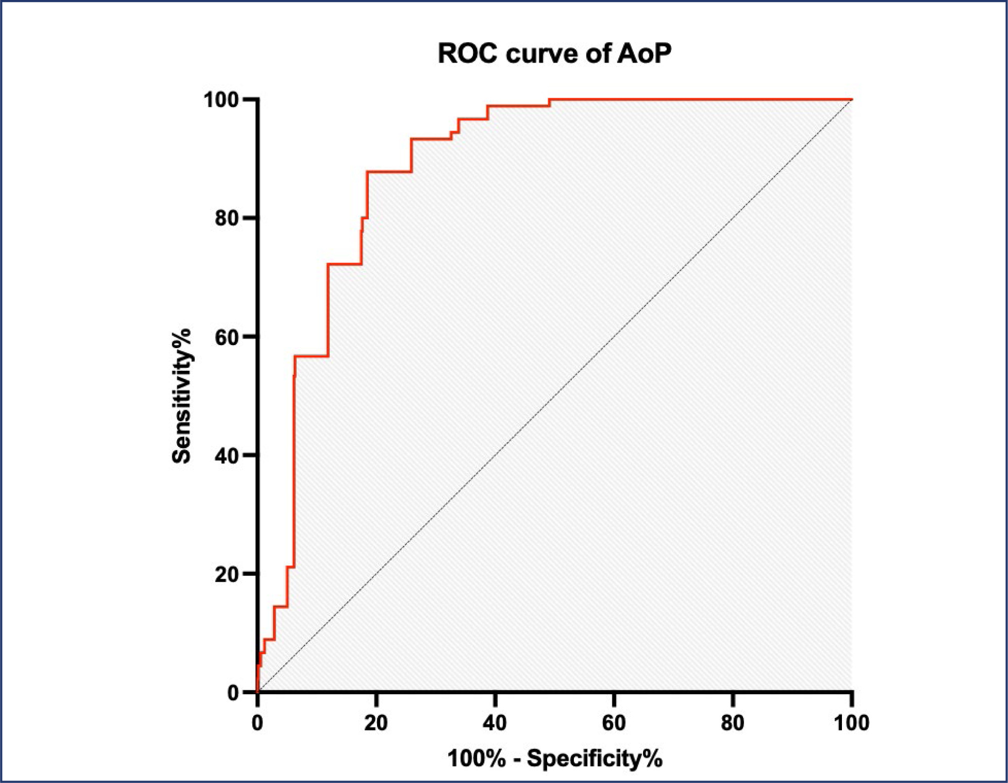
-
Original Article
A new screening of preterm birth in gestation with short cervix after pessary plus progesterone
- Marcelo Santucci França
 ,
, - Valter Lacerda de Andrade Jr. ,
- Alan Roberto Hatanaka ,
- Roberto Santos ,
- Francisco Herlanio Costa Carvalho , [ ... ],
- Rodolfo de Carvalho Pacagnella
09-06-2024
Summary
Original ArticleA new screening of preterm birth in gestation with short cervix after pessary plus progesterone
Revista Brasileira de Ginecologia e Obstetrícia. 2024;46:e-rbgo39i
09-06-2024- Marcelo Santucci França ,
- Valter Lacerda de Andrade Jr. ,
- Alan Roberto Hatanaka ,
- Roberto Santos ,
- Francisco Herlanio Costa Carvalho ,
- Maria Laura Costa ,
- Gabriela Ubeda Santucci França ,
- Rosiane Mattar ,
- Ben Willem Mol ,
- Antonio Fernandes Moron ,
- Rodolfo de Carvalho Pacagnella
Views135See moreAbstract
Objective
This study aims to create a new screening for preterm birth < 34 weeks after gestation with a cervical length (CL) ≤ 30 mm, based on clinical, demographic, and sonographic characteristics.
Methods
This is a post hoc analysis of a randomized clinical trial (RCT), which included pregnancies, in middle-gestation, screened with transvaginal ultrasound. After observing inclusion criteria, the patient was invited to compare pessary plus progesterone (PP) versus progesterone only (P) (1:1). The objective was to determine which variables were associated with severe preterm birth using logistic regression (LR). The area under the curve (AUC), sensitivity, specificity, and positive predictive value (PPV) and negative predictive value (NPV) were calculated for both groups after applying LR, with a false positive rate (FPR) set at 10%.
Results
The RCT included 936 patients, 475 in PP and 461 in P. The LR selected: ethnics white, absence of previous curettage, previous preterm birth, singleton gestation, precocious identification of short cervix, CL < 14.7 mm, CL in curve > 21.0 mm. The AUC (CI95%), sensitivity, specificity, PPV, and PNV, with 10% of FPR, were respectively 0.978 (0.961-0.995), 83.4%, 98.1%, 83.4% and 98.1% for PP < 34 weeks; and 0.765 (0.665-0.864), 38.7%, 92.1%, 26.1% and 95.4%, for P < 28 weeks.
Conclusion
Logistic regression can be effective to screen preterm birth < 34 weeks in patients in the PP Group and all pregnancies with CL ≤ 30 mm.
Views135This is an Open Access article distributed under the terms of the Creative Commons Attribution License, which permits unrestricted use, distribution, and reproduction in any medium, provided the original work is properly cited. Summary
Original ArticleA new screening of preterm birth in gestation with short cervix after pessary plus progesterone
Revista Brasileira de Ginecologia e Obstetrícia. 2024;46:e-rbgo39i
09-06-2024- Marcelo Santucci França ,
- Valter Lacerda de Andrade Jr. ,
- Alan Roberto Hatanaka ,
- Roberto Santos ,
- Francisco Herlanio Costa Carvalho ,
- Maria Laura Costa ,
- Gabriela Ubeda Santucci França ,
- Rosiane Mattar ,
- Ben Willem Mol ,
- Antonio Fernandes Moron ,
- Rodolfo de Carvalho Pacagnella
Views135See moreAbstract
Objective
This study aims to create a new screening for preterm birth < 34 weeks after gestation with a cervical length (CL) ≤ 30 mm, based on clinical, demographic, and sonographic characteristics.
Methods
This is a post hoc analysis of a randomized clinical trial (RCT), which included pregnancies, in middle-gestation, screened with transvaginal ultrasound. After observing inclusion criteria, the patient was invited to compare pessary plus progesterone (PP) versus progesterone only (P) (1:1). The objective was to determine which variables were associated with severe preterm birth using logistic regression (LR). The area under the curve (AUC), sensitivity, specificity, and positive predictive value (PPV) and negative predictive value (NPV) were calculated for both groups after applying LR, with a false positive rate (FPR) set at 10%.
Results
The RCT included 936 patients, 475 in PP and 461 in P. The LR selected: ethnics white, absence of previous curettage, previous preterm birth, singleton gestation, precocious identification of short cervix, CL < 14.7 mm, CL in curve > 21.0 mm. The AUC (CI95%), sensitivity, specificity, PPV, and PNV, with 10% of FPR, were respectively 0.978 (0.961-0.995), 83.4%, 98.1%, 83.4% and 98.1% for PP < 34 weeks; and 0.765 (0.665-0.864), 38.7%, 92.1%, 26.1% and 95.4%, for P < 28 weeks.
Conclusion
Logistic regression can be effective to screen preterm birth < 34 weeks in patients in the PP Group and all pregnancies with CL ≤ 30 mm.
This is an Open Access article distributed under the terms of the Creative Commons Attribution License, which permits unrestricted use, distribution, and reproduction in any medium, provided the original work is properly cited. - Marcelo Santucci França
-
Original Article
Agreement between frozen section and histopathology to detect malignancy in adnexal masses according to size and morphology by ultrasound
- Clarissa de Andrade Amaral ,
- Priscila Grecca Pedrão ,
- Luani Rezende Godoy ,
- Yasmin Medeiros Guimarães ,
- Cassia Arantes Petroni Macedo , [ ... ],
- Ricardo dos Reis
07-26-2024
Summary
Original ArticleAgreement between frozen section and histopathology to detect malignancy in adnexal masses according to size and morphology by ultrasound
Revista Brasileira de Ginecologia e Obstetrícia. 2024;46:e-rbgo63
07-26-2024- Clarissa de Andrade Amaral ,
- Priscila Grecca Pedrão ,
- Luani Rezende Godoy ,
- Yasmin Medeiros Guimarães ,
- Cassia Arantes Petroni Macedo ,
- Marcia Appel ,
- Guilherme Spagna Accorsi ,
- Jeferson Rodrigo Zanon ,
- Ricardo dos Reis
Views120See moreAbstract
Objective
Management of suspect adnexal masses involves surgery to define the best treatment. Diagnostic choices include a two-stage procedure for histopathology examination (HPE) or intraoperative histological analysis – intraoperative frozen section (IFS) and formalin-fixed and paraffin-soaked tissues (FFPE). Preoperative assessment with ultrasound may also be useful to predict malignancy. We aimed at determining the accuracy of IFS to evaluate adnexal masses stratified by size and morphology having HPE as the diagnostic gold standard.
Methods
A retrospective chart review of 302 patients undergoing IFS of adnexal masses at Hospital de Clínicas de Porto Alegre, between January2005 and September2011 was performed. Data were collected regarding sonographic size (≤10cm or >10cm), characteristics of the lesion, and diagnosis established in IFS and HPE. Eight groups were studied: unilocular lesions; septated/cystic lesions; heterogeneous (solid/cystic) lesions; and solid lesions, divided in two main groups according to the size of lesion, ≤10cm or >10cm. Kappa agreement between IFS and HPE was calculated for each group.
Results
Overall agreement between IFS and HPE was 96.1% for benign tumors, 96.1% for malignant tumors, and 73.3% for borderline tumors. Considering the combination of tumor size and morphology, 100% agreement between IFS and HPE was recorded for unilocular and septated tumors ≤10cm and for solid tumors.
Conclusion
Stratification of adnexal masses according to size and morphology is a good method for preoperative assessment. We should wait for final HPE for staging decision, regardless of IFS results, in heterogeneous adnexal tumors of any size, solid tumors ≤10cm, and all non-solid tumors >10cm.
Views120This is an Open Access article distributed under the terms of the Creative Commons Attribution License, which permits unrestricted use, distribution, and reproduction in any medium, provided the original work is properly cited. Summary
Original ArticleAgreement between frozen section and histopathology to detect malignancy in adnexal masses according to size and morphology by ultrasound
Revista Brasileira de Ginecologia e Obstetrícia. 2024;46:e-rbgo63
07-26-2024- Clarissa de Andrade Amaral ,
- Priscila Grecca Pedrão ,
- Luani Rezende Godoy ,
- Yasmin Medeiros Guimarães ,
- Cassia Arantes Petroni Macedo ,
- Marcia Appel ,
- Guilherme Spagna Accorsi ,
- Jeferson Rodrigo Zanon ,
- Ricardo dos Reis
Views120See moreAbstract
Objective
Management of suspect adnexal masses involves surgery to define the best treatment. Diagnostic choices include a two-stage procedure for histopathology examination (HPE) or intraoperative histological analysis – intraoperative frozen section (IFS) and formalin-fixed and paraffin-soaked tissues (FFPE). Preoperative assessment with ultrasound may also be useful to predict malignancy. We aimed at determining the accuracy of IFS to evaluate adnexal masses stratified by size and morphology having HPE as the diagnostic gold standard.
Methods
A retrospective chart review of 302 patients undergoing IFS of adnexal masses at Hospital de Clínicas de Porto Alegre, between January2005 and September2011 was performed. Data were collected regarding sonographic size (≤10cm or >10cm), characteristics of the lesion, and diagnosis established in IFS and HPE. Eight groups were studied: unilocular lesions; septated/cystic lesions; heterogeneous (solid/cystic) lesions; and solid lesions, divided in two main groups according to the size of lesion, ≤10cm or >10cm. Kappa agreement between IFS and HPE was calculated for each group.
Results
Overall agreement between IFS and HPE was 96.1% for benign tumors, 96.1% for malignant tumors, and 73.3% for borderline tumors. Considering the combination of tumor size and morphology, 100% agreement between IFS and HPE was recorded for unilocular and septated tumors ≤10cm and for solid tumors.
Conclusion
Stratification of adnexal masses according to size and morphology is a good method for preoperative assessment. We should wait for final HPE for staging decision, regardless of IFS results, in heterogeneous adnexal tumors of any size, solid tumors ≤10cm, and all non-solid tumors >10cm.
This is an Open Access article distributed under the terms of the Creative Commons Attribution License, which permits unrestricted use, distribution, and reproduction in any medium, provided the original work is properly cited. - Clarissa de Andrade Amaral
-
Original Article
Nodular image in the appendix observed on ultrasound: endometriosis or neuroendocrine neoplasia?
Revista Brasileira de Ginecologia e Obstetrícia. 2024;46:e-rbgo1
03-14-2024
Summary
Original ArticleNodular image in the appendix observed on ultrasound: endometriosis or neuroendocrine neoplasia?
Revista Brasileira de Ginecologia e Obstetrícia. 2024;46:e-rbgo1
03-14-2024Views285See moreAbstract
Objective:
To evaluate the association between clinical and imaging with surgical and pathological findings in patients with suspected neuroendocrine tumor of appendix and/or appendix endometriosis.
Methods:
Retrospective descriptive study conducted at the Teaching and Research Institute of Hospital Israelita Albert Einstein, in which medical records and databases of patients with suspected neuroendocrine tumor of appendix and/or endometriosis of appendix were analyzed by imaging.
Results:
Twenty-eight patients were included, all of which had some type of appendix alteration on the ultrasound examination. The pathological outcome of the appendix found 25 (89.3%) lesions compatible with endometriosis and three (10.7%) neuroendocrine tumors. The clinical findings of imaging and surgery were compared with the result of pathological anatomy by means of relative frequency.
Conclusion:
It was possible to observe a higher prevalence of appendix endometriosis when the patient presented more intense pain symptoms. The image observed on ultrasound obtained a high positive predictive value for appendicular endometriosis.
Views285This is an Open Access article distributed under the terms of the Creative Commons Attribution License, which permits unrestricted use, distribution, and reproduction in any medium, provided the original work is properly cited. Summary
Original ArticleNodular image in the appendix observed on ultrasound: endometriosis or neuroendocrine neoplasia?
Revista Brasileira de Ginecologia e Obstetrícia. 2024;46:e-rbgo1
03-14-2024Views285See moreAbstract
Objective:
To evaluate the association between clinical and imaging with surgical and pathological findings in patients with suspected neuroendocrine tumor of appendix and/or appendix endometriosis.
Methods:
Retrospective descriptive study conducted at the Teaching and Research Institute of Hospital Israelita Albert Einstein, in which medical records and databases of patients with suspected neuroendocrine tumor of appendix and/or endometriosis of appendix were analyzed by imaging.
Results:
Twenty-eight patients were included, all of which had some type of appendix alteration on the ultrasound examination. The pathological outcome of the appendix found 25 (89.3%) lesions compatible with endometriosis and three (10.7%) neuroendocrine tumors. The clinical findings of imaging and surgery were compared with the result of pathological anatomy by means of relative frequency.
Conclusion:
It was possible to observe a higher prevalence of appendix endometriosis when the patient presented more intense pain symptoms. The image observed on ultrasound obtained a high positive predictive value for appendicular endometriosis.
This is an Open Access article distributed under the terms of the Creative Commons Attribution License, which permits unrestricted use, distribution, and reproduction in any medium, provided the original work is properly cited. -
Original Article
Correlation of pelvic ultrasonography with pubertal development in girls
- Francine Zap Bertoncello ,
- Mariane Faccin Beust ,
- Cláudia Mendes Tagliari ,
- Liliane Diefenthaeler Herter ,
- Cristiane Kopacek
00-00-2024
Summary
Original ArticleCorrelation of pelvic ultrasonography with pubertal development in girls
Revista Brasileira de Ginecologia e Obstetrícia. 2024;46:e-rbgo5
00-00-2024- Francine Zap Bertoncello ,
- Mariane Faccin Beust ,
- Cláudia Mendes Tagliari ,
- Liliane Diefenthaeler Herter ,
- Cristiane Kopacek
Views168See moreAbstract
Objectives:
This study aims to correlate pelvic ultrasound with female puberty and evaluate the usual ultrasound parameters as diagnostic tests for the onset of puberty and, in particular, a less studied parameter: the Doppler evaluation of the uterine arteries.
Methods:
Cross-sectional study with girls aged from one to less than eighteen years old, with normal pubertal development, who underwent pelvic ultrasound examination from November 2020 to December 2021. The presence of thelarche was the clinical criterion to distinguish pubescent from non-pubescent girls. The sonographic parameters were evaluated using the ROC curve and the cutoff point defined through the Youden index (J).
Results:
60 girls were included in the study. Uterine volume ≥ 2.45mL had a sensitivity of 93%, specificity of 90%, PPV of 90%, NPV of 93% and accuracy of 91% (AUC 0.972) for predicting the onset of puberty. Mean ovarian volume ≥ 1.48mL had a sensitivity of 96%, specificity of 90%, PPV of 90%, NPV of 97% and accuracy of 93% (AUC 0.966). Mean PI ≤ 2.75 had 100% sensitivity, 48% specificity, 62% PPV, 100% NPV and 72% accuracy (AUC 0.756) for predicting the onset of puberty.
Conclusion:
Pelvic ultrasound proved to be an excellent tool for female pubertal assessment and uterine and ovarian volume, the best ultrasound parameters for detecting the onset of puberty. The PI of the uterine arteries, in this study, although useful in the pubertal evaluation, showed lower accuracy in relation to the uterine and ovarian volume.
Views168This is an Open Access article distributed under the terms of the Creative Commons Attribution License, which permits unrestricted use, distribution, and reproduction in any medium, provided the original work is properly cited. Summary
Original ArticleCorrelation of pelvic ultrasonography with pubertal development in girls
Revista Brasileira de Ginecologia e Obstetrícia. 2024;46:e-rbgo5
00-00-2024- Francine Zap Bertoncello ,
- Mariane Faccin Beust ,
- Cláudia Mendes Tagliari ,
- Liliane Diefenthaeler Herter ,
- Cristiane Kopacek
Views168See moreAbstract
Objectives:
This study aims to correlate pelvic ultrasound with female puberty and evaluate the usual ultrasound parameters as diagnostic tests for the onset of puberty and, in particular, a less studied parameter: the Doppler evaluation of the uterine arteries.
Methods:
Cross-sectional study with girls aged from one to less than eighteen years old, with normal pubertal development, who underwent pelvic ultrasound examination from November 2020 to December 2021. The presence of thelarche was the clinical criterion to distinguish pubescent from non-pubescent girls. The sonographic parameters were evaluated using the ROC curve and the cutoff point defined through the Youden index (J).
Results:
60 girls were included in the study. Uterine volume ≥ 2.45mL had a sensitivity of 93%, specificity of 90%, PPV of 90%, NPV of 93% and accuracy of 91% (AUC 0.972) for predicting the onset of puberty. Mean ovarian volume ≥ 1.48mL had a sensitivity of 96%, specificity of 90%, PPV of 90%, NPV of 97% and accuracy of 93% (AUC 0.966). Mean PI ≤ 2.75 had 100% sensitivity, 48% specificity, 62% PPV, 100% NPV and 72% accuracy (AUC 0.756) for predicting the onset of puberty.
Conclusion:
Pelvic ultrasound proved to be an excellent tool for female pubertal assessment and uterine and ovarian volume, the best ultrasound parameters for detecting the onset of puberty. The PI of the uterine arteries, in this study, although useful in the pubertal evaluation, showed lower accuracy in relation to the uterine and ovarian volume.
This is an Open Access article distributed under the terms of the Creative Commons Attribution License, which permits unrestricted use, distribution, and reproduction in any medium, provided the original work is properly cited. - Francine Zap Bertoncello
-
Review Article
Assessment of Pelvic Floor Disorders due to the Gestational Diabetes Mellitus Using Three-Dimensional Ultrasonography: A Narrative Review
- Carlos Izaias Sartorão Filho ,
- Angélica Mércia Pascon Barbosa ,
- Iracema de Mattos Paranhos Calderon ,
- Marilza Vieira Cunha Rudge
03-24-2022
Summary
Review ArticleAssessment of Pelvic Floor Disorders due to the Gestational Diabetes Mellitus Using Three-Dimensional Ultrasonography: A Narrative Review
Revista Brasileira de Ginecologia e Obstetrícia. 2022;44(12):1134-1140
03-24-2022- Carlos Izaias Sartorão Filho ,
- Angélica Mércia Pascon Barbosa ,
- Iracema de Mattos Paranhos Calderon ,
- Marilza Vieira Cunha Rudge
Views133See moreAbstract
Gestational diabetes mellitus (GDM)is an entity with evolving conceptual nuances that deserve full consideration. Gestational diabetes leads to complications and adverse effects on the mother's and infants' health during and after pregnancy. Women also have a higher prevalence of urinary incontinence (UI) related to the hyperglycemic status during pregnancy. However, the exact pathophysiological mechanism is still uncertain. We conducted a narrative review discussing the impact of GDM on the women's pelvic floor and performed image assessment using three-dimensional ultrasonography to evaluate and predict future UI.
Views133This is an Open Access article distributed under the terms of the Creative Commons Attribution License, which permits unrestricted use, distribution, and reproduction in any medium, provided the original work is properly cited. Summary
Review ArticleAssessment of Pelvic Floor Disorders due to the Gestational Diabetes Mellitus Using Three-Dimensional Ultrasonography: A Narrative Review
Revista Brasileira de Ginecologia e Obstetrícia. 2022;44(12):1134-1140
03-24-2022- Carlos Izaias Sartorão Filho ,
- Angélica Mércia Pascon Barbosa ,
- Iracema de Mattos Paranhos Calderon ,
- Marilza Vieira Cunha Rudge
Views133See moreAbstract
Gestational diabetes mellitus (GDM)is an entity with evolving conceptual nuances that deserve full consideration. Gestational diabetes leads to complications and adverse effects on the mother's and infants' health during and after pregnancy. Women also have a higher prevalence of urinary incontinence (UI) related to the hyperglycemic status during pregnancy. However, the exact pathophysiological mechanism is still uncertain. We conducted a narrative review discussing the impact of GDM on the women's pelvic floor and performed image assessment using three-dimensional ultrasonography to evaluate and predict future UI.
This is an Open Access article distributed under the terms of the Creative Commons Attribution License, which permits unrestricted use, distribution, and reproduction in any medium, provided the original work is properly cited. - Carlos Izaias Sartorão Filho
-
Original Article
Placenta Accreta Spectrum Prenatal Diagnosis Performance: Are Ultrasound False-positive Results Acceptable in Limited-resources Settings?
- Albaro José Nieto-Calvache ,
- Juan Pablo Benavides-Calvache ,
- Alejandra Hidalgo ,
- Natalia Padilla ,
- Jaime López-Tenorio , [ ... ],
- Juan Manuel Burgos-Luna
09-06-2022
Summary
Original ArticlePlacenta Accreta Spectrum Prenatal Diagnosis Performance: Are Ultrasound False-positive Results Acceptable in Limited-resources Settings?
Revista Brasileira de Ginecologia e Obstetrícia. 2022;44(9):838-844
09-06-2022- Albaro José Nieto-Calvache ,
- Juan Pablo Benavides-Calvache ,
- Alejandra Hidalgo ,
- Natalia Padilla ,
- Jaime López-Tenorio ,
- Alejandro Victoria ,
- Martin Rengifo ,
- Mauricio Mejía ,
- Lina María Vergara-Galliadi ,
- Stiven Ernesto Sinisterra-Díaz ,
- Juliana Maya ,
- María Andrea Zambrano ,
- Juan Manuel Burgos-Luna
Views272Abstract
Objective
The immediate referral of patients with risk factors for placenta accreta spectrum (PAS) to specialized centers is recommended, thus favoring an early diagnosis and an interdisciplinary management. However, diagnostic errors are frequent, even in referral centers (RCs). We sought to evaluate the performance of the prenatal diagnosis for PAS in a Latin American hospital.
Methods
A retrospective descriptive study including patients referred due to the suspicion of PAS was conducted. Data from the prenatal imaging studies were compared with the final diagnoses (intraoperative and/or histological).
Results
A total of 162 patients were included in the present study. The median gestational age at the time of the first PAS suspicious ultrasound was 29 weeks, but patients arrived at the PAS RC at 34 weeks. The frequency of false-positive results at referring hospitals was 68.5%. Sixty-nine patients underwent surgery based on the suspicion of PAS at 35 weeks, and there was a 28.9% false-positive rate at the RC. In 93 patients, the diagnosis of PAS was ruled out at the RC, with a 2.1% false-negative frequency.
Conclusion
The prenatal diagnosis of PAS is better at the RC. However, even in these centers, false-positive results are common; therefore, the intraoperative confirmation of the diagnosis of PAS is essential.
Key-words false positiveoperative surgical procedurePlacenta accretaprenatal ultrasonic diagnosisUltrasonographySee moreViews272This is an Open Access article distributed under the terms of the Creative Commons Attribution License, which permits unrestricted use, distribution, and reproduction in any medium, provided the original work is properly cited. Summary
Original ArticlePlacenta Accreta Spectrum Prenatal Diagnosis Performance: Are Ultrasound False-positive Results Acceptable in Limited-resources Settings?
Revista Brasileira de Ginecologia e Obstetrícia. 2022;44(9):838-844
09-06-2022- Albaro José Nieto-Calvache ,
- Juan Pablo Benavides-Calvache ,
- Alejandra Hidalgo ,
- Natalia Padilla ,
- Jaime López-Tenorio ,
- Alejandro Victoria ,
- Martin Rengifo ,
- Mauricio Mejía ,
- Lina María Vergara-Galliadi ,
- Stiven Ernesto Sinisterra-Díaz ,
- Juliana Maya ,
- María Andrea Zambrano ,
- Juan Manuel Burgos-Luna
Views272Abstract
Objective
The immediate referral of patients with risk factors for placenta accreta spectrum (PAS) to specialized centers is recommended, thus favoring an early diagnosis and an interdisciplinary management. However, diagnostic errors are frequent, even in referral centers (RCs). We sought to evaluate the performance of the prenatal diagnosis for PAS in a Latin American hospital.
Methods
A retrospective descriptive study including patients referred due to the suspicion of PAS was conducted. Data from the prenatal imaging studies were compared with the final diagnoses (intraoperative and/or histological).
Results
A total of 162 patients were included in the present study. The median gestational age at the time of the first PAS suspicious ultrasound was 29 weeks, but patients arrived at the PAS RC at 34 weeks. The frequency of false-positive results at referring hospitals was 68.5%. Sixty-nine patients underwent surgery based on the suspicion of PAS at 35 weeks, and there was a 28.9% false-positive rate at the RC. In 93 patients, the diagnosis of PAS was ruled out at the RC, with a 2.1% false-negative frequency.
Conclusion
The prenatal diagnosis of PAS is better at the RC. However, even in these centers, false-positive results are common; therefore, the intraoperative confirmation of the diagnosis of PAS is essential.
Key-words false positiveoperative surgical procedurePlacenta accretaprenatal ultrasonic diagnosisUltrasonographySee moreThis is an Open Access article distributed under the terms of the Creative Commons Attribution License, which permits unrestricted use, distribution, and reproduction in any medium, provided the original work is properly cited. - Albaro José Nieto-Calvache
-
Original Article
Polycystic Ovary Syndrome and Metabolic Syndrome: Clinical and Laboratory Findings and Non-Alcoholic Fatty Liver Disease Assessed by Elastography
Revista Brasileira de Ginecologia e Obstetrícia. 2022;44(3):287-294
06-27-2022
Summary
Original ArticlePolycystic Ovary Syndrome and Metabolic Syndrome: Clinical and Laboratory Findings and Non-Alcoholic Fatty Liver Disease Assessed by Elastography
Revista Brasileira de Ginecologia e Obstetrícia. 2022;44(3):287-294
06-27-2022Views162See moreAbstract
Objective
To evaluate the association between polycystic ovary syndrome (PCOS) and metabolic syndrome (MetS), adding liver assessment through elastography and ultrasound, for correlation with non-alcoholic fatty liver disease (NAFLD). Metabolic syndrome occurs in~43% of women with PCOS, and NAFLD is the hepatic expression of MetS.
Methods
One hundred women, 50 with PCOS and 50 controls, matched by age (18- 35 years) and body mass index (BMI) were included, restricted to patients with overweight and obesity grade 1, at the Assis Chateaubrian Maternity School, Universidade Federal do Ceará, Brazil. For the diagnosis of PCOS, we adopted the Rotterdam criteria, and for the diagnosis of MetS, the criteria of the National Cholesterol Education Program (NCEP/ATP III). Hepatic elastography and ultrasound were performed to assess liver stiffness and echotexture, respectively.
Results
The average ages were 29.1 (±5.3) and 30.54 (±4.39) years, for the PCOS and the control group, respectively. Patients with PCOS had a risk 4 times higher of having MetS, odds ratio (95% confidence interval)=4.14, than those in the control group. Women with PCOS had higher average of abdominal circumference (100.9±9.08 cm vs 94.96±6.99 cm) and triglycerides (162±54.63 mg/dL vs 137.54±36.91mg/dL) and lower average of HDL cholesterol (45.66±6.88 mg/dL vs 49.78±7.05 mg/dL), with statistically significant difference. Hepatic steatosis was observed on ultrasound in women with PCOS; however, with no statistically significant difference. There was no change to NAFLD at elastography in any group.
Conclusion
Women with PCOS had 4-fold higher frequency of MetS andmore hepatic steatosis, with no statistically significant difference. There was no change in liver stiffness between the groups at elastography. The results can be extended only to populations of overweight and obesity grade 1, with PCOS or not. They cannot be generalized to other untested groups.
Views162This is an Open Access article distributed under the terms of the Creative Commons Attribution License, which permits unrestricted use, distribution, and reproduction in any medium, provided the original work is properly cited. Summary
Original ArticlePolycystic Ovary Syndrome and Metabolic Syndrome: Clinical and Laboratory Findings and Non-Alcoholic Fatty Liver Disease Assessed by Elastography
Revista Brasileira de Ginecologia e Obstetrícia. 2022;44(3):287-294
06-27-2022Views162See moreAbstract
Objective
To evaluate the association between polycystic ovary syndrome (PCOS) and metabolic syndrome (MetS), adding liver assessment through elastography and ultrasound, for correlation with non-alcoholic fatty liver disease (NAFLD). Metabolic syndrome occurs in~43% of women with PCOS, and NAFLD is the hepatic expression of MetS.
Methods
One hundred women, 50 with PCOS and 50 controls, matched by age (18- 35 years) and body mass index (BMI) were included, restricted to patients with overweight and obesity grade 1, at the Assis Chateaubrian Maternity School, Universidade Federal do Ceará, Brazil. For the diagnosis of PCOS, we adopted the Rotterdam criteria, and for the diagnosis of MetS, the criteria of the National Cholesterol Education Program (NCEP/ATP III). Hepatic elastography and ultrasound were performed to assess liver stiffness and echotexture, respectively.
Results
The average ages were 29.1 (±5.3) and 30.54 (±4.39) years, for the PCOS and the control group, respectively. Patients with PCOS had a risk 4 times higher of having MetS, odds ratio (95% confidence interval)=4.14, than those in the control group. Women with PCOS had higher average of abdominal circumference (100.9±9.08 cm vs 94.96±6.99 cm) and triglycerides (162±54.63 mg/dL vs 137.54±36.91mg/dL) and lower average of HDL cholesterol (45.66±6.88 mg/dL vs 49.78±7.05 mg/dL), with statistically significant difference. Hepatic steatosis was observed on ultrasound in women with PCOS; however, with no statistically significant difference. There was no change to NAFLD at elastography in any group.
Conclusion
Women with PCOS had 4-fold higher frequency of MetS andmore hepatic steatosis, with no statistically significant difference. There was no change in liver stiffness between the groups at elastography. The results can be extended only to populations of overweight and obesity grade 1, with PCOS or not. They cannot be generalized to other untested groups.
This is an Open Access article distributed under the terms of the Creative Commons Attribution License, which permits unrestricted use, distribution, and reproduction in any medium, provided the original work is properly cited.