Summary
Rev Bras Ginecol Obstet. 2012;34(8):357-361
DOI 10.1590/S0100-72032012000800003
PURPOSE: To compare the frequency of metabolic syndrome (MetS) and the risk factors associated with this syndrome in women from the Brazilian Southeast with polycystic ovary syndrome (POS) evaluated during adolescence and adult age. METHODS: This was a cross-sectional study conducted on 147 patients with a diagnosis of POS who were divided into two groups: Adolescents, 42 adolescents aged 13 to 19 years, and Adults, 105 women aged 20 to 40 years. The following factors were evaluated: clinical characteristics (body mass index - BMI, Ferriman index, abdominal circumference - AC, and systemic arterial pressure), mean ovarian volume, laboratory variables (serum androgen profile, lipid profile, glycemia, and fasting insulin), and frequency of MetS. The results were expressed as mean±standard deviation. We used multiple logistic regression with the response variable being the presence of MetS and the predictor variables the levels of total testosterone, insulin and BMI. RESULTS: The frequency of MetS was approximately twice higher in the group of adult women compared to the adolescents with POS (Adolescents: 23.8 vs. Adults: 42.9%, p=0.04). Among the defining criteria of MetS, only the qualitative variable of systemic arterial pressure ≥130/85 mmHg was more frequent among the adult women (p=0,01). The BMI was an independent predictor of MetS among the adolescent (p=0.03) and adult women (p<0.01) with POS. Serum insulin level was a predictor of MetS only among adult women with POS (p<0.01). AC was greater among adult women (p=0.04). CONCLUSION: Adult women with POS have a twice higher frequency of MetS than adolescents with POS from the Brazilian Southeast. Although the BMI is associated with the development of MetS in any phase of life in women with POS, serum insulin level was an independent predictor of MetS only among adult women with this disorder.
Summary
Rev Bras Ginecol Obstet. 2012;34(7):323-328
DOI 10.1590/S0100-72032012000700006
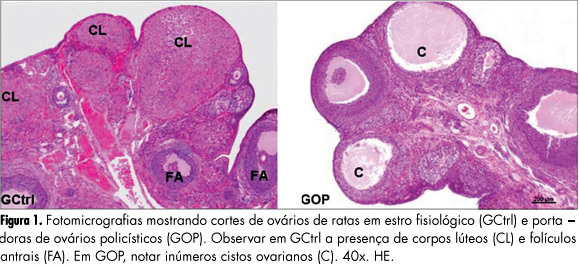
PURPOSES: To evaluate the histomorphometry of ovarian interstitial cells, as well as the blood sex steroid concentrations of female rats with polycystic ovaries induced by continuous light. METHODS: Twenty female rats were divided into two groups: Control Group - in the estrous phase (CtrlG), and a group of rats with polycystic ovaries induced by continuous illumination (POG). CtrlG animals were maintained on a light period from 07:00 a.m. to 07:00 p.m., and POG animals with continuous illumination (400 Lux) for 60 days. After this period all animals were anesthetized and blood was collected for the determination of serum estradiol (E2), progesterone (P4), and testosterone (T), followed by removal of the ovaries that were fixed in 10% formalin and processed for paraffin embedding. Five-µm histological sections were stained with hematoxylin and eosin and used for histomorphometric analysis. Morphological analyses, cyst count, determination of concentration and of the nuclear volume of interstitial cells were performed with the aid of a light microscope adapted to a high resolution camera (AxioCam), whose images were transmitted to and analyzed by the computer using AxioVision Rel 4.8 software (Carl Zeiss). Data were analyzed statistically by the Student's t-test (p<0.05). RESULTS: Morphological analysis showed the presence of ovarian cysts in POG animals and corpora lutea in CtrlG animals, as well as evidence of the origin of interstitial cells from the internal theca of these cysts. POG animals presented increased serum estradiol levels (pg/mL) compared to CtrlG animals (POG=124.9±4.2>CtrlG=73.2±6.5, p<0.05), the same occurring with testosterone levels (pg/mL) (POG=116.9±4.6>CtrlG=80.6±3.9, p<0.05). However, progesterone levels (ng/mL) were higher in CtrlG than in POG animals (CtrlG=16.3±2.0>POG=4.2±1.5, p<0.05). Morphometry showed a significant increase in nuclear volume in POG animals (POG=102.1±5.2>CtrlG=63.6±16.5, p<0.05), as well as in the area occupied (%) by interstitial cells (POG=24.4±6.9>CtrlG=6.9±3.2, p<0.05) compared to CtrlG animals. CONCLUSION: The interstitial cells of the rat polycystic ovary probably originate from ovarian cysts due to the degeneration of granulosa cells and differentiation of the internal theca cells. The elevations of serum testosterone and estradiol were probably due to the significant increase in cell activity and in the area occupied by interstitial cells.
Summary
Rev Bras Ginecol Obstet. 2012;34(7):323-328
DOI 10.1590/S0100-72032012000700006
PURPOSES: To evaluate the histomorphometry of ovarian interstitial cells, as well as the blood sex steroid concentrations of female rats with polycystic ovaries induced by continuous light. METHODS: Twenty female rats were divided into two groups: Control Group - in the estrous phase (CtrlG), and a group of rats with polycystic ovaries induced by continuous illumination (POG). CtrlG animals were maintained on a light period from 07:00 a.m. to 07:00 p.m., and POG animals with continuous illumination (400 Lux) for 60 days. After this period all animals were anesthetized and blood was collected for the determination of serum estradiol (E2), progesterone (P4), and testosterone (T), followed by removal of the ovaries that were fixed in 10% formalin and processed for paraffin embedding. Five-µm histological sections were stained with hematoxylin and eosin and used for histomorphometric analysis. Morphological analyses, cyst count, determination of concentration and of the nuclear volume of interstitial cells were performed with the aid of a light microscope adapted to a high resolution camera (AxioCam), whose images were transmitted to and analyzed by the computer using AxioVision Rel 4.8 software (Carl Zeiss). Data were analyzed statistically by the Student's t-test (p<0.05). RESULTS: Morphological analysis showed the presence of ovarian cysts in POG animals and corpora lutea in CtrlG animals, as well as evidence of the origin of interstitial cells from the internal theca of these cysts. POG animals presented increased serum estradiol levels (pg/mL) compared to CtrlG animals (POG=124.9±4.2>CtrlG=73.2±6.5, p<0.05), the same occurring with testosterone levels (pg/mL) (POG=116.9±4.6>CtrlG=80.6±3.9, p<0.05). However, progesterone levels (ng/mL) were higher in CtrlG than in POG animals (CtrlG=16.3±2.0>POG=4.2±1.5, p<0.05). Morphometry showed a significant increase in nuclear volume in POG animals (POG=102.1±5.2>CtrlG=63.6±16.5, p<0.05), as well as in the area occupied (%) by interstitial cells (POG=24.4±6.9>CtrlG=6.9±3.2, p<0.05) compared to CtrlG animals. CONCLUSION: The interstitial cells of the rat polycystic ovary probably originate from ovarian cysts due to the degeneration of granulosa cells and differentiation of the internal theca cells. The elevations of serum testosterone and estradiol were probably due to the significant increase in cell activity and in the area occupied by interstitial cells.
Summary
Rev Bras Ginecol Obstet. 2012;34(7):316-322
DOI 10.1590/S0100-72032012000700005
PURPOSE: To compare the metabolic parameters, body composition and muscle strength of women with Polycystic Ovary Syndrome (PCOS) to those of women with ovulatory menstrual cycles. METHODS: A case-control study was conducted on 27 women with PCOS and 28 control women with ovulatory cycles, aged 18 to 27 years with a body mass index of 18 to 39.9 kg/m², who did not practice regular physical activity. Serum testosterone, androstenedione, prolactin, sex hormone-binding globulin (SHBG), insulin and glycemia levels were determined. Free androgen index (FAI) and resistance to insulin (by HOMA) were calculated. The volunteers were submitted to evaluation of body composition based on skin folds and DEXA and to 1-RM maximum muscle strength tests in three exercises after familiarization procedures and handgrip isometric force was determined. RESULTS: Testosterone levels were higher in the PCOS group than in the Control Group (68.07±20.18 versus 58.20±12.82 ng/dL; p=0.02), as also were the FAI (282.51±223.86 versus 127.08±77.19; p=0.01), insulin (8.41±7.06 versus 4.05±2.73 µIU/mL; p=0.01), and HOMA (2.3±2.32 versus 1.06±0.79; p=0.01), and SBHG levels were lower (52.51±43.27 versus 65.45±27.43 nmol/L; p=0.04). No significant differences in body composition were observed between groups using the proposed methods. The PCOS group showed greater muscle strength in the 1-RM test in the bench press (31.2±4.75 versus 27.79±3.63 kg; p=0.02), and leg extension exercises (27.9±6.23 versus 23.47±4.21 kg; p=0.02) as well as handgrip isometric force (5079.61±1035.77 versus 4477.38±69.66 kgf/m², p=0.04). PCOS was an independent predictor of increase muscle strength in bench press exercises (estimate (E)=2.7) (p=0.04) and leg extension (E=3.5) (p=0.04), and BMI in the exercise of isometric handgrip (E=72.2) (p<0.01), bench press (E=0.2) (p=0.02) and arm curl (E=0.3) (p<0.01). No association was found between HOMA-IR and muscle strength. CONCLUSIONS: Women with POS showed greater muscle strength, with no difference in body composition, and IR was not associated with muscle strength performance. Muscle strength may be possibly related to high levels of androgens in these women.
Summary
Rev Bras Ginecol Obstet. 2012;34(3):128-132
DOI 10.1590/S0100-72032012000300007
PURPOSE: To evaluate the importance of the oral glucose tolerance test for the diagnosis of glucose intolerance (GI) and type 2 diabetes mellitus (DM-2) in women with PCOS. METHODS: A retrospective study was conducted on 247 patients with PCOS selected at random. The diagnosis of GI was obtained from the two-hour oral glucose tolerance test with 75 g of glucose according to the criteria of the World Health Organization (WHO) (GI: 120 minutes for plasma glucose >140 mg/dL and <200 mg/dL), and the diagnosis of DM-2 was obtained by both the oral glucose tolerance test (DM: 120 minutes for plasma glucose >200 mg/dL) and fasting glucose using the criteria of the American Diabetes Association (impaired fasting glucose: fasting plasma glucose >100 and <126 mg/dL; DM: fasting glucose >126 mg/dL). A logistic regression model for repeated measures was applied to compare the oral glucose tolerance test with fasting plasma glucose. ANOVA followed by the Tukey test was used for the analysis of the clinical and biochemical characteristics of patients with and without GI and/or DM-2. A p<0.05 was considered statistically significant. RESULTS: PCOS patients had a mean age of 24.8±6.3, and body mass index (BMI) of 18.3 to 54.9 kg/m² (32.5±7.6). The percentage of obese patients was 64%, the percentage of overweight patients was 18.6% and 17.4% had healthy weight. The oral glucose tolerance test identified 14 cases of DM-2 (5.7%), while fasting glucose detected only three cases (1.2%), and the frequency of these disorders was higher with increasing age and BMI. CONCLUSIONS: The results of this study demonstrate the superiority of the oral glucose tolerance test in relation to fasting glucose in diagnosing DM-2 in young women with PCOS and should be performed in these patients.
Summary
Rev Bras Ginecol Obstet. 2012;34(2):74-79
DOI 10.1590/S0100-72032012000200006
PURPOSE: To analyze the prevalence of insulin resistance, according to different biochemical and anthropometric measurements in women with polycystic ovary syndrome. METHODS: A total of 189 patients with polycystic ovary syndrome were retrospectively analyzed. Insulin resistance diagnosis was performed using fasting insulin, HOMA-IR, QUICKI, insulin sensibility index and glucose/fasting insulin ratio. Body mass index and lipid accumulation product were used. Data were analyzed statistically by descriptive statistics, ANOVA, Tukey post-test, and Pearson's correlation. RESULTS: The polycystic ovary syndrome patients had a mean age of 24.9±5.2 and a mean body mass index of 31.8±7.6. The percentage of obese patients was 57.14%. Among the methods of insulin resistance investigation, the insulin sensibility index was the technique that most detected (56.4%) the presence of insulin resistance in women with polycystic ovary syndrome. The insulin resistance was detected in 87% of obese patients. The fasting glucose/fasting insulin ratio and insulin sensibility index were strongly correlated with lipid accumulation product. CONCLUSION: The prevalence of insulin resistance varied according to the method used, and it was greater the higher the body mass index. Lipid accumulation product was also related to insulin resistance.
Summary
Rev Bras Ginecol Obstet. 2012;34(1):4-10
DOI 10.1590/S0100-72032012000100002
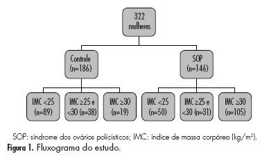
PURPOSE: To assess the prevalence of metabolic syndrome and of its defining criteria in women with polycystic ovary syndrome from the Brazilian Southeast, who were stratified according to body mass index and compared to ovulatory controls. METHODS: This was a cross-sectional study conducted on 332 women of reproductive age, who were divided into two groups: Control, consisting of 186 women with regular menstrual cycles and ovulatory symptoms and without a diagnosis of polycystic ovary syndrome or other type of chronic anovulation, and the Polycystic ovary syndrome,Group, consisting of 146 women with a diagnosis of polycystic ovary syndrome (Rotterdam Consensus ASRM/ESHRE). Each group was stratified according to the body mass index, as follows: body mass index ( < 25 ≥25 and <30, and ≥ 30 kg/m²). The frequencies of metabolic syndrome and of its defining criteria and the clinical and hormonal characteristics (follicle stimulating hormone, total testosterone, dehydroepiandrostenedione sulfate) were analyzed. RESULTS: The frequency of metabolic syndrome was six times higher in the obese Polycystic ovary syndrome Group than among control women with the same body mass index (Control with 10.5 versus Polycystic ovary syndrome with 67.9%, p<0.01); twice higher in the Polycystic ovary syndrome Group with body mass index ≥ 25 and <30 kg/m² (Control with 13.2 versus Polycystic ovary syndrome with 22.7%, p<0.01), and three times higher in the Polycystic ovary syndrome Group with body mass index <25 kg/m² (Control with 7.9 versus Polycystic ovary syndrome with 2.5%, p<0.01), compared to control women paired for the same body mass index. Regardless of the body mass index, women with polycystic ovary syndrome had a higher frequency of all the criteria defining metabolic syndrome. CONCLUSION: Women with polycystic ovary syndrome have higher frequency of metabolic syndrome and of its defining criteria regardless of the body mass index. Hyperinsulinemia and hyperandrogenism are important characteristics of the origin of these alterations, especially in obese women with polycystic ovary syndrome.
Summary
Rev Bras Ginecol Obstet. 2012;34(1):4-10
DOI 10.1590/S0100-72032012000100002
PURPOSE: To assess the prevalence of metabolic syndrome and of its defining criteria in women with polycystic ovary syndrome from the Brazilian Southeast, who were stratified according to body mass index and compared to ovulatory controls. METHODS: This was a cross-sectional study conducted on 332 women of reproductive age, who were divided into two groups: Control, consisting of 186 women with regular menstrual cycles and ovulatory symptoms and without a diagnosis of polycystic ovary syndrome or other type of chronic anovulation, and the Polycystic ovary syndrome,Group, consisting of 146 women with a diagnosis of polycystic ovary syndrome (Rotterdam Consensus ASRM/ESHRE). Each group was stratified according to the body mass index, as follows: body mass index ( < 25 ≥25 and <30, and ≥ 30 kg/m²). The frequencies of metabolic syndrome and of its defining criteria and the clinical and hormonal characteristics (follicle stimulating hormone, total testosterone, dehydroepiandrostenedione sulfate) were analyzed. RESULTS: The frequency of metabolic syndrome was six times higher in the obese Polycystic ovary syndrome Group than among control women with the same body mass index (Control with 10.5 versus Polycystic ovary syndrome with 67.9%, p<0.01); twice higher in the Polycystic ovary syndrome Group with body mass index ≥ 25 and <30 kg/m² (Control with 13.2 versus Polycystic ovary syndrome with 22.7%, p<0.01), and three times higher in the Polycystic ovary syndrome Group with body mass index <25 kg/m² (Control with 7.9 versus Polycystic ovary syndrome with 2.5%, p<0.01), compared to control women paired for the same body mass index. Regardless of the body mass index, women with polycystic ovary syndrome had a higher frequency of all the criteria defining metabolic syndrome. CONCLUSION: Women with polycystic ovary syndrome have higher frequency of metabolic syndrome and of its defining criteria regardless of the body mass index. Hyperinsulinemia and hyperandrogenism are important characteristics of the origin of these alterations, especially in obese women with polycystic ovary syndrome.
Summary
Rev Bras Ginecol Obstet. 2011;33(6):310-316
DOI 10.1590/S0100-72032011000600008
PURPOSE: To compare the metabolic characteristics of obese and non-obese young women with polycystic ovary syndrome (POS) from the Brazilian Southeast. METHODS: This was a cross-sectional study conducted on 218 women of reproductive age with a diagnosis of POS - 90 non-obese women (BMI between 18.5 and 29.9 kg/m²), and 128 obese patients (BMI >30 kg/m²) selected at the time of diagnosis. The frequency of insulin resistance (IR), glucose intolerance (GI), metabolic syndrome (MetS) and type 2 diabetes mellitus (DM2) and mean values of total cholesterol (TC), triglycerides (TG), high-density (HDL) and low-density lipoproteins (LDL), were compared between obese and non-obese patients with POS. The two groups were also compared in terms of clinical and hormonal characteristics (follicle stimulating hormone, prolactin, thyroid stimulating hormone, total testosterone, dihydroepiandrostenedione sulfate, and 17-hydroxyprogesterone). Statistical analysis was performed using the SAS 9.0 software. Quantitative variables were compared by the Student´s t-test (data with normal distribution) or by the Mann-Whitney test (non-parametric distribution). Qualitative variables were compared by the Fisher test. The level of significance was set at 5% (p<0.05) in all analyses. RESULTS: The frequency of IR, GI and MetS was significantly higher in obese than non-obese patients with POS (66.7, 29.9, and 63% versus 24.7, 12.2, and 16.4%, respectively). Obese patients had higher TC and TG levels (189.8±35.8 mg/dL and 145.4±71.1 mg/dL, respectively) than non-obese patients (172.1±38.4 mg/dL and 99.3±54 mg/dL, respectively). Both groups had mean HDL levels below 50 mg/dL. CONCLUSIONS: Young obese women with POS have a higher frequency of IR, GI and MS than non-obese. However, the occurrence of metabolic disorders is elevated also in the non-obese patients, suggesting that the presence of the syndrome may favor the development of metabolic comorbidities with potential medium- and long-term repercussions.
Summary
Rev Bras Ginecol Obstet. 2011;33(1):31-36
DOI 10.1590/S0100-72032011000100005
PURPOSE: to investigate the prevalence of elevated blood pressure (BP) in patients with polycystic ovary syndrome (PCOS) and to correlate the BP levels with other cardiovascular risk factors. METHODS: a cross-sectional study was conducted on 113 PCOS women (26.2±4.3 years) and on a Control Group of 242 healthy women from the general population (26.8±5.0 years). The variables considered were: systolic and diastolic BP, anthropometric parameters and plasma levels of glucose, total cholesterol, HDL-cholesterol, and triglycerides. The BP values were classified according to the V Brazilian Guidelines of Hypertension. Statistical analysis was performed by intergroup comparison with the Student's t-test and χ2 test, and correlation analysis was performed using Pearson's coefficient. RESULTS: the PCOS Group showed a significantly higher prevalence of altered BP (>130/85 mmHg) than the Control Group (18.6 versus 9.9%, respectively; p<0.05). PCOS women had higher mean systolic BP, body mass index (BMI), waist circumference (WC), triglycerides and fasting glucose, and lower HDL-cholesterol, compared to the Control Group (p<0.01). In the PCOS Group, the values of systolic and diastolic BP showed a significant positive correlation with age, BMI, WC, and triglycerides (p<0.05). CONCLUSIONS: according to the results obtained, it is possible to conclude that the frequency of women with BP values above the normal limit was significantly higher in the PCOS Group than in the Control Group. Additionally, the BP values also correlated with other cardiovascular risk factors. These findings underscore the importance of preventive strategies in PCOS women, in order to prevent pathological events related to the cardiovascular system.