Summary
Revista Brasileira de Ginecologia e Obstetrícia. 2022;44(1):32-39
02-28-2022
To evaluate the improvement in screening accuracy of the Fracture Risk Assessment Tool (FRAX) for the risk of developing osteoporosis among young postmenopausal women by associating with it clinical muscle mass measures.
A sample of postmenopausal women was submitted to calcaneal quantitative ultrasound (QUS), application of the FRAX questionnaire, and screening for the risk of developing sarcopenia at a health fair held in the city of São Bernardo do Campo in 2019. The sample also underwent anthropometric measurements, muscle mass, walking speed and handgrip tests. A major osteoporotic fracture (MOF) risk ≥ 8.5% on the FRAX, a classification of medium risk on the clinical guideline of the National Osteoporosis Guideline Group (NOGG), and a QUS T-score ≤ -1.8 sd were considered risks of having low bone mass, and QUS T-score ≤ -2.5sd, risk of having fractures.
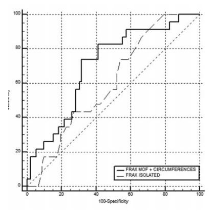
In total, 198 women were evaluated, with a median age of 64±7.7 years, median body mass index (BMI) of 27.3±5.3 kg/m2 and median QUS T-score of -1.3±1.3 sd. The accuracy of the FRAX with a MOF risk ≥ 8.5% to identify women with T-scores ≤ -1.8 sd was poor, with an area under the curve (AUC) of 0.604 (95% confidence interval [95%CI]: 0.509-0.694) for women under 65 years of age, and of 0.642 (95%CI: 0.571-0.709) when age was not considered. Including data on muscle mass in the statistical analysis led to a significant improvement for the group of women under 65 years of age, with an AUC of 0,705 (95%CI: 0.612-0.786). The ability of the high-risk NOGG tool to identify T-scores ≤ -1.8 sd was limited.
Clinical muscle mass measurements increased the accuracy of the FRAX to screen for osteoporosis in women aged under 65 years.
Summary
Revista Brasileira de Ginecologia e Obstetrícia. 2022;44(1):32-39
02-28-2022
To evaluate the improvement in screening accuracy of the Fracture Risk Assessment Tool (FRAX) for the risk of developing osteoporosis among young postmenopausal women by associating with it clinical muscle mass measures.
A sample of postmenopausal women was submitted to calcaneal quantitative ultrasound (QUS), application of the FRAX questionnaire, and screening for the risk of developing sarcopenia at a health fair held in the city of São Bernardo do Campo in 2019. The sample also underwent anthropometric measurements, muscle mass, walking speed and handgrip tests. A major osteoporotic fracture (MOF) risk ≥ 8.5% on the FRAX, a classification of medium risk on the clinical guideline of the National Osteoporosis Guideline Group (NOGG), and a QUS T-score ≤ -1.8 sd were considered risks of having low bone mass, and QUS T-score ≤ -2.5sd, risk of having fractures.
In total, 198 women were evaluated, with a median age of 64±7.7 years, median body mass index (BMI) of 27.3±5.3 kg/m2 and median QUS T-score of -1.3±1.3 sd. The accuracy of the FRAX with a MOF risk ≥ 8.5% to identify women with T-scores ≤ -1.8 sd was poor, with an area under the curve (AUC) of 0.604 (95% confidence interval [95%CI]: 0.509-0.694) for women under 65 years of age, and of 0.642 (95%CI: 0.571-0.709) when age was not considered. Including data on muscle mass in the statistical analysis led to a significant improvement for the group of women under 65 years of age, with an AUC of 0,705 (95%CI: 0.612-0.786). The ability of the high-risk NOGG tool to identify T-scores ≤ -1.8 sd was limited.
Clinical muscle mass measurements increased the accuracy of the FRAX to screen for osteoporosis in women aged under 65 years.
Summary
Revista Brasileira de Ginecologia e Obstetrícia. 2017;39(12):663-669
12-01-2017
To examine the role of the panoramic mandibular radiograph in the diagnosis of low bone mineral density (BMD) in postmenopausal women.
A cross-sectional study including volunteer women aged over 40 years in amenorrhea due to ovarian failure for at least 12 months, who were cared for at the climacteric outpatient clinic of a university hospital in the city of Cuiabá, in the state of Mato Grosso, Brazil. The panoramic radiographs were evaluated using a specific software. Two aspects were analyzed in the mandibular panoramic radiograph: a qualitative aspect regarding the shape of the mandibular cortical bone, and a quantitative aspect regarding thewidth of themandibular cortical bone. Themorphology of themandibular cortical bone in the digital panoramic radiograph was determined bilaterally by the observation of the bone structure between the mental foramen and the base of the jaw. The mandibular cortical bonewas categorized into three groups. Themental index (MI)was used to evaluate the thickness of themandibular cortical bone through a perpendicular line drawn fromthe base of the mandible at the height of the center of the mental foramen, with another line drawn tangent to the inferior border of the mandible, and a third line parallel to the line at the superior border of themandible. The MI data are expressed in millimeters, with a normal value of 3.0 mm. The densities of the lumbar spine and femur, expressed in g/cm2, were categorized as normal, osteopenia or osteoporosis.
The agreement index between the MI and the BMD of the lumbar spine was good (Kappa = 0.718), but the same index between the MI and the BMD of the femoral neck was poor (Kappa = 0.443). An excellent agreement occurred when the mandibular cortical index (MCI) was compared with the BMD of the lumbar spine (Kappa = 0.912). The agreement between MCI and the BMD in the femur was moderated (Kappa = 0.579).
The radiomorphometric indices evaluated in the mandibular panoramic radiograph are capable of identifying postmenopausal women with low mineral density in the mandible, and the results can be used to refer these women to appropriate medical investigation and/or treatment.
Summary
Revista Brasileira de Ginecologia e Obstetrícia. 2016;38(5):225-230
05-01-2016
Proper physical activity is related to the prevention and the treatment of osteoporosis.
To assess the level of physical activity (PA) in post-menopausal women with low bone mineral density ( BMD ).
This cross-sectional clinical study included 123 post-menopausal women. The inclusion criteria were: age of 45 years with last menses at least 12 months prior to the initiation of the study, and bone density scan (BDS) values measured over the preceding 12 months. Women with severe osteoarthritis were excluded. Women were allocated into three groups, according to BMD measured by BDS [osteoporosis (OP; 54 women), osteopenia (35 women), and normal bone density (NBD; 35 women)], and compared for general, clinical, and anthropometric data, and for PA level. The latter was assessed using the International Physical Activity Questionnaire (IPAQ), in metabolic equivalent of task (MET) units. Participants were classified as sedentary, active or very active. Quantitative variables were compared using ANOVA followed by Tukey's test. Associations between qualitative variables were tested by Chi-square (χ2) or Fisher's exact test. In order to check for differences among groups and IPAQ domains, a generalized linear model with Gamma distribution was adjusted for values in METs.
The OP group differed from the NBD group regarding age (61.8 10.1 and 52.9 5.4 years), percentage of participants with self-declared white ethnicity (43.9 and 28.0%), body mass index (BMI - 25.7 5.4 and 30.9 5.1 kg/m2), and time since menopause (15.5 7.5 and 5.8 4.5 years). Smoking rates were higher in the OP (55.6%) and NBD groups (33.3%) than in the osteopenia group (11.1%). Within the OP group, the rate of subjects with sedentary lifestyles was higher (42.6%), and time spent sitting was greater (344.3 204.8 METs) than in the groups with osteopenia (20.0 % and 300.9 230.6 METs) and NBD (17.7% and 303.2 187.9 METs).
The rate of sedentary lifestyles was higher in post-menopausal women with OP than in those with either osteopenia or NBD. In order to change this physical inactivity profile, strategies should be created to address this group of patients.
Summary
Revista Brasileira de Ginecologia e Obstetrícia. 2013;35(11):497-502
01-10-2013
DOI 10.1590/S0100-72032013001100004
PURPOSE: To analyze the prevalence of and factors associated with fragility fractures in Brazilian women aged 50 years and older. METHODS: This cross-sectional population survey, conducted between May 10 and October 31, 2011, included 622 women aged >50 years living in a city in southeastern Brazil. A questionnaire was administered to each woman by a trained interviewer. The associations between the occurrence of a fragility fracture after age 50 years and sociodemographic data, health-related habits and problems, self-perception of health and evaluation of functional capacity were determined by the χ2 test and Poisson regression using the backward selection criteria. RESULTS: The mean age of the 622 women was 64.1 years. The prevalence of fragility fractures was 10.8%, with 1.8% reporting hip fracture. In the final statistical model, a longer time since menopause (PR 1.03; 95%CI 1.01-1.05; p<0.01) and osteoporosis (PR 1.97; 95%CI 1.27-3.08; p<0.01) were associated with a higher prevalence of fractures. CONCLUSIONS: These findings may provide a better understanding of the risk factors associated with fragility fractures in Brazilian women and emphasize the importance of performing bone densitometry.
Summary
Revista Brasileira de Ginecologia e Obstetrícia. 2012;34(12):563-567
01-11-2012
DOI 10.1590/S0100-72032012001200006
PURPOSE: To investigate the relationship between periodontitis and osteoporosis, using a case-control study about periodontal status of postmenopausal women. METHODS: A total of 99 postmenopausal women were divided into three groups: normal bone (Gn, n=45), osteopenia (Gpenia, n=31) and osteoporosis (Gporosis, n=23). The categorization of bone mass was measured by dual energy absorptiometry with X-rays in the lumbar spine (L2 - L4), by assessing bone mineral density. Clinical attachment level (CAL), gingival bleeding index (GI), plaque index (PI), and probing depth (PD) were determined in all participants by a single examiner. The data were submitted to BioEstat 2.0 software through parametric analysis of variance (ANOVA) and the Bonferroni test, with the level of significance set at 5%. RESULTS: Women with osteoporosis presented the highest percentage of periodontal disease, with higher average CAL (2.6±0.4 mm) and PD (2.8±0.6 mm), GI (72.8±25.9 mm) and PI (72.9±24.2 mm). Statistical analysis revealed a significant difference in periodontal situation between Gn and Gporosis (p=0,01) and between Gpenia and Gporosis (p=0,03). CONCLUSION: Osteoporosis may have an influence on periodontal condition, based on the relation between periodontitis and osteoporosis in postmenopausal women.
Summary
Revista Brasileira de Ginecologia e Obstetrícia. 2012;34(7):335-342
08-28-2012
DOI 10.1590/S0100-72032012000700008
PURPOSE: To evaluate whether climacteric women undergoing liver transplantation had higher prevalence of decreased bone mass than those without any liver disease. METHODS: A cross-sectional study with 48 women receiving follow-up care at a university hospital in Southeastern Brazil, from February 4th 2009 to January 5th 2011, was conducted. Of these women, 24 were 35 years or older and had undergone liver transplantation at least one year before study entry. The remaining 24 women had no liver disease and their ages and menstrual patterns were similar to those of transplanted patients. Laboratorial tests (follicle-stimulating hormone and estradiol) and bone density measurements of the lumbar spine and femur (equipment Hologic, Discovery WI) were performed. Statistical analysis was carried out by Fisher's exact test, simple Odds Ratio (OR), and multiple logistic regression. RESULTS: Mean age of the women included in the study was 52.8 (±10.7) years-old, 27.1% were premenopausal and 72.9% were peri/postmenopausal. Approximately 14.6% of these women exhibited osteoporosis and 35.4% had low bone mass. The following items were associated with decreased bone mass: being postmenopausal (OR=71.4; 95%CI 3.8 - 1,339.7; p<0.0001), current age over 49 years-old (OR=11.4; 95%CI 2.9 - 44.0; p=0.0002), and serum estradiol levels lower than 44.5 pg/mL (OR=18.3; 95%CI 3.4 - 97.0; p<0.0001). Having a history of liver transplantation was not associated with decreased bone mass (OR=1.4; 95%CI 0.4 - 4.3; p=0.56). CONCLUSION: Liver transplantation was not associated with decreased bone mass in this group of climacteric women.
Summary
Revista Brasileira de Ginecologia e Obstetrícia. 2011;33(5):231-237
08-26-2011
DOI 10.1590/S0100-72032011000500005
PURPOSE: To investigate the relationship between quality of life and spinal fracture in women aged over 60 living in Southern Brazil. METHODS: A case-control study was conducted with the application of the WHOQOL-bref questionnaire to 100 women living in the city of Chapecó (SC), aged over 60, postmenopausal, white or Caucasian, with no important cognitive impairment or a history of diseases known to affect bone metabolism, or malignant neoplasias. The population was divided into two groups depending on the presence or absence of fractures in the spine radiography. We analyzed variables related to the current and previous medical history, life habits and family history of fractures, and the domains and facets that compose the WHOQOL-bref. All participants were informed about the objectives and methodologies adopted and gave written informed consent to participate in the study. RESULTS: The mean age of the women in the fracture group was older than that of women with fractures (p<0.05). Also women with fractures tended to belong to a higher social class, to have more years of study, a higher family income, and a greater use of alcoholic drinks (p<0.05). In the evaluation of the WHOQOL-bref domains, the fracture group had the highest average in the psychological field (..=63.6± 3.0) and the lowest in the environment field (..=9.3±58.8). In the group without fracture, the highest average also occurred in the psychological domain (..=67.2± 9.3) and the lowest in the field of social relations (..=57.5±7.7). Statistical analysis showed no significant correlation between the averages of the facets that make up the areas between the groups with and without fractures. CONCLUSIONS: This study suggests that there is no impairment of quality of life among older women with vertebral fractures, but the relation between QL and time of occurrence and severity of the fractures should be better evaluated. Both groups had higher scores in the psychological domain, showing that the respondents rely on personal beliefs, spirituality and religion, accept their physical appearance while maintaining self-esteem and the ability to think, to learn and to concentrate despite the presence of this disease. There was no statistically significant difference between groups or between domains in the same group.
Summary
Revista Brasileira de Ginecologia e Obstetrícia. 2009;31(10):496-502
11-19-2009
DOI 10.1590/S0100-72032009001000005
PURPOSE: to evaluate the rate of fall and its association with stabilometric parameters in postmenopause women, with or without osteoporosis. METHODS: transversal cohort study including 266 over 60-year-old women with and without osteoporosis, with at least 12 months of amenorrhea. The women were interviewed about the occurrence of falls in the previous 12 months, and about clinical and sociodemographic information. The osteoporosis diagnosis was done through bone densitometry and the postural stability evaluated through a stabilometric platform. For statistical analysis, mean, standard deviation, percentage, Mann-Whitney test, χ2 and Odds Ratio, and Spearman's correlation coefficient have been calculated. RESULTS: women with osteoporosis presented lower body mass index (BMI), lower schooling, shorter hormonal therapy and sooner menopause onset. The rate of fall was significantly higher in the group of women with osteoporosis (51.1%) (p<0.01), that presented an adjusted risk of 1.9 (1.3 to 3.4) times higher of falls and 3.2 (1.2 a 8.2) times higher of recurrent falls than the group without osteoporosis. Women with osteoporosis presented higher amplitude of Y axis oscillation in the open-eye test, than women without osteoporosis. The adjusted correlation analysis between stabilometric parameters and falls has not shown any significant correlation. CONCLUSIONS: women with post-menopausal osteoporosis present higher rate of falls and higher risk of recurrent falls, as compared with women without osteoporosis.