-
Case Report
Fine-needle Aspiration Cytology to Identify a Rare Mimicker of Breast Cancer: Plasma Cell Mastitis
Revista Brasileira de Ginecologia e Obstetrícia. 2018;40(8):491-493
08-01-2018
Summary
Case ReportFine-needle Aspiration Cytology to Identify a Rare Mimicker of Breast Cancer: Plasma Cell Mastitis
Revista Brasileira de Ginecologia e Obstetrícia. 2018;40(8):491-493
08-01-2018Views114See moreAbstract
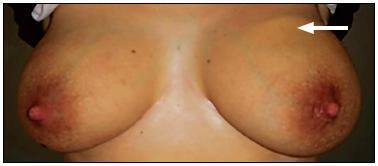
There are rare benign diseases that can mimic malignant breast neoplasms in the clinical exam and in mammography. We evaluated the contribution of an accessible procedure to most clinicians, the fine-needle aspiration cytology, to identify a rare mimicker of malignant breast neoplasms. A type 2 diabetic 85-year-old female presented with a 6-month history of a left breast lump. The physical exam and mammography were compatible with breast cancer. Nevertheless, after fine-needle aspiration cytology, the diagnosis was plasma cellmastitis. Once this rare diagnosis was established, the tumor was extirpated, and the final histologic diagnosis corroborated chronic plasma cellmastitis. The patient’s postoperative evolution was uneventful, and no other treatment was needed. Fine-needle aspiration cytology could be a valuable tool to identify rare mimickers of malignant breast neoplasms.
Views114

This is an Open Access article distributed under the terms of the Creative Commons Attribution License, which permits unrestricted use, distribution, and reproduction in any medium, provided the original work is properly cited. Summary
Case ReportFine-needle Aspiration Cytology to Identify a Rare Mimicker of Breast Cancer: Plasma Cell Mastitis
Revista Brasileira de Ginecologia e Obstetrícia. 2018;40(8):491-493
08-01-2018Views114See moreAbstract
There are rare benign diseases that can mimic malignant breast neoplasms in the clinical exam and in mammography. We evaluated the contribution of an accessible procedure to most clinicians, the fine-needle aspiration cytology, to identify a rare mimicker of malignant breast neoplasms. A type 2 diabetic 85-year-old female presented with a 6-month history of a left breast lump. The physical exam and mammography were compatible with breast cancer. Nevertheless, after fine-needle aspiration cytology, the diagnosis was plasma cellmastitis. Once this rare diagnosis was established, the tumor was extirpated, and the final histologic diagnosis corroborated chronic plasma cellmastitis. The patient’s postoperative evolution was uneventful, and no other treatment was needed. Fine-needle aspiration cytology could be a valuable tool to identify rare mimickers of malignant breast neoplasms.
This is an Open Access article distributed under the terms of the Creative Commons Attribution License, which permits unrestricted use, distribution, and reproduction in any medium, provided the original work is properly cited. 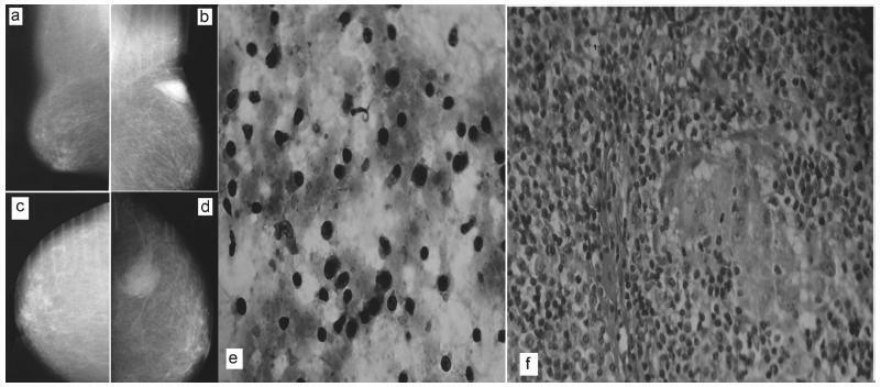
-
Case Report
Mondor’s disease in puerperium: case report
Revista Brasileira de Ginecologia e Obstetrícia. 2014;36(3):139-141
03-01-2014
Summary
Case ReportMondor’s disease in puerperium: case report
Revista Brasileira de Ginecologia e Obstetrícia. 2014;36(3):139-141
03-01-2014DOI 10.1590/S0100-72032014000300008
Views161See moreMondor's disease is a rare entity characterized by sclerosing thrombophlebitis classically involving one or more of the subcutaneous veins of the breast and anterior chest wall. It is usually a self-limited, benign condition, despite of rare cases of association to cancer. We present the case of a 32 year-old female, breast-feeding, who went to emergency due to left mastalgia for the past week. She was taking antibiotics and non-steroidal anti-inflammatory drugs, previously prescribed for suspicious of mastitis, for three days, with no clinical improvement. Physical examination showed an enlarged left breast, an axillary lump and a painful cord-like structure in the upper outer quadrant of the same breast. Ultrasound scan showed a markedly dilated superficial vein in the upper outer quadrant of left breast. The patient was given a ventropic therapy and was kept in anti-inflammatory, with progressive pain improvement. Ultrasound control was performed after four weeks, showing reperfusion.
Views161This is an Open Access article distributed under the terms of the Creative Commons Attribution License, which permits unrestricted use, distribution, and reproduction in any medium, provided the original work is properly cited. Summary
Case ReportMondor’s disease in puerperium: case report
Revista Brasileira de Ginecologia e Obstetrícia. 2014;36(3):139-141
03-01-2014DOI 10.1590/S0100-72032014000300008
Views161See moreMondor's disease is a rare entity characterized by sclerosing thrombophlebitis classically involving one or more of the subcutaneous veins of the breast and anterior chest wall. It is usually a self-limited, benign condition, despite of rare cases of association to cancer. We present the case of a 32 year-old female, breast-feeding, who went to emergency due to left mastalgia for the past week. She was taking antibiotics and non-steroidal anti-inflammatory drugs, previously prescribed for suspicious of mastitis, for three days, with no clinical improvement. Physical examination showed an enlarged left breast, an axillary lump and a painful cord-like structure in the upper outer quadrant of the same breast. Ultrasound scan showed a markedly dilated superficial vein in the upper outer quadrant of left breast. The patient was given a ventropic therapy and was kept in anti-inflammatory, with progressive pain improvement. Ultrasound control was performed after four weeks, showing reperfusion.
This is an Open Access article distributed under the terms of the Creative Commons Attribution License, which permits unrestricted use, distribution, and reproduction in any medium, provided the original work is properly cited.