Summary
Revista Brasileira de Ginecologia e Obstetrícia. 2024;46:e-rbgo81
To verify the prevalence and factors associated with Non-Alcoholic Fatty Liver Disease (NAFLD) among women with Polycystic Ovary Syndrome (PCOS).
A cross-sectional study was conducted with 53 patients with PCOS. The diagnosis of PCOS followed the Rotterdam criteria. The diagnosis of NAFLD was made through US showing hepatic steatosis, excluding significant alcohol consumption and chronic liver disease. The following variables were compared between the groups of women with and without NAFLD: age, race, anthropometric data, blood pressure levels, liver enzymes, glycemic and lipid profiles, total testosterone, presence of hirsutism, and metabolic syndrome (MS). Variables were compared between the groups using T-test, Mann-Whitney, and Chi-square tests.
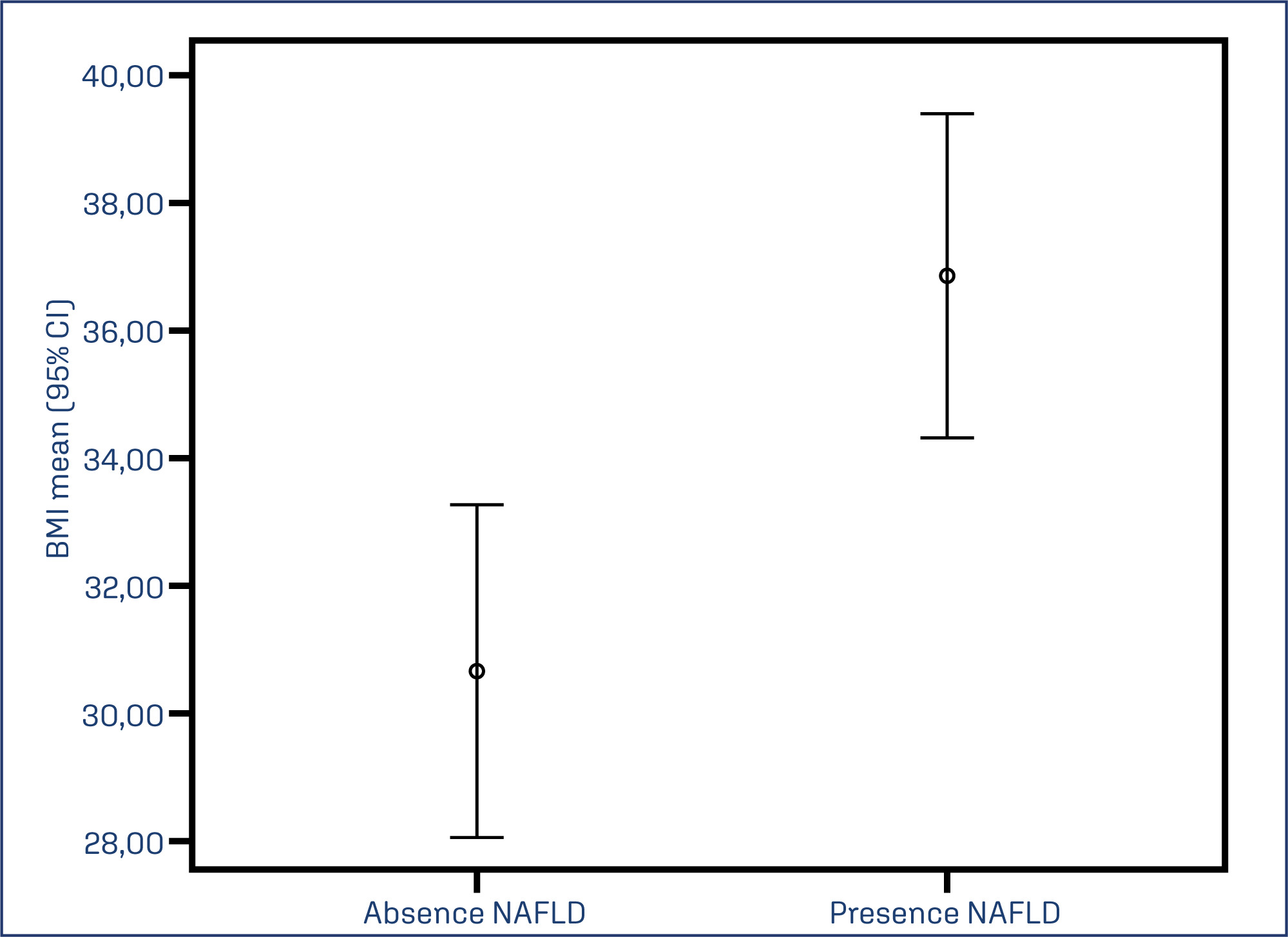
Among 53 patients with PCOS, 50.9% had NAFLD. The NAFLD group had higher weight (p=0.003), BMI (p=0.001), waist circumference (p≤0.001), fasting glucose (p=0.021), HbA1C% (p=0.028), triglycerides (p=0.023), AST (p=0.004), ALT (p=0.001), higher prevalence of MS (p=0.004), and lower levels of HDL cholesterol (p=0.043). The other variables did not differ between the groups. Both groups were predominantly of caucasian race, and there was no significant difference in age.
The prevalence of NAFLD among patients with PCOS was 50.9%. Metabolic and hepatic enzyme abnormalities were more prevalent in this group compared to the group without the disease. Obesity tripled the prevalence of NAFLD.
Summary
Revista Brasileira de Ginecologia e Obstetrícia. 2024;46:e-rbgo81
To verify the prevalence and factors associated with Non-Alcoholic Fatty Liver Disease (NAFLD) among women with Polycystic Ovary Syndrome (PCOS).
A cross-sectional study was conducted with 53 patients with PCOS. The diagnosis of PCOS followed the Rotterdam criteria. The diagnosis of NAFLD was made through US showing hepatic steatosis, excluding significant alcohol consumption and chronic liver disease. The following variables were compared between the groups of women with and without NAFLD: age, race, anthropometric data, blood pressure levels, liver enzymes, glycemic and lipid profiles, total testosterone, presence of hirsutism, and metabolic syndrome (MS). Variables were compared between the groups using T-test, Mann-Whitney, and Chi-square tests.
Among 53 patients with PCOS, 50.9% had NAFLD. The NAFLD group had higher weight (p=0.003), BMI (p=0.001), waist circumference (p≤0.001), fasting glucose (p=0.021), HbA1C% (p=0.028), triglycerides (p=0.023), AST (p=0.004), ALT (p=0.001), higher prevalence of MS (p=0.004), and lower levels of HDL cholesterol (p=0.043). The other variables did not differ between the groups. Both groups were predominantly of caucasian race, and there was no significant difference in age.
The prevalence of NAFLD among patients with PCOS was 50.9%. Metabolic and hepatic enzyme abnormalities were more prevalent in this group compared to the group without the disease. Obesity tripled the prevalence of NAFLD.
Summary
Revista Brasileira de Ginecologia e Obstetrícia. 2024;46:e-rbgo37
To identify the impact of redox imbalance on the clinical evolution of patients with polycystic ovary syndrome and carry out a qualitative and quantitative projection of the benefits of vitamin D supplementation.
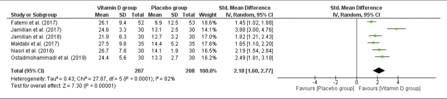
Combinations of the keywords polycystic ovary syndrome, vitamin D, oxidative stress, reactive oxygen species, antioxidant, and free radicals were used in PubMed, Cochrane Library, LILACS, EMBASE, and Web of Science databases. The last search was conducted on August 22, 2023.Selection of studies: Based on the inclusion and exclusion criteria, studies were selected considering a low risk of bias, published in the last 5 years in English, which investigated the effects of vitamin D supplementation in women with PCOS, focusing on oxidative stress markers. Of the 136 articles retrieved, 6 intervention studies (445 women) were included.
The risk of bias in included studies was assessed using the Jadad scale, and analysis and visualization of continuous data were performed using Review Manager 5.4.1, summarized as standardized mean differences (SMD) with confidence intervals (CI) of 95%.
Vitamin D effectively reduced malondialdehyde (P=0.002) and total testosterone (P=0.0004) levels and increased total antioxidant capacity levels (P=0.01). Although possible improvements in the modified Ferriman–Gallwey hirsutism score, levels of sex hormone-binding globulin, and free androgen index were identified and the results were not statistically significant.
Vitamin D is a promising alternative for the treatment of PCOS with a positive influence on the oxidative, metabolic, and endocrine disorders of this syndrome.
Summary
Revista Brasileira de Ginecologia e Obstetrícia. 2024;46:e-rbgo37
To identify the impact of redox imbalance on the clinical evolution of patients with polycystic ovary syndrome and carry out a qualitative and quantitative projection of the benefits of vitamin D supplementation.
Combinations of the keywords polycystic ovary syndrome, vitamin D, oxidative stress, reactive oxygen species, antioxidant, and free radicals were used in PubMed, Cochrane Library, LILACS, EMBASE, and Web of Science databases. The last search was conducted on August 22, 2023.Selection of studies: Based on the inclusion and exclusion criteria, studies were selected considering a low risk of bias, published in the last 5 years in English, which investigated the effects of vitamin D supplementation in women with PCOS, focusing on oxidative stress markers. Of the 136 articles retrieved, 6 intervention studies (445 women) were included.
The risk of bias in included studies was assessed using the Jadad scale, and analysis and visualization of continuous data were performed using Review Manager 5.4.1, summarized as standardized mean differences (SMD) with confidence intervals (CI) of 95%.
Vitamin D effectively reduced malondialdehyde (P=0.002) and total testosterone (P=0.0004) levels and increased total antioxidant capacity levels (P=0.01). Although possible improvements in the modified Ferriman–Gallwey hirsutism score, levels of sex hormone-binding globulin, and free androgen index were identified and the results were not statistically significant.
Vitamin D is a promising alternative for the treatment of PCOS with a positive influence on the oxidative, metabolic, and endocrine disorders of this syndrome.
Summary
Revista Brasileira de Ginecologia e Obstetrícia. 2022;44(2):142-153
To examine the possible effects of adrenal prohormones in the prediction of clinical and metabolic abnormalities in women with polycystic ovary syndrome (PCOS).
The present study enrolled 299 normal cycling non-PCOS, 156 normoandrogenemic, and 474 hyperandrogenemic women with PCOS. Baseline characteristics were compared using a chi-squared test or analysis of variance (ANOVA) as appropriate. The roles of adrenal prohormones and their ratios with total testosterone in predicting co-occurring morbidities in women PCOS were evaluated using univariate and multivariate logistic regression analyses.
Adrenal hyperandrogenism per dehydroepiandrosterone sulfate (DHEAS) levels were found in 32% of women with PCOS. In non-PCOS women, dehydroepiandrosterone (DHEA) and its sulfate had no predictive role concerning clinical, anthropometric, and metabolic parameters. In PCOS women, mainly in the hyperandrogenemic group, DHEA showed to be a significant predictor against most anthropometric-metabolic index abnormalities (odds ratio [OR]=0.36-0.97; p<0.05), and an increase in triglycerides (TG) levels (OR=0.76; p=0.006). Dehydroepiandrosterone sulfate presented a few predictive effects regarding PCOS-associated disorders. In controls, DHEAS predicted against the increase in estimated average glucose (OR= 0.38; p=0.036). In the normoandrogenic group, it predicted against elevation in the waist/hip ratio (WHR) (OR= 0.59; p=0.042), and in hyperandrogenemic PCOS women, it predicted against abnormality in the conicity index (CI) (OR=0.31; p=0.028).
Dehydroepiandrosterone was shown to be a better predictor of abnormal anthropometric and biochemical parameters in women with PCOS than DHEAS. Thus, regarding adrenal prohormones, DHEA measurement, instead of DHEAS, should be preferred in PCOS management. The effects of androgen prohormones on the prediction of PCOS abnormalities are weak.
Summary
Revista Brasileira de Ginecologia e Obstetrícia. 2020;42(12):811-819
The present study aimed to investigate the physical performance of handgrip strength (HGS) in women with polycystic ovary syndrome (PCOS).
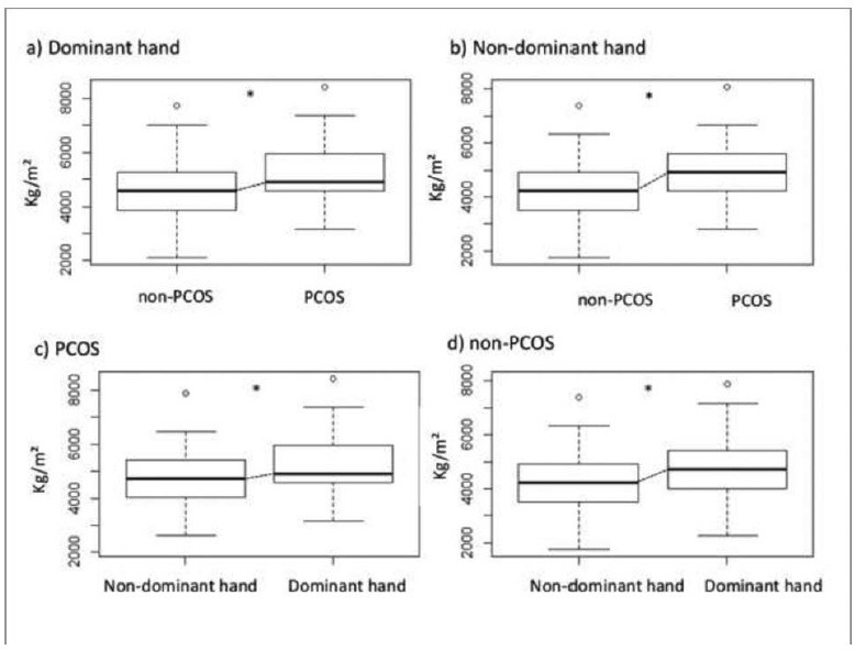
A case-control study that included 70 women with PCOS and 93 agematched healthy women aged between 18 and 47 years with body mass index (BMI) between 18 Kg/m2-39.9 Kg/m2. The serum levels of total testosterone, androstenedione, insulin, estradiol, thyroid-stimulating hormone (TSH), prolactin, sex hormonebinding globulin (SHBG), and 17-hydroxyprogesterone (17-OHP) were measured. The free androgen index (FAI) and the homeostatic model assessment of insulin resistance (HOMA-IR) were calculated. The body composition regions of interest (ROIs) were assessed by dual-energy X-ray absorptiometry (DXA), and the handgrip strength (HGS) was evaluated for both the dominant and the non-dominant hands with a manual Sammons Preston (Bolingbrook, IL, US) bulb dynamometer.
Women with PCOS had high serum levels of total testosterone (p < 0.01), androstenedione (p = 0.03), and insulin (p < 0.01), as well as high FAI (p < 0.01) and HOMA-IR (p = 0.01) scores. Compared with the non-PCOS group, the PCOS group had greater total lean mass in the dominant hand (p < 0.03) and greater HGS in both the dominant and the non-dominant hands (p < 0.01). The HGS was correlated with lean mass (p < 0.01).
Women with PCOS have greater HGS. This may be associated with age and BMI, and it may be related to lean mass. In addition, the dominance effect on muscle mass may influence the physical performance regarding HGS in women with PCOS.
Summary
Revista Brasileira de Ginecologia e Obstetrícia. 2020;42(12):811-819
The present study aimed to investigate the physical performance of handgrip strength (HGS) in women with polycystic ovary syndrome (PCOS).
A case-control study that included 70 women with PCOS and 93 agematched healthy women aged between 18 and 47 years with body mass index (BMI) between 18 Kg/m2-39.9 Kg/m2. The serum levels of total testosterone, androstenedione, insulin, estradiol, thyroid-stimulating hormone (TSH), prolactin, sex hormonebinding globulin (SHBG), and 17-hydroxyprogesterone (17-OHP) were measured. The free androgen index (FAI) and the homeostatic model assessment of insulin resistance (HOMA-IR) were calculated. The body composition regions of interest (ROIs) were assessed by dual-energy X-ray absorptiometry (DXA), and the handgrip strength (HGS) was evaluated for both the dominant and the non-dominant hands with a manual Sammons Preston (Bolingbrook, IL, US) bulb dynamometer.
Women with PCOS had high serum levels of total testosterone (p < 0.01), androstenedione (p = 0.03), and insulin (p < 0.01), as well as high FAI (p < 0.01) and HOMA-IR (p = 0.01) scores. Compared with the non-PCOS group, the PCOS group had greater total lean mass in the dominant hand (p < 0.03) and greater HGS in both the dominant and the non-dominant hands (p < 0.01). The HGS was correlated with lean mass (p < 0.01).
Women with PCOS have greater HGS. This may be associated with age and BMI, and it may be related to lean mass. In addition, the dominance effect on muscle mass may influence the physical performance regarding HGS in women with PCOS.
Summary
Revista Brasileira de Ginecologia e Obstetrícia. 2020;42(2):81-89
The present study aimed to analyze cardiac autonomic modulation via spectral and symbolic analysis of heart rate variability (HRV) in women with polycystic ovary syndrome (PCOS) who were subjected to two consecutive tilt tests.
A total of 64 women were selected and divided into 2 groups: control (without PCOS), and PCOS. Concentrations of follicle-stimulating hormone, luteinizing hormone, prolactin, estradiol, homocysteine, sex hormone-binding globulin, thyroid stimulating hormone, fasting insulin, testosterone, androstenedione, and 17-hydroxyprogesterone levels, triglycerides, free androgen index (FAI), and homeostasis assessment model (HOMA-IR) were assessed. Cardiac autonomic modulation was evaluated by spectral and symbolic analyses during two consecutive tilt tests (two moments) and supine moments before, between and after (three moments) the tilt tests.
Women with PCOS had higher fasting insulin, HOMA-IR indexes, testosterone and FAI. Additionally, we observed that the PCOS group had greater sympathetic autonomic cardiac modulation in supine 2, tilt 1, and supine 3 moments compared with controls.
Women with PCOS had higher autonomic sympathetic cardiac modulation even after a second tilt test. No adaptation to this provocative test was observed. Spectral analysis was more sensitive for identifying differences between groups than the symbolic analysis.
Summary
Revista Brasileira de Ginecologia e Obstetrícia. 2013;35(12):562-568
DOI 10.1590/S0100-72032013001200006
PURPOSE: To assess the contribution of hyperandrogenism to the development of metabolic syndrome (MetS) in obese women with polycystic ovary syndrome (PCOS). METHODS: Retrospective cross-sectional study conducted on 60 obese women with classic PCOS phenotype - Rotterdam Consensus - and 70 non-PCOS obese women. MetS was diagnosed by the NCEP-ATP III criteria and obesity was defined by body mass index. The Ferriman-Gallwey score (mFG) was used to evaluate hirsutism. The following measurements were performed: total testosterone, dehydroepiandrosterone sulfate (DHEA-S), glucose and insulin, total cholesterol, HDL, and triglycerides. Insulin resistance was measured using the HOMA-IR and insulin sensitivity index of Matsuda and De Fronzo (ISI). Statistical analysis was performed using the Student's t-test, χ² test and multivariate logistic regression analysis (p<0.05). RESULTS: Obese women with PCOS had significantly higher mFG (15.4±6.1), waist circunference (105.6±11.4 cm), DHEA-S (200.8±109.2 µg/dL), testosterone (135.8±71.4 ng/dL), and HOMA-IR (8.4±8.5) values and lower ISI values (2.0±1.8) than non-obese PCOS women (3.2±2.1; 101.4±9.2 cm; 155.0±92.7 µg/dL; 50.0±18.2 ng/dL; 5.1±4.7 and 3.3±2.7, respectively) (p<0.05). The frequency of MetS was higher in PCOS obese (75%) than non-PCOS obese (52.8%) women (p=0.015). Multivariate analysis did not reveal the contribution of the variables IFG, testosterone, and DHEAS to the development of MetS (p>0.05). CONCLUSION: Obese women with PCOS have a higher frequency of metabolic syndrome than non-PCOS obese women, and hyperandrogenism does not contribute to the development of metabolic syndrome in this group of women.
Summary
Revista Brasileira de Ginecologia e Obstetrícia. 2013;35(6):249-254
DOI 10.1590/S0100-72032013000600003
PURPOSE: To evaluate the clinical, ultrasonographic, biochemical and metabolic alterations of adolescents with polycystic ovary syndrome (PCOS). METHODS: Retrospective observational study conducted on 44 adolescents aged 12 to 19 years, diagnosed with PCOS according to the Rotterdam Consensus. Metabolic changes were assessed according to the recommendations of the International Diabetes Federation, considering: waist circumference (WC) >90th percentile (10-15 years of age) or >80 cm (age >16 years), fasting glucose >100 mg/dL, triglycerides >150 mg/dL, HDL <40 mg/dL, and blood pressure >Hg 130/85 mm. RESULTS: Mean age was 16.7±2.2 years and age at menarche was 11.8±1.4 years. The menstrual irregularity most frequently observed was amenorrhea (72.7%) followed by oligomenorrhea (27.3%); hirsutism was observed in 86.4% and acne in 56.8%. Polycystic ovaries were observed by ultrasound only in 27.3%. Mean BMI was 30.3±6.6 kg/m². According to BMI, 52.3% of adolescents were obese, 13.6% were overweight and 6.8% had a healthy weight. Increased waist circumference (63.6%, 28/44) and the reduction of HDL-C (34.1%, 15/44) were the metabolic changes most frequently observed. Increased triglycerides were observed in 27.3% (12/44) and increased blood pressure and impaired fasting glucose were found in 9.1% (4/44) and 4.5% (2/44) of cases, respectively. Acanthosis nigricans was observed in 52.3% and insulin resistance in 62.8% of the adolescents with PCOS. Metabolic syndrome was identified in six children (13.6%), all of them obese or overweight. CONCLUSION: In the adolescents with PCOS studied here, menstrual irregularity and hirsutism were the most common clinical manifestations, while the sonographic findings consistent with polycystic ovaries were less prevalent. Obesity associated with insulin resistance predisposes these adolescents to a higher frequency of metabolic disorders.
Summary
Revista Brasileira de Ginecologia e Obstetrícia. 2012;34(7):316-322
DOI 10.1590/S0100-72032012000700005
PURPOSE: To compare the metabolic parameters, body composition and muscle strength of women with Polycystic Ovary Syndrome (PCOS) to those of women with ovulatory menstrual cycles. METHODS: A case-control study was conducted on 27 women with PCOS and 28 control women with ovulatory cycles, aged 18 to 27 years with a body mass index of 18 to 39.9 kg/m², who did not practice regular physical activity. Serum testosterone, androstenedione, prolactin, sex hormone-binding globulin (SHBG), insulin and glycemia levels were determined. Free androgen index (FAI) and resistance to insulin (by HOMA) were calculated. The volunteers were submitted to evaluation of body composition based on skin folds and DEXA and to 1-RM maximum muscle strength tests in three exercises after familiarization procedures and handgrip isometric force was determined. RESULTS: Testosterone levels were higher in the PCOS group than in the Control Group (68.07±20.18 versus 58.20±12.82 ng/dL; p=0.02), as also were the FAI (282.51±223.86 versus 127.08±77.19; p=0.01), insulin (8.41±7.06 versus 4.05±2.73 µIU/mL; p=0.01), and HOMA (2.3±2.32 versus 1.06±0.79; p=0.01), and SBHG levels were lower (52.51±43.27 versus 65.45±27.43 nmol/L; p=0.04). No significant differences in body composition were observed between groups using the proposed methods. The PCOS group showed greater muscle strength in the 1-RM test in the bench press (31.2±4.75 versus 27.79±3.63 kg; p=0.02), and leg extension exercises (27.9±6.23 versus 23.47±4.21 kg; p=0.02) as well as handgrip isometric force (5079.61±1035.77 versus 4477.38±69.66 kgf/m², p=0.04). PCOS was an independent predictor of increase muscle strength in bench press exercises (estimate (E)=2.7) (p=0.04) and leg extension (E=3.5) (p=0.04), and BMI in the exercise of isometric handgrip (E=72.2) (p<0.01), bench press (E=0.2) (p=0.02) and arm curl (E=0.3) (p<0.01). No association was found between HOMA-IR and muscle strength. CONCLUSIONS: Women with POS showed greater muscle strength, with no difference in body composition, and IR was not associated with muscle strength performance. Muscle strength may be possibly related to high levels of androgens in these women.