-
Review Article
Efficacy of Hormonal and Nonhormonal Approaches to Vaginal Atrophy and Sexual Dysfunctions in Postmenopausal Women: A Systematic Review
Revista Brasileira de Ginecologia e Obstetrícia. 2022;44(10):986-994
01-23-2022
Summary
Review ArticleEfficacy of Hormonal and Nonhormonal Approaches to Vaginal Atrophy and Sexual Dysfunctions in Postmenopausal Women: A Systematic Review
Revista Brasileira de Ginecologia e Obstetrícia. 2022;44(10):986-994
01-23-2022Views239See moreAbstract
Objective
To evaluate the efficacy of the hormonal and nonhormonal approaches to symptoms of sexual dysfunction and vaginal atrophy in postmenopausal women.
Data Sources
We conducted a search on the PubMed, Embase, Scopus, Web of Science, SciELO, the Cochrane Central Register of Controlled Trials (CENTRAL), and Cumulative Index to Nursing and Allied Health Literature (CINAHL) databases, as well as on clinical trial databases. We analyzed studies published between 1996 and May 30, 2020. No language restrictions were applied.
Selection of Studies
We selected randomized clinical trials that evaluated the treatment of sexual dysfunction in postmenopausal women.
Data Collection
Three authors (ACAS, APFC, and JL) reviewed each article based on its title and abstract. Relevant data were subsequently taken from the full-text article. Any discrepancies during the review were resolved by consensus between all the listed authors.
Data Synthesis
A total of 55 studies were included in the systematic review. The approaches tested to treat sexual dysfunction were as follows: lubricants and moisturizers (18 studies); phytoestrogens (14 studies); dehydroepiandrosterone (DHEA; 8 studies); ospemifene (5 studies); vaginal testosterone (4 studies); pelvic floor muscle exercises (2 studies); oxytocin (2 studies); vaginal CO2 laser (2 studies); lidocaine (1 study); and vitamin E vaginal suppository (1 study).
Conclusion
We identified literature that lacks coherence in terms of the proposed treatments and selected outcome measures. Despite the great diversity in treatment modalities and outcome measures, the present systematic review can shed light on potential targets for the treatment, which is deemed necessary for sexual dysfunction, assuming that most randomized trials were evaluated with a low risk of bias according to the Cochrane Collaboration risk of bias tool. The present review is registered with the International Prospective Register of Systematic Reviews (PROSPERO; CRD42018100488).
-
Case Report
Short-term Prophylaxis for Delivery in Pregnant Women with Hereditary Angioedema with Normal C1-Inhibitor
Revista Brasileira de Ginecologia e Obstetrícia. 2020;42(12):845-848
01-11-2020
Summary
Case ReportShort-term Prophylaxis for Delivery in Pregnant Women with Hereditary Angioedema with Normal C1-Inhibitor
Revista Brasileira de Ginecologia e Obstetrícia. 2020;42(12):845-848
01-11-2020Views148See moreAbstract
Objective
To verify the efficacy of short-term prophylaxis for vaginal or cesarean section childbirth with plasma-derived C1-inhibitor concentrate in pregnant women. They should have hereditary angioedema (HAE) and normal plasma C1-inhibitor.
Methods
Case report of pregnant women diagnosed with HAE with normal C1- inhibitor who had been treated with intravenous C1-inhibitor concentrate for prophylaxis of angioedema attacks when hospitalized for delivery. The exon 9 of the Factor 12 (F12) genotyping gene was performed by automatic sequencing in all patients.
Results
Three cases of pregnant women with HAE with normal serum level of C1- inhibitor are reported. The genetic test detected the presence of a pathogenic mutation in the F12 gene. Deliveries occurred uneventfully and patients had no HAE symptoms in the following 72 hours.
Conclusion
C1-inhibitor concentrate could be useful to prevent angioedema attacks during and after delivery.
-
Original Articles
The Influence of CYP3A4 Polymorphism in Sex Steroids as a Risk Factor for Breast Cancer
Revista Brasileira de Ginecologia e Obstetrícia. 2018;40(11):699-704
11-01-2018
Summary
Original ArticlesThe Influence of CYP3A4 Polymorphism in Sex Steroids as a Risk Factor for Breast Cancer
Revista Brasileira de Ginecologia e Obstetrícia. 2018;40(11):699-704
11-01-2018Views162See moreAbstract
Objective
Epidemiological studies have shown evidence of the effect of sex hormones in the pathogenesis of breast cancer, and have suggested a relationship of the disease with variations in genes involved in estrogen synthesis and/or metabolism. The aim of the present study was to evaluate the association between the CYP3A4*1B gene polymorphism (rs2740574) and the risk of developing breast cancer.
Methods
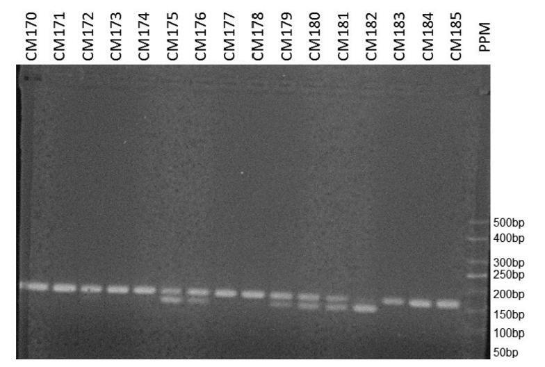
In the present case-control study, the frequency of the CYP3A4*1B gene polymorphism was determined in 148 women with breast cancer and in 245 women without the disease. The DNA of the participants was extracted from plasma samples, and the gene was amplified by polymerase chain reaction. The presence of the polymorphism was determined using restriction enzymes.
Results
After adjusting for confounding variables, we have found that the polymorphism was not associated with the occurrence of breast cancer (odds ratio = 1.151; 95% confidence interval: 0.714–1.856; p= 0.564). We have also found no association with the presence of hormone receptors, with human epidermal growth factor receptor 2 (HER2) overexpression, or with the rate of tumor cell proliferation.
Conclusion
We have not observed a relationship between the CYP3A4*1B gene polymorphism and the occurrence of breast cancer.

-
Case Report
Postpartum Genital Melanoma – A Case Report
Revista Brasileira de Ginecologia e Obstetrícia. 2018;40(3):163-167
03-01-2018
Summary
Case ReportPostpartum Genital Melanoma – A Case Report
Revista Brasileira de Ginecologia e Obstetrícia. 2018;40(3):163-167
03-01-2018Views126See moreAbstract
Melanomas of the female genital tract may occur in the vulva, the vagina, the ovary or the cervix.Pregnancy has been considered an aggravating factor in the evolution and prognosis of melanoma. A 35-year-old female presented with vaginal bleeding 2 months after a term cesarean delivery. An endovaginal ultrasound revealed a lesion in the uterine cervix. The pathological report revealed a small round-cell neoplasm, and the immunohistochemistry confirmed the diagnosis of malignant melanoma. A positron emission tomography revealed an expansive hypermetabolic lesion centered on the cervix, and hypermetabolic lesions in the liver and right kidney. Non-surgical treatment was provided, with biochemotherapy followed by ipilimumab and nivolumab. The patient died one year later. Postpartum vaginal bleeding, even if late-onset, should be investigated, as it may be a pregnancy-associated malignant melanoma, which has a poor prognosis.
-
Original Article
Combined Effect of the PGR + 331C > T, CYP17A1 -34A > G and CYP19A1 1531G > A Polymorphisms on the Risk of Developing Endometriosis
Revista Brasileira de Ginecologia e Obstetrícia. 2017;39(6):273-281
06-01-2017
Summary
Original ArticleCombined Effect of the PGR + 331C > T, CYP17A1 -34A > G and CYP19A1 1531G > A Polymorphisms on the Risk of Developing Endometriosis
Revista Brasileira de Ginecologia e Obstetrícia. 2017;39(6):273-281
06-01-2017Views111See moreAbstract
Purpose
To evaluate the magnitude of the association of the polymorphisms of the genes PGR, CYP17A1 and CYP19A1 in the development of endometriosis.
Methods
This is a retrospective case-control study involving 161 women with endometriosis (cases) and 179 controls. The polymorphisms were genotyped by real-time polymerase chain reaction using the TaqMan system. The association of the polymorphisms with endometriosis was evaluated using the multivariate logistic regression.
Results
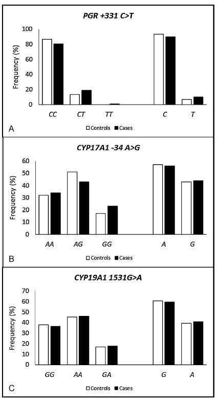
The endometriosis patients were significantly younger than the controls (36.0±7.3 versus 38.0±8.5 respectively, p = 0.023), and they had a lower body mass index (26.3±4.8 versus 27.9±5.7 respectively, p = 0.006), higher average duration of the menstrual flow (7.4±4.9 versus 6.1±4.4 days respectively, p = 0.03), and lower average time intervals between menstrual periods (25.2±9.6 versus 27.5±11.1 days respectively, p = 0.05). A higher prevalence of symptoms of dysmenorrhea, dyspareunia, chronic pelvic pain, infertility and intestinal or urinary changes was observed in the case group when compared with the control group. The interval between the onset of symptoms and the definitive diagnosis of endometriosis was 5.2±6.9 years. When comparing both groups, significant differences were not observed in the allelic and genotypic frequencies of the polymorphisms PGR + 331C > T, CYP17A1 -34A > G and CYP19A1 1531G > A, even when considering the symptoms, classification and stage of the endometriosis. The combined genotype PGR + 331TT/CYP17A1 -34AA/CYP19A11531AA is positively associated with endometriosis (odds ratio [OR] = 1.72; 95% confidence interval [95%CI] = 1.09-2.72).
Conclusions
The combined analysis of the polymorphisms PGR-CYP17A1-CYP19A1 suggests a gene-gene interaction in the susceptibility to endometriosis. These results may contribute to the identification of biomarkers for the diagnosis and/or prognosis of the disease and of possible molecular targets for individualized treatments.

-
Artigos Originais
Effects of high doses of genistein on mammary gland of female rat
Revista Brasileira de Ginecologia e Obstetrícia. 2011;33(9):264-269
12-20-2011
Summary
Artigos OriginaisEffects of high doses of genistein on mammary gland of female rat
Revista Brasileira de Ginecologia e Obstetrícia. 2011;33(9):264-269
12-20-2011DOI 10.1590/S0100-72032011000900008
Views80See morePURPOSE: to evaluate the effects of high doses of genistein on the mammary glands of adult female rats. METHODS: Twenty-eight days after oophorectomy, 50 adult female rats were divided into five groups, as follows: a control group (Ctrl), three rats that received genistein (GEN) at the doses of 46 mg/kg (GEN46;), 125 mg/kg (GEN125) and 250 mg/kg (GEN250); one group received conjugated equine estrogen at the dose of 50 µg/g (ECE50). The substances were administered daily for 30 consecutive days by gavage and in the last week of the period of treatment, colpocytological exams were carried out for seven consecutive days. After treatment, the animals were anesthetized, blood samples were collected for estradiol and progesterone determination and the first pair of inguinal mammary glands was removed and processed for histomorphometric analysis. Collected data were subjected to analysis of variance supplemented by the Tukey-Kramer test (p<0.05). RESULTS: the ctrl group and the ones treated with different doses of GEN showed atrophic mammary glands, whereas the glands were more developed in the ECE group, where numerous mammary ducts and alveoli were observed. Morphometry showed a larger area of mammary parenchyma in the ECE group (98.870.1±550.4 µm²* per mm²; p<0.05) compared with other groups (Ctrl=36.875.6±443.4; GEN46=37.001.7±557.4; GEN125=36.480.8±658.3 and GEN250=37.502.8±669.3). The same occurred in the number of alveoli in the ECE group (33.2±6.9* per mm²; p<0.05) compared to the other groups (Ctrl=10.4±2.1, GEN46=11.2±3.1; GEN125=11.6±2.1 and GEN250=12.3±2.3). The estradiol level was higher in the ECE group compared to the other groups (9.4±1.7 pg/mL; p<0.05), whereas serum levels of progesterone were similar in all groups. CONCLUSION: the administration of genistein at high doses had no trophic effect on the mammary glands of rats.
-
Artigos Originais
Effect of trimegestone on mammary gland of castrated rats
Revista Brasileira de Ginecologia e Obstetrícia. 2011;33(7):137-142
10-11-2011
Summary
Artigos OriginaisEffect of trimegestone on mammary gland of castrated rats
Revista Brasileira de Ginecologia e Obstetrícia. 2011;33(7):137-142
10-11-2011DOI 10.1590/S0100-72032011000700004
Views81See morePURPOSE: To evaluate the efect of trimegestone on the histological changes of the mammary tissue of castrated rats. METHODS: Forty-five virgin female Wistar rats were used after oophorectomy. Sixty days after surgery, with hypoestrogenisms confirmed, the experimental rats were randomly assigned to three groups of 15 animals each, when then the specific treatment for each group was started. The control group (C) and experimental groups 1 and 2 respectively received 0.9% saline solution, 17-beta-estradiol and 17-beta-estradiol in combination with trimegestone for 60 consecutive days. After the end of treatment , the inguinal mammary glands were removed, stained with hematoxylin and eosin (HE) for morphometry and examined by immunohistochemistry for the quantification of anti-PCNA antibody in the mammary tissue, followed by euthanasia. The morphometric parameters evaluated were: epithelium cell-proliferation, secretor activity and mammary stroma changes. There were nine deaths during the experiment. The variables were submitted to statistical analysis adopting the 0.05 level of significance. RESULTS:Histological changes were observed in 16/36 rats, mild epithelial hyperplasia in 13/36, moderate epithelial hyperplasia in 3/36, with no cases of severe epithelial hyperplasia. Stromal fibrosis was found in 10/36 and secretory activity in 5/36 rats. All morphometric variables were significant in the estrogen group compared to control (p=0.0361), although there were no difference between the group receiving combined treatment and the controls (p=0.405). The immunohistochemical analysis showed no difference between groups. CONCLUSIONS:The hormones administered to castrated rats, i.e., 17 beta-estradiol alone or in combination with trimegestone, increased the proliferation of breast cells, but this effect appeared to be lower in the combined treatment, the same occurring regarding fibrosis of the mammary stroma.
-
Revisão
Current aspects on diagnosis and treatment of endometriosis
Revista Brasileira de Ginecologia e Obstetrícia. 2010;32(6):298-307
09-28-2010
Summary
RevisãoCurrent aspects on diagnosis and treatment of endometriosis
Revista Brasileira de Ginecologia e Obstetrícia. 2010;32(6):298-307
09-28-2010DOI 10.1590/S0100-72032010000600008
Views67See moreEndometriosis is characterized by the presence of endometrial tissue, localized outside the uterine cavity, such as peritoneal surface, ovaries, and rectum-vaginal septum. The prevalence is about 6 to 10%. Concerning the etiopathogenesis, the retrograde menstruation theory is accepted, although disruption in endometrial molecular biology seems to be fundamental to the development of endometriosis ectopic focuses. Women with endometriosis may be asymptomatic or may present complaints of dysmenorrhea, dispareunia, chronic pelvic pain and/or infertility. Although the definitive diagnosis of endometriosis needs a surgical intervention, mainly by laparoscopy, many findings obtained by physicalexamination and imaging and laboratory tests can predict, with a high degree of reliability, that the patient has endometriosis. The most common current treatments include surgery, ovarian suppression therapy or both. Pharmacological treatments that do not inhibit ovarian function are under investigation.


