-
Case Report05-01-2018
Hemoptysis and Endometriosis: An Unusual Association – Case Report and Review of the Literature
Revista Brasileira de Ginecologia e Obstetrícia. 2018;40(5):300-303
Abstract
Case ReportHemoptysis and Endometriosis: An Unusual Association – Case Report and Review of the Literature
Revista Brasileira de Ginecologia e Obstetrícia. 2018;40(5):300-303
Views142See moreAbstract
Thoracic endometriosis syndrome is a rare condition that includes four entities: catamenial pneumothorax, catamenial hemothorax, catamenial hemoptysis and lung nodules. We describe the case of a 23-year-old woman with complaints of hemoptysis during menstrual period in the two years prior to the appointment. Initially, a treatment for tuberculosis was established with no success. Further investigation showed a 4 mmnodule in the right lung, and the transvaginal ultrasonography indicated the presence of deep endometriosis. Considering the occurrence of symptoms only during menses, an empirical therapy was instituted with remission of the complaints.
-
Review Article03-01-2018
Ascites and Encapsulating Peritonitis in Endometriosis: a Systematic Review with a Case Report
Revista Brasileira de Ginecologia e Obstetrícia. 2018;40(3):147-155
Abstract
Review ArticleAscites and Encapsulating Peritonitis in Endometriosis: a Systematic Review with a Case Report
Revista Brasileira de Ginecologia e Obstetrícia. 2018;40(3):147-155
Views195See moreAbstract
Endometriosis can have several different presentations, including overt ascites and peritonitis; increased awareness can improve diagnostic accuracy and patient outcomes. We aimto provide a systematic review and report a case of endometriosis with this unusual clinical presentation. The PubMed/MEDLINE database was systematically reviewed until October 2016. Women with histologically-proven endometriosis presenting with clinically significant ascites and/or frozen abdomen and/or encapsulating peritonitis were included; thosewith potentially confounding conditionswere excluded.Our search yielded 37 articles describing 42 women, all of reproductive age. Ascites was mostly hemorrhagic, recurrent and not predicted by cancer antigen 125 (CA-125) levels. In turn, dysmenorrhea, dyspareunia and infertility were not consistently reported. The treatment choices and outcomes were different across the studies, and are described in detail. Endometriosis should be a differential diagnosis of massive hemorrhagic ascites in women of reproductive age.
-
Original Article06-01-2017
Combined Effect of the PGR + 331C > T, CYP17A1 -34A > G and CYP19A1 1531G > A Polymorphisms on the Risk of Developing Endometriosis
Revista Brasileira de Ginecologia e Obstetrícia. 2017;39(6):273-281
Abstract
Original ArticleCombined Effect of the PGR + 331C > T, CYP17A1 -34A > G and CYP19A1 1531G > A Polymorphisms on the Risk of Developing Endometriosis
Revista Brasileira de Ginecologia e Obstetrícia. 2017;39(6):273-281
Views166See moreAbstract
Purpose
To evaluate the magnitude of the association of the polymorphisms of the genes PGR, CYP17A1 and CYP19A1 in the development of endometriosis.
Methods
This is a retrospective case-control study involving 161 women with endometriosis (cases) and 179 controls. The polymorphisms were genotyped by real-time polymerase chain reaction using the TaqMan system. The association of the polymorphisms with endometriosis was evaluated using the multivariate logistic regression.
Results
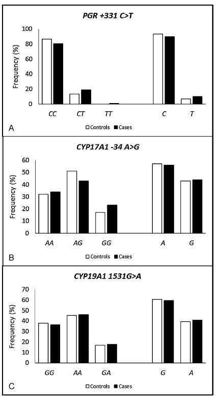
The endometriosis patients were significantly younger than the controls (36.0±7.3 versus 38.0±8.5 respectively, p = 0.023), and they had a lower body mass index (26.3±4.8 versus 27.9±5.7 respectively, p = 0.006), higher average duration of the menstrual flow (7.4±4.9 versus 6.1±4.4 days respectively, p = 0.03), and lower average time intervals between menstrual periods (25.2±9.6 versus 27.5±11.1 days respectively, p = 0.05). A higher prevalence of symptoms of dysmenorrhea, dyspareunia, chronic pelvic pain, infertility and intestinal or urinary changes was observed in the case group when compared with the control group. The interval between the onset of symptoms and the definitive diagnosis of endometriosis was 5.2±6.9 years. When comparing both groups, significant differences were not observed in the allelic and genotypic frequencies of the polymorphisms PGR + 331C > T, CYP17A1 -34A > G and CYP19A1 1531G > A, even when considering the symptoms, classification and stage of the endometriosis. The combined genotype PGR + 331TT/CYP17A1 -34AA/CYP19A11531AA is positively associated with endometriosis (odds ratio [OR] = 1.72; 95% confidence interval [95%CI] = 1.09-2.72).
Conclusions
The combined analysis of the polymorphisms PGR-CYP17A1-CYP19A1 suggests a gene-gene interaction in the susceptibility to endometriosis. These results may contribute to the identification of biomarkers for the diagnosis and/or prognosis of the disease and of possible molecular targets for individualized treatments.

-
Original Article05-01-2017
The Effect of Mesenchymal Stem Cells on Fertility in Experimental Retrocervical Endometriosis
Revista Brasileira de Ginecologia e Obstetrícia. 2017;39(5):217-223
Abstract
Original ArticleThe Effect of Mesenchymal Stem Cells on Fertility in Experimental Retrocervical Endometriosis
Revista Brasileira de Ginecologia e Obstetrícia. 2017;39(5):217-223
Views166See moreAbstract
Purpose
To evaluate the effect of mesenchymal stem cells (MSCs) on fertility in experimental retrocervical endometriosis.
Methods
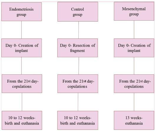
A total of 27 New Zealand rabbits were divided into three groups: endometriosis, in which endometrial implants were created; mesenchymal, in which MSCs were applied in addition to the creation of endometrial implants; and control, the group without endometriosis. Fisher’s exact test was performed to compare the dichotomous qualitative variables among the groups. The quantitative variables were compared by the nonparametric Mann-Whitney and Kruskal-Wallis tests. The MannWhitney test was used for post-hoc multiple comparison with Boniferroni correction.
Results
Regarding the beginning of the fertile period, the three groups had medians of 14±12.7, 40±5, and 33±8.9 days respectively (p = 0.005). With regard to fertility (number of pregnancies), the endometriosis and control groups showed a rate of 77.78%, whereas the mesenchymal group showed a rate of 11.20% (p = 0.015). No differences in Keenan’s histological classification were observed among the groups (p = 0.730). With regard to the macroscopic appearance of the lesions, the mesenchymal group showed the most pelvic adhesions.
Conclusion
The use of MSCs in endometriosis negatively contributed to fertility, suggesting the role of these cells in the development of this disease.

-
Case Report01-01-2017
Tubocutaneous Fistula due to Endometriosis – A Differential Diagnosis in Cutaneous Fistulas with Cyclic Secretion
Revista Brasileira de Ginecologia e Obstetrícia. 2017;39(1):31-34
Abstract
Case ReportTubocutaneous Fistula due to Endometriosis – A Differential Diagnosis in Cutaneous Fistulas with Cyclic Secretion
Revista Brasileira de Ginecologia e Obstetrícia. 2017;39(1):31-34
Views90See moreABSTRACT
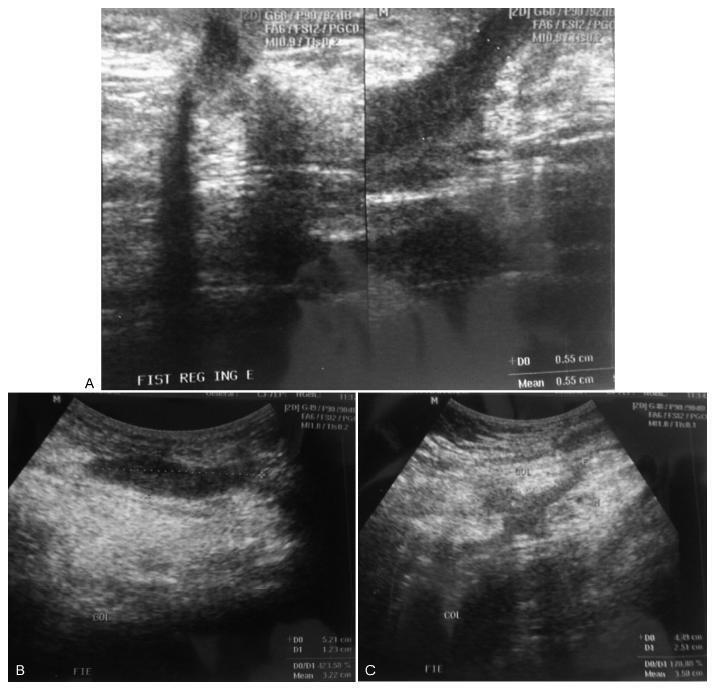
The development of a tubocutaneous fistula due to endometriosis in a post-cesarean section surgical scar is a rare complication that generates significant morbidity in the affected women. Surgery is the treatment of choice in these cases. Hormonal therapies may lead to an improvement in symptoms, but do not eradicate such lesions. In this report, we present a 34-year-old patient with a cutaneous fistula in the left iliac fossa with cyclic secretion. Anamnesis, a physical examination, and supplementary tests led us to suggest endometriosis as the main diagnosis, which was confirmed after surgical intervention.

-
Original Article05-01-2016
Endometriosis, Ovarian Reserve and Live Birth Rate Following In Vitro Fertilization/Intracytoplasmic Sperm Injection
Revista Brasileira de Ginecologia e Obstetrícia. 2016;38(5):218-224
Abstract
Original ArticleEndometriosis, Ovarian Reserve and Live Birth Rate Following In Vitro Fertilization/Intracytoplasmic Sperm Injection
Revista Brasileira de Ginecologia e Obstetrícia. 2016;38(5):218-224
Views126See moreAbstract
Purpose
To evaluate whether women with endometriosis have different ovarian reserves and reproductive outcomes when compared with women without this diagnosis undergoing in vitro fertilization/intracytoplasmic sperm injection ( IVF/ ICSI), and to compare the reproductive outcomes between women with and without the diagnosis considering the ovarian reserve assessed by antral follicle count ( AFC ).
Methods
This retrospective cohort study evaluated all women who underwent IVF/ ICSI in a university hospital in Brazil between January 2011 and December 2012. All patients were followed up until a negative pregnancy test or until the end of the pregnancy. The primary outcomes assessed were number of retrieved oocytes and live birth. Women were divided into two groups according to the diagnosis of endometriosis, and each group was divided again into a group that had AFC 6 (poor ovarian reserve) and another that had AFC 7 (normal ovarian reserve). Continuous variables with normal distribution were compared using unpaired t-test, and those without normal distribution, using Mann-Whitney test. Binary data were compared using either Fisher's exact test or Chi-square (2) test. The significance level was set as p < 0.05.
Results
787 women underwent IVF/ICSI (241 of which had endometriosis). Although the mean age has been similar between women with and without the diagnosis of endometriosis (33.8 4 versus 33.7 4.4 years, respectively), poor ovarian reserves were much more common in women with endometriosis (39.8 versus 22.7%). The chance of achieving live birth was similar between women with the diagnosis of endometriosis and those without it (19.1 versus 22.5%), and also when considering only women with a poor ovarian reserve (9.4 versus 8.9%) and only those with a normal ovarian reserve (25.5 versus 26.5%).
Conclusions
Women diagnosed with endometriosis are more likely to have a poor ovarian reserve; however, their chance of conceiving by IVF/ICSI is similar to the one observed in patients without endometriosis and with a comparable ovarian reserve.
-
Review Article05-01-2016
Common Dysregulated Genes in Endometriosis and Malignancies
Revista Brasileira de Ginecologia e Obstetrícia. 2016;38(5):253-262
Abstract
Review ArticleCommon Dysregulated Genes in Endometriosis and Malignancies
Revista Brasileira de Ginecologia e Obstetrícia. 2016;38(5):253-262
Views153See moreAbstract
Several authors have investigated the malignant transformation of endometriosis, which supports the hypothesis of the pre-neoplastic state of endometriotic lesions, but there are few data about the pathways and molecular events related to this phenomenon. This review provides current data about deregulated genes that may function as key factors in the malignant transition of endometriotic lesions. In order to do so, we first searched for studies that have screened differential gene expression between endometriotic tissues and normal endometrial tissue of women without endometriosis, and found only two articles with 139 deregulated genes. Further, using the PubMed database, we crossed the symbol of each gene with the terms related to malignancies, such as cancer and tumor, and obtained 9,619 articles, among which 444 were studies about gene expression associated with specific types of tumor. This revealed that more than 68% of the analyzed genes are also deregulated in cancer. We have also found genes functioning as tumor suppressors and an oncogene. In this study, we present a list of 95 informative genes in order to understand the genetic components that may be responsible for endometriosis' malignant transformation.
However, future studies should be conducted to confirm these findings.


