Summary
Revista Brasileira de Ginecologia e Obstetrícia. 2007;29(8):396-401
DOI 10.1590/S0100-72032007000800003
PURPOSE: to translate and to validate the Female Sexual Function Index (FSFI) for Brazilian pregnant women. METHODS: ninety-two pregnant women attended at a low risk prenatal clinic, with diagnosis of the pregnancy confirmed by precocious ultrasonography, participated in the research. Initially, we translated the FSFI questionnaire for Portuguese language (of Brazil) in agreement with the international criteria. Cultural, conceptual and semantics adaptations of FSFI were accomplished, because of the differences of the language, so that the pregnant women understood the subjects. All the patients answered FSFI twice, in the same day, with two different interviewers, with an hour interval from one to other interview. After 7 to 14 days, the questionnaire was applied again in a second interview. Reliability (internal intra and interobserver consistence) and the validity of the constructo (to demonstrate that questionnaire measures the sexual function) were appraised. RESULTS: Cultural adaptations were necessary for us to obtain the final version. The internal intra-observer (alpha of Chronbach) consistence of the several domains oscillated from moderate to strong (0,791 to 0,911) and the interobserver consistence varied from 0,791 to 0,914. In the validation of the constructo, were obtained moderate correlations to fort among the final scores (general) of FSFI and of Female Sexual Quotient (QS-F) that has the capacity to evaluate the feminine sexual function. CONCLUSIONS: FSFI was adapted to the Portuguese language and to the Brazilian culture, presenting significant reliability and validity; it could be included and used in future studies of the Brazilian pregnant sexual function.
Summary
Revista Brasileira de Ginecologia e Obstetrícia. 1999;21(5):287-290
DOI 10.1590/S0100-72031999000500007
Purpose: to evaluate the knowledge and practice of breast self-examination among medical students and to determine possible factors associated with this practice. Method: the authors used a questionnaire to gather information about the students and their knowledge of this self-examination. This questionnaire also allowed the authors to verify the frequency with which the female students performed breast self-examination. The chi² test and Student's "t" test were used, when applicable, to check the association of certain factors. Results: of the 348 questionnaires which were answered, 16% (55) were submitted by 5th year medical students, who had already attended the Gynecology course; 43% were answered by females, 62% of the students had medical doctors among their relatives, and 17% had a family history of breast cancer. In terms of breast self-examination, 95% knew about the method. Of the 149 females who answered the questionnaire, only 64% checked their breasts regularly. The reasons given for not performing self-examination varied: 24% considered themselves to be too young, 4% thought they would not have cancer, 9% listed fear as the reason, 19% reported they were too lazy, and 44% of the female students had no clear reason for not performing breast self-examination. Neither the knowledge nor the practice of the breast self-examination were associated with the subjects the students had or had not yet taken in medical school, with a family history of breast cancer or with the fact that one or more relatives were medical doctors. Conclusion: breast self-examination is known by practically all the medical students; nevertheless, only one third of the female students performed it regularly. This fact highlights the importance of emphasizing breast self-examination among medical students, so that they can help to disseminate this practice among the general population, rather than delegating this responsibility to the midia.
Summary
Revista Brasileira de Ginecologia e Obstetrícia. 2004;26(4):295-298
DOI 10.1590/S0100-72032004000400005
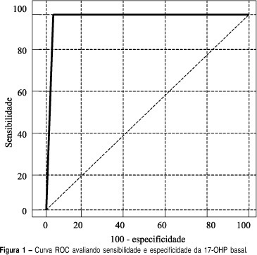
INTRODUCTION: adrenal hyperplasia is a common genetic disorder and 95% of the cases are due to a 21-hydroxylase deficiency. Clinical presentation varies from life-threatening salt-losing adrenal hyperplasia to simple androgenic states, which can be of late-onset and very similar to polycystic ovary syndrome. Diagnosis is usually made by synthetic ACTH provocative tests but efforts are being made to simplify this investigation. OBJECTIVE: to evaluate basal 17-hydroxyprogesterone as a predictor of the provocative test for the diagnosis of late-onset congenial adrenal hyperplasia. METHODS: A total of 122 patients under clinical suspicion of diagnosis of late-onset congenial adrenal hyperplasia were included and retrospectively evaluated in the study. Such suspicion included signs and/or symptoms of hyperandrogenism (hirsutism, acne, oily skin, menstrual irregularity etc.). All the patients were submitted to the 0.25mg synthetic ACTH provocative test (Synacthen®). After resting for 60 minutes, the samples were taken in the basal time and 60 minutes after the administration of 0.25mg synthetic ACTH, in order to assay 17-hydroxiprogesteron, the venous access being kept through a heparinized catheter. Radioimmuoessay was the method used to accomplish the assay of seric 17-hydroxiprogesteron. The sensitivity and specificity of the basal 17-hydroxiprogesteron were measured, assessing several cutoff points. ROC curves were made to analyze the test performance, using the software Medcalc®. RESULTS: ROC curve analysis showed that the best cutoff point was 181 ng/dl, which was very similar to the most common recommendation of 200 ng/dl of the literature. The cutoff point of 200 ng/dl shows positive and negative predictive values of 75 and 100%, and accuracy of 98,4% as a diagnostic test for late-onset adrenal hyperplasia. CONCLUSIONS: considering our data, we suggest that all hyperandrogenic patients should start the investigation with basal 17-hydroxyprogesteron and in case it is above 181 ng/dl, then they should do the synthetic 17-hydroxyprogesteron provocative test.

Summary
Revista Brasileira de Ginecologia e Obstetrícia. 2002;24(3):195-199
DOI 10.1590/S0100-72032002000300008
Purpose: to evaluate, in a prospective way, the importance of ultrasound features of solid breast lesions in the differentiation between benign and malignant lumps. Methods: one hundred and forty-two patients with solid breast lesions, from the Department of Gynecology and Obstetrics of the Federal University of Goias (Brazil), were included in the trial. All ultrasound examinations were performed by a training doctor, always supervised by an experienced professional. The characteristics of the lesions studied were: shape, retrotumoral echoes, internal echoes, oriented diameter, halo of bright echoes and Cooper ligaments. Each of the ultrasound features was compared to the results of the histological examination. Results: among the 142 patients included in the trial, 90 (63%) had their lesions excised, and 77 (86%) had pathologic diagnoses of benign tumors and 13 (14%) of malignant tumors. The following characteristics were statistically significant in the diagnosis of the breast cancer (c²): masses with retrotumoral shadowing (p=0.0001), irregular shape (p=0.0007), heterogeneous internal echoes (p=0.0015) and vertically oriented - taller than wide (p<0.0001). The presence of halo of bright echoes anterior to the lump and the presence of wider Cooper ligaments were not related to the correct diagnosis of malignancy in this trial. Conclusion: ultrasound is a diagnostic method that can help physicians between the differentiation of benign and malignant breast lumps. The presence of retrotumoral shadowing, irregular shape, heterogeneous internal echoes and vertical orientation - lesions taller than wide - were related to the pathologic diagnosis of breast malignancies.