-
Relato de Caso
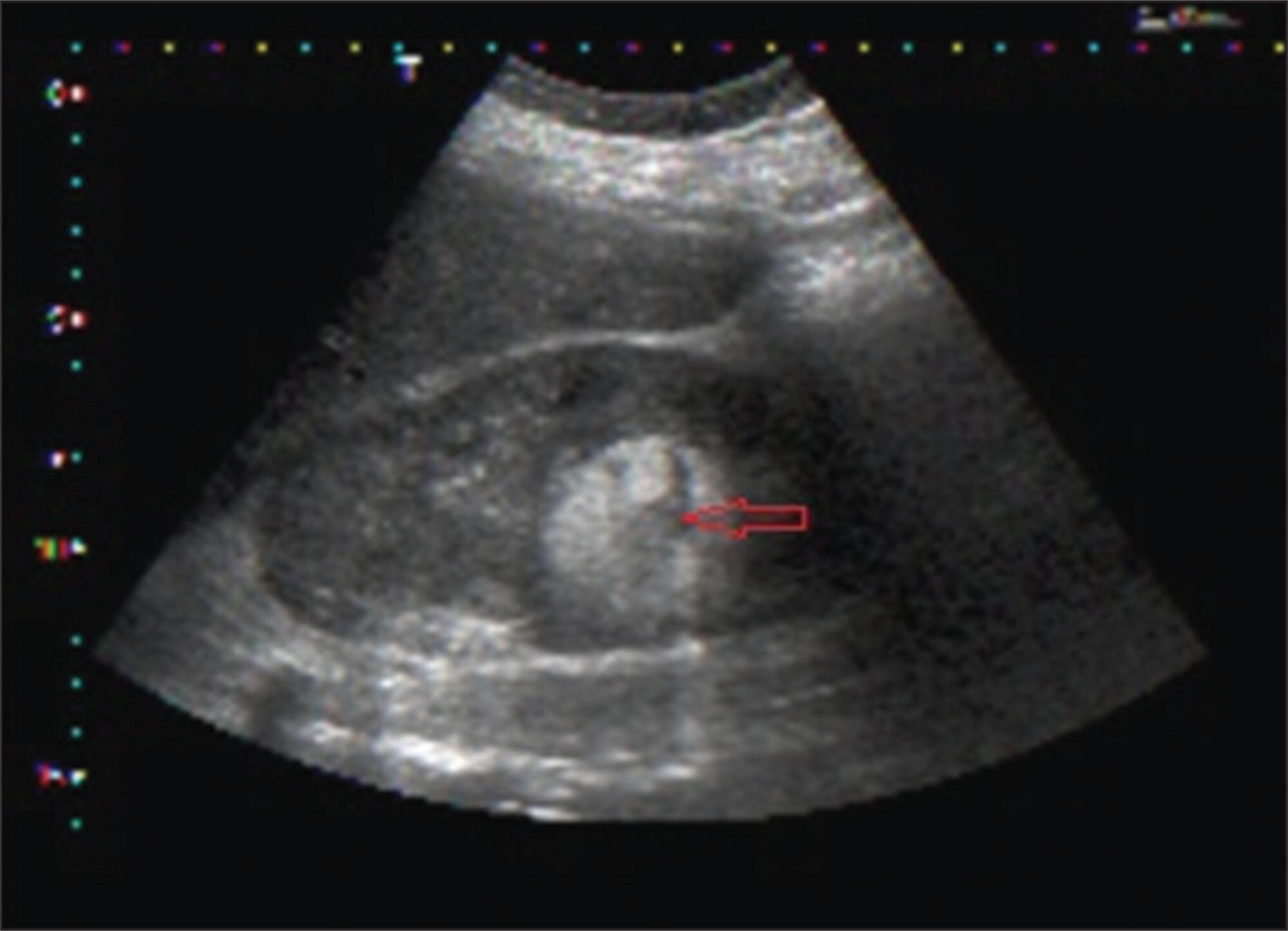
Renal vein thrombosis in the puerperium: case report
Revista Brasileira de Ginecologia e Obstetrícia. 2015;37(12):593-597
12-01-2015
Summary
Relato de CasoRenal vein thrombosis in the puerperium: case report
Revista Brasileira de Ginecologia e Obstetrícia. 2015;37(12):593-597
12-01-2015DOI 10.1590/S0100-720320150005455
Views67Abstract
Pregnancy and puerperium are periods of blood hypercoagulability and, therefore, of risk for thromboembolic events. Renal vein thrombosis is a serious and infrequent condition of difficult diagnosis. This study reported a case of renal vein thrombosis in the puerperium, and described the clinical case, risk factors, diagnostic methods, and treatment instituted.
Key-words Case reportsPostpartum periodPregnancy complications, hematologicThrombophiliaVenous thrombosisSee more -
Relato de Caso
Beta thalassemia major and pregnancy during adolescence: report of two cases
Revista Brasileira de Ginecologia e Obstetrícia. 2015;37(6):291-296
06-01-2015
Summary
Relato de CasoBeta thalassemia major and pregnancy during adolescence: report of two cases
Revista Brasileira de Ginecologia e Obstetrícia. 2015;37(6):291-296
06-01-2015DOI 10.1590/SO100-720320150005169
Views136See moreBeta thalassemia major is a rare hereditary blood disease in which impaired synthesis
of beta globin chains causes severe anemia. Medical treatment consists of chronic
blood transfusions and iron chelation. We describe two cases of adolescents with beta
thalassemia major with unplanned pregnancies and late onset of prenatal care. One had
worsening of anemia with increased transfusional requirement, fetal growth
restriction, and placental senescence. The other was also diagnosed with
hypothyroidism and low maternal weight, and was admitted twice during pregnancy due
to dengue shock syndrome and influenza H1N1-associated respiratory infection. She
also developed fetal growth restriction and underwent vaginal delivery at term
complicated by uterine hypotonia. Both patients required blood transfusions after
birth and chose medroxyprogesterone as a contraceptive method afterwards. This report
highlights the importance of medical advice on contraceptive methods for these women
and the role of a specialized prenatal follow-up in association with a
hematologist. -
Case Report
Spontaneous rupture of renal angiomyolipoma during pregnancy
Revista Brasileira de Ginecologia e Obstetrícia. 2014;36(8):377-380
08-01-2014
Summary
Case ReportSpontaneous rupture of renal angiomyolipoma during pregnancy
Revista Brasileira de Ginecologia e Obstetrícia. 2014;36(8):377-380
08-01-2014DOI 10.1590/SO100-720320140005019
Views136Renal angiomyolipoma is a benign tumor, composed of adipocytes, smooth muscle cells and blood vessels. The association with pregnancy is rare and related with an increased risk of complications, including rupture with massive retroperitoneal hemorrhage. The follow-up is controversial because of the lack of known cases, but the priorities are: timely diagnosis in urgent cases and a conservative treatment when possible. The mode of delivery is not consensual and should be individualized to each case. We report a case of a pregnant woman with 18 weeks of gestation admitted in the emergency room with an acute right low back pain with no other symptoms. The diagnosis of rupture of renal angiomyolipoma was established by ultrasound and, due to hemodinamically stability, conservative treatment with imaging and clinical monitoring was chosen. At 35 weeks of gestation, it was performed elective cesarean section without complications for both mother and fetus.
Key-words Angiomyolipoma/complicationsAngiomyolipoma/diagnosisCase reportsPregnancy complications, neoplasticRupture, spontaneousSee more
-
Relato de Caso
Endocervical schistosomiasis: case report
Revista Brasileira de Ginecologia e Obstetrícia. 2014;36(6):276-280
06-01-2014
Summary
Relato de CasoEndocervical schistosomiasis: case report
Revista Brasileira de Ginecologia e Obstetrícia. 2014;36(6):276-280
06-01-2014DOI 10.1590/S0100-720320140004827
Views83See moreSchistosomiasis mansoni is found in different endemic areas of Brazil. It is a serious public health problem in Brazil and worldwide. Ectopic forms of the disease may affect the female reproductive system, representing a rare type of Schistosoma mansoni infection. A 26-year-old patient complained of vaginal discharge, dyspareunia and pain on palpation of the hypogastrium. Gynecological examination revealed an endocervical polyp. A biopsy was performed. Under microscopy, several granulomas surrounding degenerate and viable eggs of Schistosoma mansoni were seen. Treated with praziquantel, she was asymptomatic after four weeks of treatment. Vaginal discharge and dyspareunia may be secondary causes of cervicitis caused by Schistosoma mansoni. The search for eggs in routine vaginal smear or histological examination should be part of the gynecologic evaluation of patients from endemic areas, with the purpose of tracking ectopic schistosomiasis of the female genital tract.
-
Case Report
Mondor’s disease in puerperium: case report
Revista Brasileira de Ginecologia e Obstetrícia. 2014;36(3):139-141
03-01-2014
Summary
Case ReportMondor’s disease in puerperium: case report
Revista Brasileira de Ginecologia e Obstetrícia. 2014;36(3):139-141
03-01-2014DOI 10.1590/S0100-72032014000300008
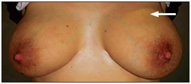
Views163See moreMondor's disease is a rare entity characterized by sclerosing thrombophlebitis classically involving one or more of the subcutaneous veins of the breast and anterior chest wall. It is usually a self-limited, benign condition, despite of rare cases of association to cancer. We present the case of a 32 year-old female, breast-feeding, who went to emergency due to left mastalgia for the past week. She was taking antibiotics and non-steroidal anti-inflammatory drugs, previously prescribed for suspicious of mastitis, for three days, with no clinical improvement. Physical examination showed an enlarged left breast, an axillary lump and a painful cord-like structure in the upper outer quadrant of the same breast. Ultrasound scan showed a markedly dilated superficial vein in the upper outer quadrant of left breast. The patient was given a ventropic therapy and was kept in anti-inflammatory, with progressive pain improvement. Ultrasound control was performed after four weeks, showing reperfusion.

-
Article
Aggressive angiomyxoma of the vagina: a case report
Revista Brasileira de Ginecologia e Obstetrícia. 2013;35(12):575-582
02-03-2013
Summary
ArticleAggressive angiomyxoma of the vagina: a case report
Revista Brasileira de Ginecologia e Obstetrícia. 2013;35(12):575-582
02-03-2013DOI 10.1590/S0100-72032013001200008
Views115See moreAggressive angiomyxoma is a rare, slow-growing soft tissue tumor that usually arises in the pelvis and perineal regions of women in reproductive age, with a marked tendency to local recurrence. Because of its rarity, it is often initially misdiagnosed. Surgical resection is the main treatment modality of aggressive angiomyxoma. We describe a case of a vaginal aggressive angiomyxoma in a 47-year-old woman in which the diagnosis was only made after histological examination. The etiology, presentation, diagnosis and management of this rare tumor are outlined. Angiomyxoma of vulva and vagina refers to a rare disease. Pre-operative diagnosis is difficult due to rarity and absence of diagnostic features, but it should be considered in every mass in genital, perianal and pelvic region in a woman in the reproductive age. Thus, these cases should have complete radiological workup before excision, as pre-diagnosis can change the treatment modality and patient prognosis'.
-
Case Report
Gunshot wound to the pregnant uterus: case report
Revista Brasileira de Ginecologia e Obstetrícia. 2013;35(9):427-431
11-06-2013
Summary
Case ReportGunshot wound to the pregnant uterus: case report
Revista Brasileira de Ginecologia e Obstetrícia. 2013;35(9):427-431
11-06-2013DOI 10.1590/S0100-72032013000900008
Views111See moreCrime and violence have become a public health problem. Pregnant women have not been the exception and gunshot injuries occupy an important place as a cause of trauma. An important fact is that pregnant women, who suffer trauma, are special patients because pregnancy causes physiological and anatomical changes. Management of these patients should be multidisciplinary, by the general surgeon, the obstetrician and the neonatologist. However, even trauma referral centers could neither have the staff nor the ideal training for these specific cases. In this context we present the following case.
-
Relato de Caso
Sclerosing stromal tumor of the ovary associated with a Meigs’ syndrome and pregnancy: a case report
Revista Brasileira de Ginecologia e Obstetrícia. 2013;35(7):331-335
09-27-2013
Summary
Relato de CasoSclerosing stromal tumor of the ovary associated with a Meigs’ syndrome and pregnancy: a case report
Revista Brasileira de Ginecologia e Obstetrícia. 2013;35(7):331-335
09-27-2013DOI 10.1590/S0100-72032013000700008
Views101The sclerosing stromal tumor of the ovary is an extremely rare benign tumor more common in young women and without specific symptoms in most cases. Less than 150 cases have been described, of which 8 were diagnosed during pregnancy. In this report, we describe the association between sclerosing stromal tumor of the ovary, Meigs' syndrome and elevated levels of CA-125 in term pregnancy.
Key-words AscitesCase reportsHydrothoraxMeigs syndromeOvarian neoplasmsOvaryPregnancySex cord-gonadal stromal tumorsSee more


