Summary
Revista Brasileira de Ginecologia e Obstetrícia. 2013;35(2):71-77
DOI 10.1590/S0100-72032013000200006
PURPOSE: To evaluate the anthropometric characteristics of morbidity and mortality of premature newborns (NB) of hypertensive mothers according to the presence or absence of flow (DZ) or reverse (DR) diastolic flow in the dopplervelocimetry of the umbilical artery. METHODS: A prospective study was conducted on preterm newborns of pregnant women with hypertension between 25 and 33 weeks of gestational age, submitted to umbilical artery Doppler study during the five days before delivery. Delivery occurred at Hospital Regional da Asa Sul, Brasília - Federal District, between November 1st, 2009 and October 31st, 2010. The infants were stratified into two groups according to the results of Doppler velocimetry: Gdz/dr=absent end-diastolic velocity waveform or reversed end-diastolic velocity waveform, and Gn=normal Doppler velocimetry. Anthropometric measurements at birth, neonatal morbidity, and mortality were compared between the two groups. RESULTS: We studied 92 infants, as follows: Gdz/dr=52 infants and Gn=40 infants. In Gdz/dr, the incidence of infants small for gestational age was significantly greater, with a relative risk of 2.5 (95%CI 1.7 - 3.7). In Gdz/dr, infants remained on mechanical ventilation for a longer time: median 2 (0‒28) and Gn median 0.5 (0‒25) p=0.03. The need for oxygen at 28 days was higher in G dz/dr comparing to Gn (33 versus 10%; p=0.01). Neonatal mortality was higher in Gdz/dr compared to Gn (36 versus 10%; p=0.03; relative risk of 1.6; 95%CI 1.2‒2.2). Logistic regression showed that, with each 100 grams lower birth weight, the chance of death increased 6.7 times in G dz/dr (95%CI 2.0 - 11.3; p<0.01). CONCLUSION: In preterm infants of mothers with hypertensive changes in Doppler velocimetry of the umbilical artery, intrauterine growth restriction and neonatal prognosis are often worse, with a high risk of death related to birth weight.
Summary
Revista Brasileira de Ginecologia e Obstetrícia. 2012;34(10):473-477
DOI 10.1590/S0100-72032012001000007
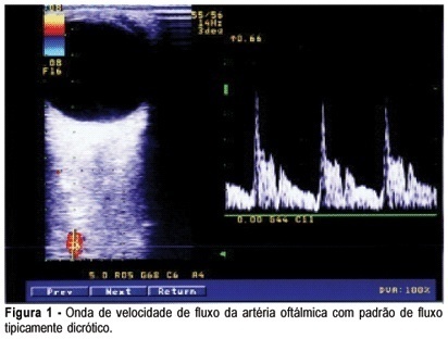
PURPOSES: To evaluate the hemodynamic patterns of the ophthalmic artery by Doppler analysis in women with gestational diabetes mellitus (GDM), comparing them to normal pregnant women. METHODS: A prospective case-control study that analyzed the ophthalmic artery Doppler indices in two groups: one consisting of 40 women diagnosed with GDM and the other of 40 normal pregnant women. Included were pregnant women with GDM criteria of the American Diabetes Association - 2012, with 27 weeks of pregnancy to term, and excluded were women with hypertension, use of vasoactive drugs on or previous diagnosis of diabetes. Doppler analysis was performed in one eye with a 10 MHz linear transducer and the Sonoace 8000 Live Medison® equipment . The following variables were analyzed: pulsatility index (PI), resistance index (RI), peak velocity ratio (PVR), peak systolic velocity (PSV) and end diastolic velocity (EDV). To analyze the normality of the samples we used the Lillefors test, and to compare means and medians we used the Student's t-test and Mann-Whitney test according to data normality, with the level of significance set at 95%. RESULTS: The median and mean values with standard deviation of the variables of the ophthalmic artery Dopplervelocimetry group GDM and normal pregnant women were: IP=1.7±0.6 and 1.6±0.4 (p=0.7); IR=0.7 and 0.7 (p=0.9); RPV=0.5±0.1 and 0.5±0.1 (p=0.1), PSV=33.6 and 31.9 cm/sec (p=0.7); VDF=6.3 and 7.9 cm/sec (p=0.4). There was no significant difference in the means and medians of these variables between the two groups of pregnant women. CONCLUSIONS: The ophthalmic artery hemodynamic patterns, analyzed by means of a Doppler technique remained unchanged in the group of pregnant women with GDM compared to the group of normal pregnant women, suggesting that the time of exposure to the disease during pregnancy was too short to cause significant vascular disorders in the central territory.
Summary
Revista Brasileira de Ginecologia e Obstetrícia. 2009;31(11):534-539
DOI 10.1590/S0100-72032009001100002
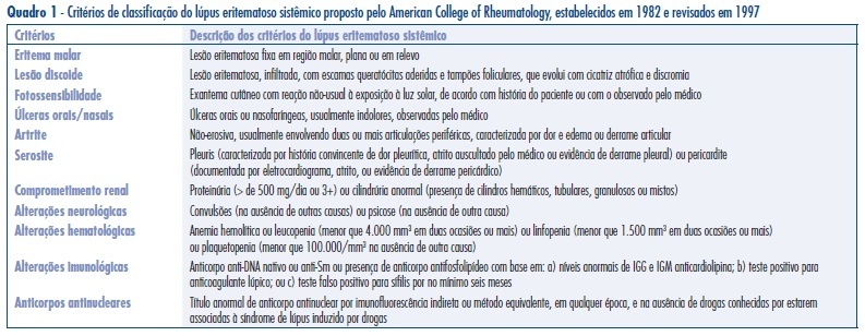
PURPOSE: to analyze the ophthalmic artery functioning in pregnant women with systemic lupus erythematosus (PL) without active renal disease as compared to non-pregnant women with lupus (NPL) without active renal disease, and to normal pregnant women (PN). METHODS: observational study that analyzed ophthalmic artery dopplervelocimetric variables of 20 PN, 10 PL and 17 NPL women. The variables analyzed were: pulsatility index (PI), final diastolic velocity (FDV) and velocity peak ratio (VPR). Mean and standard deviation of these indexes were calculated. For group mean comparison, analysis of variance (ANOVA) and the post-hoc Tukey test have been used, with confidence interval of 95% (p<0.05). RESULTS: the PN group showed the following means and standard deviations of ophthalmic artery parameters: PI=2,4±0,3; VPR=0,5±0,1 e FDV=5,1±2,1 cm/s. The PL and NPL groups showed the following values, respectively: PI=2,0±0,4 and 1,9±0,4; VPR=0,6±0,1 and 0,6±0,1; FDV=9,7±3,9 cm/s and 8,1±4,3 cm/s. There was not significant mean difference between the PL and NPL groups for PI, VPR or FDV. However, statistically significant mean differences were observed between PN and PL for PI, VPR and FDV, with higher values of FDV and VPR in the PL group. CONCLUSIONS: there was a reduction of ophthalmic artery vascular impedance with orbital hyperfusion in the two groups of women with lupus erythematosus as compared to normal pregnant women. These results may help to improve the understanding on pathophysiology of systemic lupus erythematosus. In addition, the present method may be applied in future studies as a complementary procedure for the differential diagnosis between pre-eclampsia and renal failure due to lupus.

Summary
Revista Brasileira de Ginecologia e Obstetrícia. 2008;30(10):494-498
DOI 10.1590/S0100-72032008001000003
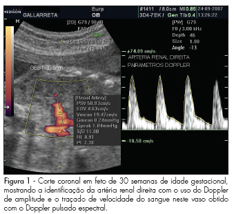
PURPOSE: to describe values found for the resistance index (RI), pulsatility index (PI) and the systole/diastole (S/D) ratio of fetal renal arteries in non-complicated gestations between the 22nd and the 38th week, and to evaluate whether those values vary along that period. METHODS: observational study, where 45 fetuses from non-complicated gestations have been evaluated in the 22nd, 26th, 30th and 38th weeks of gestational age. Doppler ultrasonography has been performed by the same observer, using a device with 4 to 7 MHz transducer. For the acquisition of the renal arteries velocity record, a 1 mm to 2 mm probe has been placed in the mean third of the renal artery for the evaluation through pulsed Doppler ultrasonography. The measurement of RI, PI and S/D ratio from three consecutive waves was performed with the automatic mode. To detect significant differences in the indexes' values along gestation, we have compared values obtained at the different gestational ages, through repeated measures ANOVA, followed by Tukey's post-hoc test. RESULTS: There were no significant differences between the right and left renal arteries, when the RI, IP and S/D ratio were compared. Nevertheless, a change in the values of these parameters has been observed between the 22nd week (RI=0.9 ± 0.02; PI=2.4 ± 0.02; S/D ratio=11.6 ± 2.2; mean ± standard deviation of the combined mean values of the right and left renal artery) and the 38th week (RI=0.8 ± 0.03; PI=2.1 ± 0.2; S/D ratio=8.7 ± 2.3) of gestation. CONCLUSIONS: the parameters evaluated (RI, PI and S/D ratio) have presented decreasing values between the 22nd and 38th, with no difference between the fetus's right and left sides.

Summary
Revista Brasileira de Ginecologia e Obstetrícia. 2008;30(1):5-11
DOI 10.1590/S0100-72032008000100002
PURPOSE: to study the value of Doppler velocimetry of the ductus venosus, between the 11th and 14th weeks of pregnancy, associated to the nuchal translucency thickness measurement, in the detection of adverse fetal outcome. METHODS: a transversal and prospective study in which a total of 1,268 fetuses were studied consecutively. In 56 cases, a cytogenetic study was performed on material obtained from a biopsy of the chorionic villus and, in 1,181 cases, the postnatal phenotype was used as a basis for the result. In addition to the routine ultrasonographic examination, all the fetuses were submitted to measurement of the nuchal translucency thickness and to Doppler velocimetry of the ductus venosus. Aiming at prevalence and accuracy indices, sensitivity, specificity, positive predictive value, negative predictive value, probability of false-positive, probability of false-negative, reason of positive probability and reason of negative probability were calculated and analyzed. RESULTS: from the total of 1,268 fetuses, 1,183 cases were selected for analysis. From this number, 1,170 fetuses were normal (98.9%) and 13 fetuses presented adverse outcome at birth (1.1%), including fetal death (trisomy 21 and 22) in two cases; genetic syndrome (Nooman) in one case; two cases of polymalformed fetuses; cardiopathy in three cases; and other structural defects in five cases. The prevalence of the modified ductus venosus (wave A zero/reverse) in the studied population was of 14 cases (1.2%), with a false-positive rate of 0.7%. CONCLUSIONS: there is a significant correlation between the alteration of the ductus venosus Doppler velocimetry and the thickness of the nuchal translucency as an ultrasonographic marker for the first trimester of gestation, in the detection of adverse fetal outcome, especially serious malformations. The ductus venosus was able to diminish the false-positive result in comparison to the isolated use of the nuchal translucency thickness, improving considerably the positive predictive value of the test.
Summary
Revista Brasileira de Ginecologia e Obstetrícia. 2005;27(7):387-392
DOI 10.1590/S0100-72032005000700004
PURPOSE: to assess peak systolic velocity (PSV) and the resistance index (RI) in the middle cerebral artery (MCA), suprarenal aorta (SRA) and infrarenal aorta (IRA) of the fetus and in the umbilical artery (UA) between the 22nd and 38th week of gestation. METHODS: a prospective study which evaluated the parameters of 33 normal fetuses in the 22nd, 26th, 30th, and 38th week of gestation. Pregnant women with a singleton fetus with no diseases or complications and who agreed to participate were included in the study. Exclusion criteria were fetal malformations, discontinuation of prenatal care visits and mothers who smoked, used alcohol or illicit drugs. Ultrasound examinations were performed by a single observer. For the acquisition of the Doppler velocimetry tracing in the MCA, SRA, IRA and UA, the sample volume was 1 to 2 mm, placed in the center of the arteries. The insonation angle was 5º to 19º in the MCA, below 45º in the SRA and IRA, and less than 60º in the UA. We used a wall filter of 50-100 Hz. The parameters were calculated automatically with the frozen image, three measurements being made. The final result was obtained by the arithmetic mean of the three values. Data were analyzed by analysis of variance (ANOVA), post hoc Bonferroni test, Pearson's correlation, and regression analysis. The level of significance was set at p<0.05 in all analyses. RESULTS: PSV increased from 26.3 to 57.7 cm/s in the MCA between the 22nd and the 38th week of gestation (p<0.05). In the SRA and in the IRA, PSV increased between the 22nd and 34th week of gestation, from 74.6 and 59.0 cm/s to 106.0 and 86.6 cm/s, respectively (p<0.05). In the UA, PSV increased between the 22nd and the 34th week of gestation, but decreased from 55.5 to 46.2 cm/s between the 34th and the 38th week of gestation. In the MCA, the RI was lower in the 22nd (0.81) and 38th week of gestation (0.75) and higher (0.85) in the 26th week (p<0.05). In the SRA, the RI values were stable in all weeks and in the IRA they were stable in most weeks (p>0.05). In the UA, RI decreased from 0.69 to 0.56 between the 22nd and 38th week of gestation (p<0.05). CONCLUSION: in normal fetuses, in the second half of gestation PSV increased in the MCA, SRA and IRA, decreasing in the UA between the 34th and 38th week of gestation. RI was lower in the 22nd and 38th weeks of gestation in the MCA, decreased between the 22nd and the 38th week in the UA, and was constant in most of the gestational weeks in the SRA and IRA.
Summary
Revista Brasileira de Ginecologia e Obstetrícia. 2005;27(4):168-173
DOI 10.1590/S0100-72032005000400002
PURPOSE: to evaluate ophthalmic and retinal central artery Doppler indices during the second and third trimesters of normal pregnancy and to compare the right with left eye Doppler indices of normotensive women. METHODS: a cross-sectional study which evaluated central retinal and ophthalmic artery Doppler velocimetry values of 51 normal pregnant women, in the 20th to 38th week of gestation. The following values were analyzed: pulsatility and resistance indexes (PI, RI), peak systolic and end-diastolic flow velocity (PSV, EDFV) and peak velocity ratio (PVR). The Doppler indices in the right and left eyes were studied by the median. The paired Student's t test was used to confront the right and left eye values and the Pearson linear correlation analysis was performed to study the value changes throughout the gestation, with the level of significance set at 5%. RESULTS: Doppler velocimetry indices of ophthalmic and central retinal arteries (median values) were, respectively: PI=1.83; RI=0.78; PSV=34.20; EDFV=6.80; PVR=0.48 and PI=1.34; RI=0.70; PSV=7.40; EDFV=2.10. There was no significant difference between the right and left side Doppler values. Linear correlation analysis showed no association between the arterial values and pregnancy age. CONCLUSION: the unilateral analysis of ophthalmic and central retinal artery Doppler velocimetry values can be used in systemic maternal disease. There is no significant change in ophthalmic and central retinal artery Doppler velocimetry values throughout normal pregnancy.
