Summary
Revista Brasileira de Ginecologia e Obstetrícia. 2008;30(11):573-582
DOI 10.1590/S0100-72032008001100008
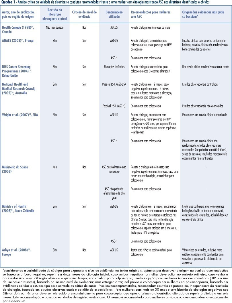
PURPOSE: to identify valid guidelines for the approach of women with cytopathological diagnosis of undetermined significance atypias in squamous cells (ASC), discussing its applicability to the Brazilian scenario. METHODS: an electronic search of publications at PubMed, National Guidelines Clearinghouse and Scholar Google was carried out, as well as a manual search of references from the texts found. The guidelines identified, and specifically related to the theme, were evaluated according to its validity and the recommendations were criticized and summarized. RESULTS: guidelines for the United Kingdom, France, Australia, the USA and New Zealand have been considered as valid. These documents recommend repeating the cytology in six or twelve months, in ASCs of undetermined significance (ASC-US) before referring to colposcopy, and immediate referral to colposcopy in ASCs, when it is not possible to disregard high degree lesions (ASC-H). We have also found valid colposcopy recommendations for women with ASC-US in special situations (immune deficient women requiring specialist assistance) and the use of oncogenic HPV test, which, when present in women over 20, should motivate referral to colposcopy. CONCLUSIONS: the clinical guidelines recommended for the Programa Nacional de Controle do Cancer do Colo do Útero in Brazil can be improved with the referral to colposcopy in special situations (immune deficient women requiring specialist assistance), the use of test for the detection of oncogenic HPV in women over 20 (when present, refer to colposcopy), the investigation of vaginal lesions, the use of estrogens before the colposcopy in post-menopausal women, and disregard biopsia in case of slighter alterations.
Summary
Revista Brasileira de Ginecologia e Obstetrícia. 2008;30(11):573-582
DOI 10.1590/S0100-72032008001100008
PURPOSE: to identify valid guidelines for the approach of women with cytopathological diagnosis of undetermined significance atypias in squamous cells (ASC), discussing its applicability to the Brazilian scenario. METHODS: an electronic search of publications at PubMed, National Guidelines Clearinghouse and Scholar Google was carried out, as well as a manual search of references from the texts found. The guidelines identified, and specifically related to the theme, were evaluated according to its validity and the recommendations were criticized and summarized. RESULTS: guidelines for the United Kingdom, France, Australia, the USA and New Zealand have been considered as valid. These documents recommend repeating the cytology in six or twelve months, in ASCs of undetermined significance (ASC-US) before referring to colposcopy, and immediate referral to colposcopy in ASCs, when it is not possible to disregard high degree lesions (ASC-H). We have also found valid colposcopy recommendations for women with ASC-US in special situations (immune deficient women requiring specialist assistance) and the use of oncogenic HPV test, which, when present in women over 20, should motivate referral to colposcopy. CONCLUSIONS: the clinical guidelines recommended for the Programa Nacional de Controle do Cancer do Colo do Útero in Brazil can be improved with the referral to colposcopy in special situations (immune deficient women requiring specialist assistance), the use of test for the detection of oncogenic HPV in women over 20 (when present, refer to colposcopy), the investigation of vaginal lesions, the use of estrogens before the colposcopy in post-menopausal women, and disregard biopsia in case of slighter alterations.
Summary
Revista Brasileira de Ginecologia e Obstetrícia. 2004;26(7):573-578
DOI 10.1590/S0100-72032004000700010
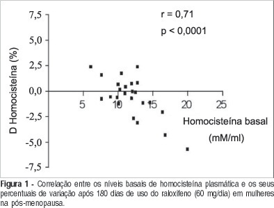
PURPOSE: to evaluate the effects of raloxifene on plasma homocysteine concentration and lipid profile in postmenopausal women. METHODS: twenty-four healthy postmenopausal women, aged 50 to 70 years, with osteopenia and/or osteoporosis, were submitted to raloxifene therapy, 60 mg/day, for six months. Plasma homocysteine concentration was determined before and after three and six months of therapy, as well as total cholesterol, HDL-cholesterol LDL-cholesterol and triglyceride levels. Plasma homocysteine was measured by a polarized immunofluorescence assay and serum lipids by the enzymatic and colorimetric method. Data were analyzed statistically by ANOVA, Newman-Keuls test and Pearson's correlation test. RESULTS: a significant decrease in total cholesterol of 15.3% (227.6±56.3 vs 200.6±29.8 vs 192.8±32.1 mg/dl; p<0.001) and LDL-cholesterol of 21.4% (151.4±46.3 vs 122.7±29.4 vs 119.0±28.6 mg/dl; p<0.001), and a significant increase in HDL-cholesterol of 9.5% (44.7±10.8 vs 52.2±12.6 vs 49.0±10.8 mg/dl; p<0.05) were observed. There was no reduction in triglyceride levels (134.9±50.3 vs 127.5±50.0 vs 121.0±36.0 mg/dl; p>0.05). Although not significant, a decrease in homocysteine by 4.5% (11.7±3.0 vs 11.0±2.9 vs 11.2±2.1 muM/l; p>0.05) was observed between the pre-and posttreatment periods, with a significant negative correlation between basal levels and posttreatment percentual reduction (r=-0.71; p<0.0001). CONCLUSIONS: raloxifene treatment, 60 mg/day, for six months caused a significant decrease in total and LDL-cholesterol and an increase in HDL-cholesterol in postmenopausal women. Plasma homocysteine concentration tended to decrease, this effect being more favorable in patients with elevated baseline levels.
Summary
Revista Brasileira de Ginecologia e Obstetrícia. 2004;26(7):573-578
DOI 10.1590/S0100-72032004000700010
PURPOSE: to evaluate the effects of raloxifene on plasma homocysteine concentration and lipid profile in postmenopausal women. METHODS: twenty-four healthy postmenopausal women, aged 50 to 70 years, with osteopenia and/or osteoporosis, were submitted to raloxifene therapy, 60 mg/day, for six months. Plasma homocysteine concentration was determined before and after three and six months of therapy, as well as total cholesterol, HDL-cholesterol LDL-cholesterol and triglyceride levels. Plasma homocysteine was measured by a polarized immunofluorescence assay and serum lipids by the enzymatic and colorimetric method. Data were analyzed statistically by ANOVA, Newman-Keuls test and Pearson's correlation test. RESULTS: a significant decrease in total cholesterol of 15.3% (227.6±56.3 vs 200.6±29.8 vs 192.8±32.1 mg/dl; p<0.001) and LDL-cholesterol of 21.4% (151.4±46.3 vs 122.7±29.4 vs 119.0±28.6 mg/dl; p<0.001), and a significant increase in HDL-cholesterol of 9.5% (44.7±10.8 vs 52.2±12.6 vs 49.0±10.8 mg/dl; p<0.05) were observed. There was no reduction in triglyceride levels (134.9±50.3 vs 127.5±50.0 vs 121.0±36.0 mg/dl; p>0.05). Although not significant, a decrease in homocysteine by 4.5% (11.7±3.0 vs 11.0±2.9 vs 11.2±2.1 muM/l; p>0.05) was observed between the pre-and posttreatment periods, with a significant negative correlation between basal levels and posttreatment percentual reduction (r=-0.71; p<0.0001). CONCLUSIONS: raloxifene treatment, 60 mg/day, for six months caused a significant decrease in total and LDL-cholesterol and an increase in HDL-cholesterol in postmenopausal women. Plasma homocysteine concentration tended to decrease, this effect being more favorable in patients with elevated baseline levels.
Summary
Revista Brasileira de Ginecologia e Obstetrícia. 2022;44(6):573-577
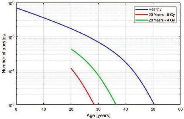
The present study aimed to develop a useful mathematical model that predicts the age at which premature ovarian insufficiency might occur after teletherapy radiation. A diagnosis of premature or early menopause has physical and psychological consequences, so women may need support and long-term medical follow-up.
To correlate ovarian radiation dose with ovarian function, we used the formula described by Wallace et al.: √g(z) = 10(2-0,15z), where “g(z)” and “z” represent oocyte survival rate and the radiation dose (in Gray), respectively. By simulating different ages and doses, we observed a pattern that could be used to simplify the relationship between radiation dose and remaining time of ovarian function.
We obtained a linear function between ovarian radiation dose and loss of ovarian function (LOF) that is the percentage of decrease in the time to the ovarian failure compared with the time expected for a woman at the same age without irradiation exposition. For patients <40 years old and with ovarian radiation doses < 5 Gy, the equation LOF = 2.70 + (11.08 × Dose) can be applied to estimate the decrease in time to premature ovarian insufficiency.
The present study reports a practicable theoretical method to estimate the loss of ovarian function. These findings can potentially improve the management and counseling of young women patients submitted to radiotherapy during their reproductive years.
Summary
Revista Brasileira de Ginecologia e Obstetrícia. 2022;44(6):573-577
The present study aimed to develop a useful mathematical model that predicts the age at which premature ovarian insufficiency might occur after teletherapy radiation. A diagnosis of premature or early menopause has physical and psychological consequences, so women may need support and long-term medical follow-up.
To correlate ovarian radiation dose with ovarian function, we used the formula described by Wallace et al.: √g(z) = 10(2-0,15z), where “g(z)” and “z” represent oocyte survival rate and the radiation dose (in Gray), respectively. By simulating different ages and doses, we observed a pattern that could be used to simplify the relationship between radiation dose and remaining time of ovarian function.
We obtained a linear function between ovarian radiation dose and loss of ovarian function (LOF) that is the percentage of decrease in the time to the ovarian failure compared with the time expected for a woman at the same age without irradiation exposition. For patients <40 years old and with ovarian radiation doses < 5 Gy, the equation LOF = 2.70 + (11.08 × Dose) can be applied to estimate the decrease in time to premature ovarian insufficiency.
The present study reports a practicable theoretical method to estimate the loss of ovarian function. These findings can potentially improve the management and counseling of young women patients submitted to radiotherapy during their reproductive years.
Summary
Revista Brasileira de Ginecologia e Obstetrícia. 2000;22(9):573-577
DOI 10.1590/S0100-72032000000900006
Purpose: to verify the frequency of cervical intraepithelial neoplasia in human immunodeficiency virus (HIV) ¾ infected women. Methods: ninety-nine HIV-seropositive women were studied. The diagnosis of the HIV infection was established through two ELISA tests complemented by Western blot test or indirect immunofluorescence test. As control group, 104 women whose ELISA test was not positive were analyzed. The investigation of cervical intraepithelial neoplasia was achieved by association of Pap smear and colposcopy in both groups. In the cases where colposcopy revealed existence of abnormal transformation zones, NIC diagnosis was obtained through colposcopy-guided biopsy complemented or not by conization. Results: cervical intraepithelial neoplasia was found in 15 of the 99 patients (15.2%), and among them there were ten NIC I, one NIC II and four NIC III. Among the 104 women of the control group, four presented cervical intraepithelial neoplasia (3.8%), one being NIC I and three NIC III. Conclusion: the comparative analysis of the results showed that the frequency of cervical intraepithelial neoplasia was significantly higher among those patients infected with HIV.
Summary
Revista Brasileira de Ginecologia e Obstetrícia. 2009;31(11):574-580
DOI 10.1590/S0100-72032009001100008
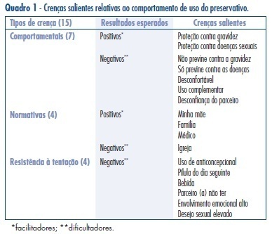
PURPOSE: to investigate factors that motivate safe sex practice, searching for antecedents of the intention to use condom among the population of young students in Belo Horizonte. METHODS: a survey based on the Theory of Planned Behavior (TPB) has been carried out in a sample of 732 students, with ages from 18 to 19 years old. Using the multiple regression analysis on data obtained from an anonymous questionnaire, the importance of antecedents of the intention to use condom, such as: attitude, subjective norm, moral norm, resistance to temptation and perceived control, was investigated. Differences in behavior and attitudes between high and low social classes and between men and women were also assessed, through the t-test for means' comparison between independent samples. RESULTS: in the overall sample, the significant association of attitude and behavioral intention was not detected In the TPB, a higher percent of the intention variance was explained when only one of the partners was responsible for the decision of using the condom (intention-me), than when it was a joint decision of the couple (intention-us). There has been no significant difference between high and low social class groups, but differences have been found between men and women. Men have shown less resistance to the temptation of not using condom. In the evaluation of social pressure (subjective norm), medical doctors and mothers seem to have more influence on the intention to use condom, especially among women. The inclusion of the moral norm antecedent has increased the explained variance in the intention to use condom from 22 to 31%. CONCLUSIONS: attitude differences between men, less resistant to the temptation of not using condom, and women, who highlight the importance of gynecologists and parents' influence in advising about safe sex, may guide campaigns to promote the regular use of condoms.
Summary
Revista Brasileira de Ginecologia e Obstetrícia. 2009;31(11):574-580
DOI 10.1590/S0100-72032009001100008
PURPOSE: to investigate factors that motivate safe sex practice, searching for antecedents of the intention to use condom among the population of young students in Belo Horizonte. METHODS: a survey based on the Theory of Planned Behavior (TPB) has been carried out in a sample of 732 students, with ages from 18 to 19 years old. Using the multiple regression analysis on data obtained from an anonymous questionnaire, the importance of antecedents of the intention to use condom, such as: attitude, subjective norm, moral norm, resistance to temptation and perceived control, was investigated. Differences in behavior and attitudes between high and low social classes and between men and women were also assessed, through the t-test for means' comparison between independent samples. RESULTS: in the overall sample, the significant association of attitude and behavioral intention was not detected In the TPB, a higher percent of the intention variance was explained when only one of the partners was responsible for the decision of using the condom (intention-me), than when it was a joint decision of the couple (intention-us). There has been no significant difference between high and low social class groups, but differences have been found between men and women. Men have shown less resistance to the temptation of not using condom. In the evaluation of social pressure (subjective norm), medical doctors and mothers seem to have more influence on the intention to use condom, especially among women. The inclusion of the moral norm antecedent has increased the explained variance in the intention to use condom from 22 to 31%. CONCLUSIONS: attitude differences between men, less resistant to the temptation of not using condom, and women, who highlight the importance of gynecologists and parents' influence in advising about safe sex, may guide campaigns to promote the regular use of condoms.
Summary
Revista Brasileira de Ginecologia e Obstetrícia. 2019;41(9):575-578
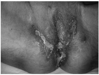
Tuberculosis is an infectious disease caused by Mycobacterium tuberculosis. According to data from the World Health Organization, this disease remains one of the leading causes of death worldwide. Although it most commonly affects the lungs, tuberculosis can compromise any organ. The present study reports a rare case of vulvar tuberculosis in a postmenopausal woman with a history of asymptomatic pulmonary and pleural tuberculosis, with no prior documented contact with the bacillus. Diagnosis was based on vulvar lesion biopsies, with histological findings suggestive of infection and isolation of M. tuberculosis by microbiological culture and polymerase chain reaction (PCR) essays. The lesions reverted to normal after tuberculostatic therapy.
Summary
Revista Brasileira de Ginecologia e Obstetrícia. 2019;41(9):575-578
Tuberculosis is an infectious disease caused by Mycobacterium tuberculosis. According to data from the World Health Organization, this disease remains one of the leading causes of death worldwide. Although it most commonly affects the lungs, tuberculosis can compromise any organ. The present study reports a rare case of vulvar tuberculosis in a postmenopausal woman with a history of asymptomatic pulmonary and pleural tuberculosis, with no prior documented contact with the bacillus. Diagnosis was based on vulvar lesion biopsies, with histological findings suggestive of infection and isolation of M. tuberculosis by microbiological culture and polymerase chain reaction (PCR) essays. The lesions reverted to normal after tuberculostatic therapy.
Summary
Revista Brasileira de Ginecologia e Obstetrícia. 2013;35(12):575-582
DOI 10.1590/S0100-72032013001200008
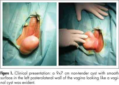
Aggressive angiomyxoma is a rare, slow-growing soft tissue tumor that usually arises in the pelvis and perineal regions of women in reproductive age, with a marked tendency to local recurrence. Because of its rarity, it is often initially misdiagnosed. Surgical resection is the main treatment modality of aggressive angiomyxoma. We describe a case of a vaginal aggressive angiomyxoma in a 47-year-old woman in which the diagnosis was only made after histological examination. The etiology, presentation, diagnosis and management of this rare tumor are outlined. Angiomyxoma of vulva and vagina refers to a rare disease. Pre-operative diagnosis is difficult due to rarity and absence of diagnostic features, but it should be considered in every mass in genital, perianal and pelvic region in a woman in the reproductive age. Thus, these cases should have complete radiological workup before excision, as pre-diagnosis can change the treatment modality and patient prognosis'.
Summary
Revista Brasileira de Ginecologia e Obstetrícia. 2013;35(12):575-582
DOI 10.1590/S0100-72032013001200008
Aggressive angiomyxoma is a rare, slow-growing soft tissue tumor that usually arises in the pelvis and perineal regions of women in reproductive age, with a marked tendency to local recurrence. Because of its rarity, it is often initially misdiagnosed. Surgical resection is the main treatment modality of aggressive angiomyxoma. We describe a case of a vaginal aggressive angiomyxoma in a 47-year-old woman in which the diagnosis was only made after histological examination. The etiology, presentation, diagnosis and management of this rare tumor are outlined. Angiomyxoma of vulva and vagina refers to a rare disease. Pre-operative diagnosis is difficult due to rarity and absence of diagnostic features, but it should be considered in every mass in genital, perianal and pelvic region in a woman in the reproductive age. Thus, these cases should have complete radiological workup before excision, as pre-diagnosis can change the treatment modality and patient prognosis'.
Summary
Revista Brasileira de Ginecologia e Obstetrícia. 2012;34(12):575-581
DOI 10.1590/S0100-72032012001200008
PURPOSE: To compare serum anti-Mullerian hormone (AMH) levels on the seventh day of ovarian stimulation between normal and poor responders. METHODS: Nineteen women aged ≥35, presenting with regular menses, submitted to ovarian stimulation for assisted reproduction were included. Women with endometriosis, polycystic ovarian syndrome or those who were previously submitted to ovarian surgery were excluded. On the basal and seventh day of ovarian stimulation, a peripheral blood sample was drawn for the determination of AMH, FSH and estradiol levels. AMH levels were assessed by ELISA and FSH, and estradiol by immunochemiluminescence. At the end of the stimulation cycle patients were classified as normal responders (if four or more oocytes were obtained during oocyte retrieval) or poor responders (if less than four oocytes were obtained during oocyte retrieval or if the cycle was cancelled due to failure of ovulation induction) and comparatively analyzed by the t-test for hormonal levels, length of ovarian stimulation, number of follicles retrieved, and number of produced and transferred embryos. The association between AMH and these parameters was also analyzed by the Spearman correlation test. RESULTS: There was no significant difference between groups for basal or the seventh day as to AMH, FSH and estradiol levels. There was a significant correlation between seventh day AMH levels and the total amount of exogenous FSH used (p=0.02). CONCLUSIONS: AMH levels on the seventh day of the ovarian stimulation cycle do not seem to predict the pattern of ovarian response and their determination is not recommended for this purpose.