You searched for:"Edmund Chada Baracat"
We found (71) results for your search.Summary
Rev Bras Ginecol Obstet. 1999;21(5):281-284
DOI 10.1590/S0100-72031999000500006
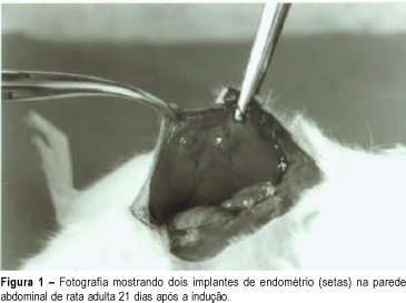
Purpose: to demonstrate the experimental endometriosis induction in animals. Method: we used adult female Wistar rats weighing 200 - 250 g anesthetized with ethyl ether to open the abdominal cavity. After identifying the uterine horns, we removed an approximately 4 cm fragment from the right uterine horn. This fragment was placed in physiological saline and, with the aid of a stereoscopic magnifying glass, the endometrium was separated from the myometrium and cut into rectangles of approximately 4 x 5 mm. These rectangles were fastened to the lateral abdominal wall near great blood vessels, taking care that the free portion of the endometrium was directed towards the lumen of the abdominal cavity. After 21 days the animals were again operated to observe the size of the implants and to remove the ectopic endometrium for microscopic analysis. Results: we macroscopically observed a significant growth of the endometrial implants. Microscopic examination showed presence of glandular epithelium and stroma similar to topic epithelium. Conclusion: this model reproduces endometriosis in the female rat allowing a better study of this pathology, mainly the action of drugs on these implants.
Summary
Rev Bras Ginecol Obstet. 1999;21(5):281-284
DOI 10.1590/S0100-72031999000500006
Purpose: to demonstrate the experimental endometriosis induction in animals. Method: we used adult female Wistar rats weighing 200 - 250 g anesthetized with ethyl ether to open the abdominal cavity. After identifying the uterine horns, we removed an approximately 4 cm fragment from the right uterine horn. This fragment was placed in physiological saline and, with the aid of a stereoscopic magnifying glass, the endometrium was separated from the myometrium and cut into rectangles of approximately 4 x 5 mm. These rectangles were fastened to the lateral abdominal wall near great blood vessels, taking care that the free portion of the endometrium was directed towards the lumen of the abdominal cavity. After 21 days the animals were again operated to observe the size of the implants and to remove the ectopic endometrium for microscopic analysis. Results: we macroscopically observed a significant growth of the endometrial implants. Microscopic examination showed presence of glandular epithelium and stroma similar to topic epithelium. Conclusion: this model reproduces endometriosis in the female rat allowing a better study of this pathology, mainly the action of drugs on these implants.
Summary
Rev Bras Ginecol Obstet. 2004;26(4):289-294
DOI 10.1590/S0100-72032004000400004
OBJECTIVE: to study the relationship of biobehavioral factors, such as age, menarche, number of gestations, and age of first sexual intercourse, with changes in Langerhans'cells in women with negative hybrid capture for HPV. METHODS: thirty women referred due to abnormal cervical cytology or premalignant cervical lesions were studied and underwent colposcopy, guided biopsy and histopathological exams. The Langerhans' cells were identified by immunohistochemical (S100+) exams. Langerhans' cells visualized in brown color were counted using the software Cytoviewer. The nonparametric Wilcoxon rank-sum test was employed for statistical analysis. RESULTS: the number of Langerhans' cells in women who had menarche after 13 years old presented statistically significant difference (173.34 cell/mm²) compared to the group whose menarche occurred before 13 (271.41 cell/mm²). The age at the first sexual intercourse was associated with the low number of Langerhans' cells, 127.15 cell/mm² and 250.14 cell/mm², respectively, for the beginning of the sexual activity up to 17 years old and after 17 (p=0.03). Previous cauterizations of the uterine cervix have been related to a lower number of Langerhans' cells in the epithelium, with the average 120.30 cell/mm² as compared to 236.06 cell/mm² for those women who never underwent that procedure (p=0.05). Other factors such as the patient's age and the number of gestations showed no statistically significant differences in the density of Langerhans' cells. CONCLUSIONS: the present study reports the association of biobehavioral factors with decrease in the number of Langerhans' cell.
Summary
Rev Bras Ginecol Obstet. 2022;44(3):295-303
Endometriosis is an inflammatory disease that affects women of reproductive age, causing pain and the possibility of infertility. Endometriosis was associated to low life quality and research shows the impact of endometriosis in several areas of life, justifying how these patients are more likely to develop depression, anxiety, and stress.
The aim of the present systematic review was to explore the field of psychology in endometriosis, identifying studies that used the cognitive behavioral therapy technique as a treatment for endometriosis and chronic pelvic pain.
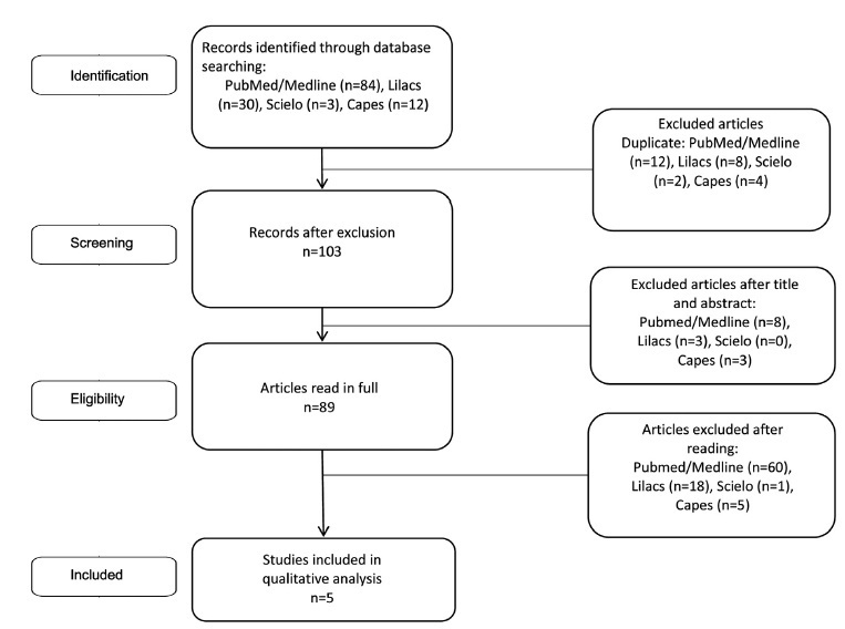
The keywords used were Endometriosis and Behavioral Therapy; Behavioral Disciplines and Activities; Cognitive Behavioral Therapy; Mental Health; Psychological Techniques; Psychology; Psychotherapy; Mental Health Services; and the search was performed in the following databases: PubMed/Medline, Scielo, Lilacs, and Capes. The study followed the PRISMA guidelines and all studies whose intervention strategy used was related to cognitive-behavioral therapy were considered.
Of the 129 articles found, only 5 were selected, and it was possible to identify that the psychological intervention whose approach brought cognitive-behavioral therapy techniques promoted a decrease in the sensation of pain, improvements in the scores of depression and stress, and significant changes in aspects of quality of life such as vitality, physical and social functioning, emotional well-being, control, and autonomy.
Cognitive-behavioral therapy can be very promising to take care of the emotional side of those who have endometriosis However, the present systematic review highlights the need to develop more structured studies with consistent, clear and replicablemethods to reach a psychological intervention protocol for patients who live with this gynecological-physical-emotional condition.
Summary
Rev Bras Ginecol Obstet. 2022;44(3):295-303
Endometriosis is an inflammatory disease that affects women of reproductive age, causing pain and the possibility of infertility. Endometriosis was associated to low life quality and research shows the impact of endometriosis in several areas of life, justifying how these patients are more likely to develop depression, anxiety, and stress.
The aim of the present systematic review was to explore the field of psychology in endometriosis, identifying studies that used the cognitive behavioral therapy technique as a treatment for endometriosis and chronic pelvic pain.
The keywords used were Endometriosis and Behavioral Therapy; Behavioral Disciplines and Activities; Cognitive Behavioral Therapy; Mental Health; Psychological Techniques; Psychology; Psychotherapy; Mental Health Services; and the search was performed in the following databases: PubMed/Medline, Scielo, Lilacs, and Capes. The study followed the PRISMA guidelines and all studies whose intervention strategy used was related to cognitive-behavioral therapy were considered.
Of the 129 articles found, only 5 were selected, and it was possible to identify that the psychological intervention whose approach brought cognitive-behavioral therapy techniques promoted a decrease in the sensation of pain, improvements in the scores of depression and stress, and significant changes in aspects of quality of life such as vitality, physical and social functioning, emotional well-being, control, and autonomy.
Cognitive-behavioral therapy can be very promising to take care of the emotional side of those who have endometriosis However, the present systematic review highlights the need to develop more structured studies with consistent, clear and replicablemethods to reach a psychological intervention protocol for patients who live with this gynecological-physical-emotional condition.
Summary
Rev Bras Ginecol Obstet. 2015;37(7):308-313
DOI 10.1590/S0100-720320150005343
To estimate the likelihood of axillary lymph node involvement for patients with early-stage breast cancer, based on a variety of clinical and pathological factors.
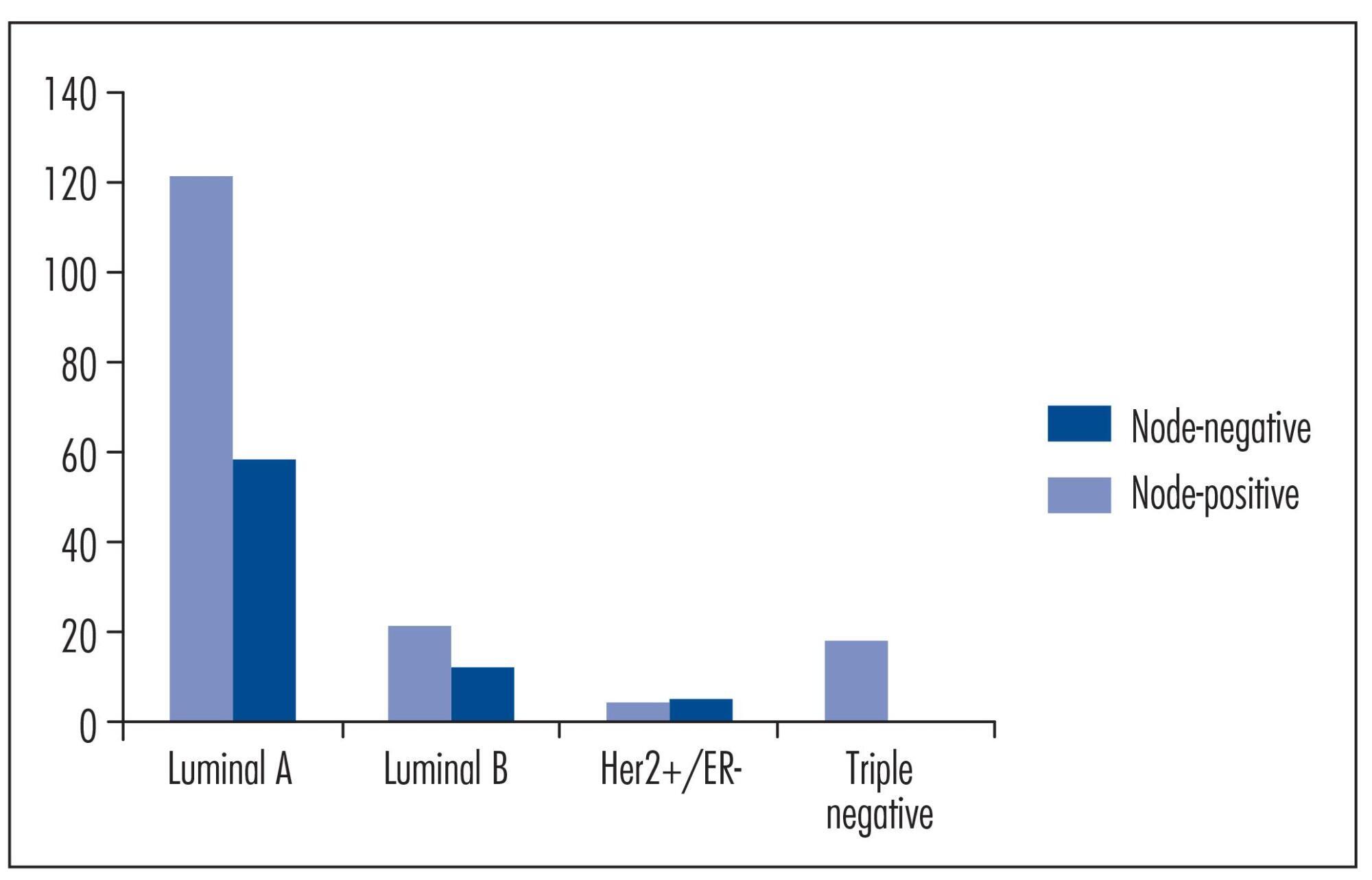
A retrospective analysis was done in hospital databases from 1999 to 2007. Two hundred thirty-nine patients were diagnosed with early-stage breast cancer. Predictive factors, such as patient age, tumor size, lymphovascular invasion, histological grade and immunohistochemical subtype were analyzed to identify variables that may be associated with axillary lymph node metastasis.
Patients with tumors that are negative for estrogen receptor, progesterone receptor, and HER2 had approximately a 90% lower chance of developing lymph node metastasis than those with luminal A tumors (e.g., ER+ and/or PR+ and HER2-) - Odds Ratio: 0.11; 95% confidence interval: 0.01-0.88; p=0.01. Furthermore, the risk for lymph node metastasis of luminal A tumors seemed to decrease as patient age increased, and it was directly correlated with tumor size.
The molecular classification of early-stage breast cancer using immunohistochemistry may help predicting the probability of developing axillary lymph node metastasis. Further studies are needed to optimize predictions for nodal involvement, with the aim of aiding the decision-making process for breast cancer treatment.
Summary
Rev Bras Ginecol Obstet. 2015;37(7):308-313
DOI 10.1590/S0100-720320150005343
To estimate the likelihood of axillary lymph node involvement for patients with early-stage breast cancer, based on a variety of clinical and pathological factors.
A retrospective analysis was done in hospital databases from 1999 to 2007. Two hundred thirty-nine patients were diagnosed with early-stage breast cancer. Predictive factors, such as patient age, tumor size, lymphovascular invasion, histological grade and immunohistochemical subtype were analyzed to identify variables that may be associated with axillary lymph node metastasis.
Patients with tumors that are negative for estrogen receptor, progesterone receptor, and HER2 had approximately a 90% lower chance of developing lymph node metastasis than those with luminal A tumors (e.g., ER+ and/or PR+ and HER2-) - Odds Ratio: 0.11; 95% confidence interval: 0.01-0.88; p=0.01. Furthermore, the risk for lymph node metastasis of luminal A tumors seemed to decrease as patient age increased, and it was directly correlated with tumor size.
The molecular classification of early-stage breast cancer using immunohistochemistry may help predicting the probability of developing axillary lymph node metastasis. Further studies are needed to optimize predictions for nodal involvement, with the aim of aiding the decision-making process for breast cancer treatment.
Summary
Rev Bras Ginecol Obstet. 2001;23(5):313-319
DOI 10.1590/S0100-72032001000500007
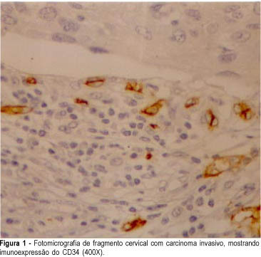
Purpose: to compare the efficiency of anti-factor VIII and anti-CD34 antibodies as vascular makers in cervical cancer, in cervical intraepithelial neoplasia and in normal cervix. Methods: using an immunohistochemical method, factor VIII-related antigen and leukocyte antigen CD34, we performed microvascular counts in 18 squamous cell carcinomas, in 15 cervical high-grade intraepithelial neoplasia, in 15 low-grade intraepithelial lesions and in 10 normal cervices. Using light microscopy we counted microvessels per 400X field in the most active areas of neovascularization with higher microvessel density in each case. Results: the average of microvessels stained with anti-CD34 in invasive carcinoma, high-grade intraepithelial lesions, low-grade intraepithelial lesions and in the normal cervices was 154, 134, 112 and 93, respectively. When we used anti-factor VIII the average was 56, 44, 33 and 30 vessels, following the same order. High-grade intraepithelial lesions and invasive carcinomas showed greater means number of vessels than normal tissue. Conclusions: the use of anti-CD34 allowed the detection of a greater number of vessels when compared to anti-factor VIII. However, we could observe that anti-factor VIII staining was able to significantly discriminate high-grade from low-grade lesions.
Summary
Rev Bras Ginecol Obstet. 2001;23(5):313-319
DOI 10.1590/S0100-72032001000500007
Purpose: to compare the efficiency of anti-factor VIII and anti-CD34 antibodies as vascular makers in cervical cancer, in cervical intraepithelial neoplasia and in normal cervix. Methods: using an immunohistochemical method, factor VIII-related antigen and leukocyte antigen CD34, we performed microvascular counts in 18 squamous cell carcinomas, in 15 cervical high-grade intraepithelial neoplasia, in 15 low-grade intraepithelial lesions and in 10 normal cervices. Using light microscopy we counted microvessels per 400X field in the most active areas of neovascularization with higher microvessel density in each case. Results: the average of microvessels stained with anti-CD34 in invasive carcinoma, high-grade intraepithelial lesions, low-grade intraepithelial lesions and in the normal cervices was 154, 134, 112 and 93, respectively. When we used anti-factor VIII the average was 56, 44, 33 and 30 vessels, following the same order. High-grade intraepithelial lesions and invasive carcinomas showed greater means number of vessels than normal tissue. Conclusions: the use of anti-CD34 allowed the detection of a greater number of vessels when compared to anti-factor VIII. However, we could observe that anti-factor VIII staining was able to significantly discriminate high-grade from low-grade lesions.
Summary
Rev Bras Ginecol Obstet. 1999;21(6):317-321
DOI 10.1590/S0100-72031999000600003
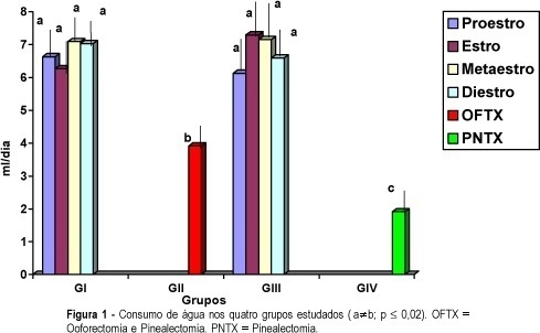
Purpose: to evaluate the effects of oophorectomy and pinealectomy on the ingestion of water and NaCl solution by adults female rats. Methods: forty-eight adult virgin female rats (Wistar EPM 1) weighing 200 g were kept on routine laboratory care and fed water and Purina rat chow ad libitum. The animals were random by divided into four groups: GI - maintained without manipulation as a control group (n = 20); GII - submitted to bilateral oophorectomy (n = 8); GIII - submitted to pinealectomy (n = 12); GIV - submitted to bilateral oophorectomy and pinealectomy (n = 8). All animals were maintained in individual cages. After three weeks the cycle phase was daily determined by vaginal smears and the volume of water and NaCl (0.25 M) solution was daily recorded for approximately three weeks. Results: the main results were: 1) rats submitted to pinealectomy alone presented a greater frequency of the estrous phase, some of these undergoing persistent estrus; 2) the liquid ingestion (water and saline solution) did not alter during the phases of the estrous cycle; 3) rats submitted to oophorectomy presented greater water ingestion and after pinealectomy water consumption returned to normal levels; 4) the oophorectomized and pinealectomized animals and those only oophorectomized showed reduction in the average consumption of saline solution. Conclusions: the data suggest that the ovaries and the pineal gland could have effects on the ingestion of salt and water in adult rats.
Summary
Rev Bras Ginecol Obstet. 1999;21(6):317-321
DOI 10.1590/S0100-72031999000600003
Purpose: to evaluate the effects of oophorectomy and pinealectomy on the ingestion of water and NaCl solution by adults female rats. Methods: forty-eight adult virgin female rats (Wistar EPM 1) weighing 200 g were kept on routine laboratory care and fed water and Purina rat chow ad libitum. The animals were random by divided into four groups: GI - maintained without manipulation as a control group (n = 20); GII - submitted to bilateral oophorectomy (n = 8); GIII - submitted to pinealectomy (n = 12); GIV - submitted to bilateral oophorectomy and pinealectomy (n = 8). All animals were maintained in individual cages. After three weeks the cycle phase was daily determined by vaginal smears and the volume of water and NaCl (0.25 M) solution was daily recorded for approximately three weeks. Results: the main results were: 1) rats submitted to pinealectomy alone presented a greater frequency of the estrous phase, some of these undergoing persistent estrus; 2) the liquid ingestion (water and saline solution) did not alter during the phases of the estrous cycle; 3) rats submitted to oophorectomy presented greater water ingestion and after pinealectomy water consumption returned to normal levels; 4) the oophorectomized and pinealectomized animals and those only oophorectomized showed reduction in the average consumption of saline solution. Conclusions: the data suggest that the ovaries and the pineal gland could have effects on the ingestion of salt and water in adult rats.
Summary
Rev Bras Ginecol Obstet. 2012;34(7):323-328
DOI 10.1590/S0100-72032012000700006
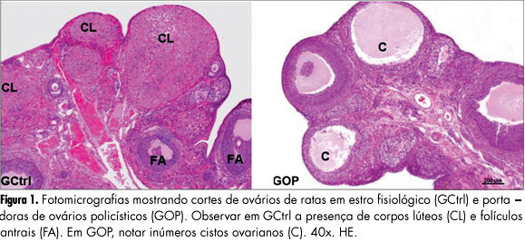
PURPOSES: To evaluate the histomorphometry of ovarian interstitial cells, as well as the blood sex steroid concentrations of female rats with polycystic ovaries induced by continuous light. METHODS: Twenty female rats were divided into two groups: Control Group - in the estrous phase (CtrlG), and a group of rats with polycystic ovaries induced by continuous illumination (POG). CtrlG animals were maintained on a light period from 07:00 a.m. to 07:00 p.m., and POG animals with continuous illumination (400 Lux) for 60 days. After this period all animals were anesthetized and blood was collected for the determination of serum estradiol (E2), progesterone (P4), and testosterone (T), followed by removal of the ovaries that were fixed in 10% formalin and processed for paraffin embedding. Five-µm histological sections were stained with hematoxylin and eosin and used for histomorphometric analysis. Morphological analyses, cyst count, determination of concentration and of the nuclear volume of interstitial cells were performed with the aid of a light microscope adapted to a high resolution camera (AxioCam), whose images were transmitted to and analyzed by the computer using AxioVision Rel 4.8 software (Carl Zeiss). Data were analyzed statistically by the Student's t-test (p<0.05). RESULTS: Morphological analysis showed the presence of ovarian cysts in POG animals and corpora lutea in CtrlG animals, as well as evidence of the origin of interstitial cells from the internal theca of these cysts. POG animals presented increased serum estradiol levels (pg/mL) compared to CtrlG animals (POG=124.9±4.2>CtrlG=73.2±6.5, p<0.05), the same occurring with testosterone levels (pg/mL) (POG=116.9±4.6>CtrlG=80.6±3.9, p<0.05). However, progesterone levels (ng/mL) were higher in CtrlG than in POG animals (CtrlG=16.3±2.0>POG=4.2±1.5, p<0.05). Morphometry showed a significant increase in nuclear volume in POG animals (POG=102.1±5.2>CtrlG=63.6±16.5, p<0.05), as well as in the area occupied (%) by interstitial cells (POG=24.4±6.9>CtrlG=6.9±3.2, p<0.05) compared to CtrlG animals. CONCLUSION: The interstitial cells of the rat polycystic ovary probably originate from ovarian cysts due to the degeneration of granulosa cells and differentiation of the internal theca cells. The elevations of serum testosterone and estradiol were probably due to the significant increase in cell activity and in the area occupied by interstitial cells.
Summary
Rev Bras Ginecol Obstet. 2012;34(7):323-328
DOI 10.1590/S0100-72032012000700006
PURPOSES: To evaluate the histomorphometry of ovarian interstitial cells, as well as the blood sex steroid concentrations of female rats with polycystic ovaries induced by continuous light. METHODS: Twenty female rats were divided into two groups: Control Group - in the estrous phase (CtrlG), and a group of rats with polycystic ovaries induced by continuous illumination (POG). CtrlG animals were maintained on a light period from 07:00 a.m. to 07:00 p.m., and POG animals with continuous illumination (400 Lux) for 60 days. After this period all animals were anesthetized and blood was collected for the determination of serum estradiol (E2), progesterone (P4), and testosterone (T), followed by removal of the ovaries that were fixed in 10% formalin and processed for paraffin embedding. Five-µm histological sections were stained with hematoxylin and eosin and used for histomorphometric analysis. Morphological analyses, cyst count, determination of concentration and of the nuclear volume of interstitial cells were performed with the aid of a light microscope adapted to a high resolution camera (AxioCam), whose images were transmitted to and analyzed by the computer using AxioVision Rel 4.8 software (Carl Zeiss). Data were analyzed statistically by the Student's t-test (p<0.05). RESULTS: Morphological analysis showed the presence of ovarian cysts in POG animals and corpora lutea in CtrlG animals, as well as evidence of the origin of interstitial cells from the internal theca of these cysts. POG animals presented increased serum estradiol levels (pg/mL) compared to CtrlG animals (POG=124.9±4.2>CtrlG=73.2±6.5, p<0.05), the same occurring with testosterone levels (pg/mL) (POG=116.9±4.6>CtrlG=80.6±3.9, p<0.05). However, progesterone levels (ng/mL) were higher in CtrlG than in POG animals (CtrlG=16.3±2.0>POG=4.2±1.5, p<0.05). Morphometry showed a significant increase in nuclear volume in POG animals (POG=102.1±5.2>CtrlG=63.6±16.5, p<0.05), as well as in the area occupied (%) by interstitial cells (POG=24.4±6.9>CtrlG=6.9±3.2, p<0.05) compared to CtrlG animals. CONCLUSION: The interstitial cells of the rat polycystic ovary probably originate from ovarian cysts due to the degeneration of granulosa cells and differentiation of the internal theca cells. The elevations of serum testosterone and estradiol were probably due to the significant increase in cell activity and in the area occupied by interstitial cells.
Summary
Rev Bras Ginecol Obstet. 2000;22(1):33-36
DOI 10.1590/S0100-72032000000100006
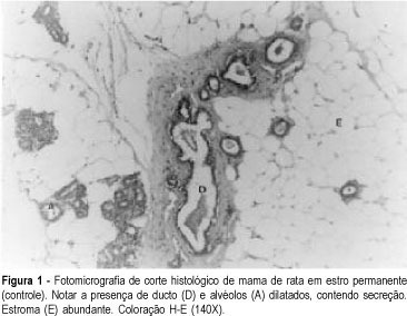
Purpose: to evaluate the morphologic and morphometric alterations produced by tamoxifen and conjugated estrogens in the mammary epithelium of rats in persistent estrus. Methods: thirty-three adult female rats in persistent estrus induced with 1.25 mg testosterone propionate were divided at random into three groups: GI -- which received only water, control group (n = 12); GII -- treated with 500 mug tamoxifen daily (n = 10); GIII -- treated with 30 mug conjugated estrogens per day (n = 11). The first inguinal-abdominal pair of mammary glands of the animals was extirpated and processed for morphologic and morphometric study. Data were analyzed statistically by the Kruskal-Wallis rank analysis of variance (p < 0.05). Results: the morphologic study revealed signs of epithelial atrophy and the morphometric study showed a statistically significant reduction in the mean number of ducts and alveoli in groups II (10.1 and 1.9, respectively) and III (11.1 and 3.5, respectively) compared to group I (25.0 and 6.6, respectively). There was no significant difference between groups II and III. Conclusions: the results of this study indicate that tamoxifen as well as conjugated estrogens at the tested doses produced mammary epithelial atrophy in rats in persistent estrus.
Summary
Rev Bras Ginecol Obstet. 2000;22(1):33-36
DOI 10.1590/S0100-72032000000100006
Purpose: to evaluate the morphologic and morphometric alterations produced by tamoxifen and conjugated estrogens in the mammary epithelium of rats in persistent estrus. Methods: thirty-three adult female rats in persistent estrus induced with 1.25 mg testosterone propionate were divided at random into three groups: GI -- which received only water, control group (n = 12); GII -- treated with 500 mug tamoxifen daily (n = 10); GIII -- treated with 30 mug conjugated estrogens per day (n = 11). The first inguinal-abdominal pair of mammary glands of the animals was extirpated and processed for morphologic and morphometric study. Data were analyzed statistically by the Kruskal-Wallis rank analysis of variance (p < 0.05). Results: the morphologic study revealed signs of epithelial atrophy and the morphometric study showed a statistically significant reduction in the mean number of ducts and alveoli in groups II (10.1 and 1.9, respectively) and III (11.1 and 3.5, respectively) compared to group I (25.0 and 6.6, respectively). There was no significant difference between groups II and III. Conclusions: the results of this study indicate that tamoxifen as well as conjugated estrogens at the tested doses produced mammary epithelial atrophy in rats in persistent estrus.