You searched for:"Antonio Alberto Nogueira"
We found (31) results for your search.Summary
Rev Bras Ginecol Obstet. 2020;42(8):486-492
To determine the average body composition (percentage of body fat), the anthropometric markers, and the intensity of clinical pain in women with a clinical diagnosis of chronic pelvic pain (CPP) secondary to endometriosis.
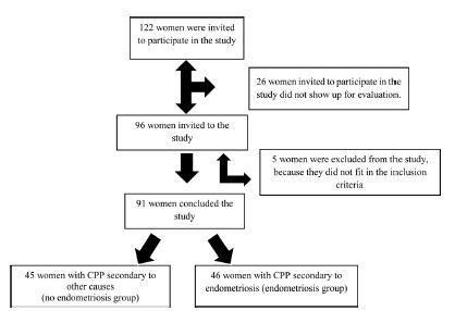
A case-control study performed with 91 women, 46 of whom with CPP secondary to endometriosis and 45 of whom with CPP secondary to other causes. They underwent an evaluation of the anthropometric parameters by means of the body mass index (BMI), the perimeters (waist, abdomen, hip), and the percentage of body fat (%BF), which were assessed on a body composition monitor by bioimpedance; the intensity of the clinical pain was evaluated using the visual analog scale (VAS), and the symptoms of anxiety and depression, using the hospital’s anxiety and depression scale (HAD).
The groups did not differ in terms of mean age, BMI, %BF or regarding the available waist-to-hip ratio (WHR). The mean intensity of the clinical pain by the VAS was of 7.2 ± 2.06 in the group with CPP secondary to endometriosis, and of 5.93 ± 2.64 in the group with CPP secondary to other causes (p = 0.03), revealing significant differences between the groups.
We concluded that, despite the difference in the pain score assessed between the two groups, there was no difference regarding body composition and anthropometry.
Summary
Rev Bras Ginecol Obstet. 2020;42(8):486-492
To determine the average body composition (percentage of body fat), the anthropometric markers, and the intensity of clinical pain in women with a clinical diagnosis of chronic pelvic pain (CPP) secondary to endometriosis.
A case-control study performed with 91 women, 46 of whom with CPP secondary to endometriosis and 45 of whom with CPP secondary to other causes. They underwent an evaluation of the anthropometric parameters by means of the body mass index (BMI), the perimeters (waist, abdomen, hip), and the percentage of body fat (%BF), which were assessed on a body composition monitor by bioimpedance; the intensity of the clinical pain was evaluated using the visual analog scale (VAS), and the symptoms of anxiety and depression, using the hospital’s anxiety and depression scale (HAD).
The groups did not differ in terms of mean age, BMI, %BF or regarding the available waist-to-hip ratio (WHR). The mean intensity of the clinical pain by the VAS was of 7.2 ± 2.06 in the group with CPP secondary to endometriosis, and of 5.93 ± 2.64 in the group with CPP secondary to other causes (p = 0.03), revealing significant differences between the groups.
We concluded that, despite the difference in the pain score assessed between the two groups, there was no difference regarding body composition and anthropometry.
Summary
Rev Bras Ginecol Obstet. 2002;24(8):521-526
DOI 10.1590/S0100-72032002000800004
Purpose: to assess the evolution of epileptic seizures during pregnancy and the occurrence of malformations in neonates born to epileptic mothers who used anticonvulsant drugs during pregnancy, as well as the perinatal characteristics of the newborns. Methods: a total of 126 medical records of epileptic patients seen at the high-risk pregnancy outpatient clinic were analyzed retrospectively in terms of the following variables: age, parity, diagnosis of the type of epileptic seizure, anticonvulsant drug used during the prenatal period, evolution of epileptic seizures during the prenatal period, type of delivery, gestational age at resolution, and perinatal characteristics of the newborns. Results: the incidence of pregnant women with epilepsy was 0.2% in relation to prenatal patients, with simple partial epilepsy being the most frequent type (40% of cases). Monotherapy was applied to 75% of the patients and carbamazepine was the most frequently used drug. Among the 111 patients evaluated in terms of course of the disease during pregnancy, 53% showed no change, 31% became worse and 16% improved. Normal delivery was performed in 62.5% of cases, with a satisfactory perinatal result in terms of Apgar score, and with a rate of low birth weight neonates above the values for low-risk populations. No fetal malformations were observed. Conclusion: epilepsy showed a favorable course during pregnancy and was not aggravated by the latter, with cases of worsening of signs and symptoms being associated with epilepsy of difficult control before pregnancy. Evaluation of the perinatal characteristics of the neonates showed satisfactory Apgar scores and evolution, indicating that epilepsy and anticonvulsant drugs do not cause severe impairment of intrapartum vitality. No cases of malformations or hemorrhagic complications were detected in the present study.
Summary
Rev Bras Ginecol Obstet. 2001;23(8):523-527
DOI 10.1590/S0100-72032001000800007
Purpose: to study perinatal outcome in asthmatic pregnant patients who required hospital admission to control acute exacerbations. Patients and Method: retrospective study of 12 pregnant asthmatic patients admitted at the Hospital das Clínicas, Department of Obstetrics and Gynecology, FMRP-USP, during the period between 1992 and 1996. The analyzed data included: maternal age, prenatal care, length of hospitalization, gestational age at delivery, type of delivery, neonatal weight and Apgar score. Results: among the 12 asthmatic pregnant patients 7 did not have prenatal care for acute exacerbation treatment before hospitalization. Three of 12 developed preeclampsia (one with premature rupture of membranes and infection of the amniotic cavity and one with premature separation of the placenta); 2 of 12 were diagnosed with premature placental aging (one with premature labor and twin-to-twin transfusion syndrome and one with oligohydramnios); 1 of 12 was diagnosed with oligohydramnios and fetal death and had pneumonia, and 1 of 12 was diagnosed with polyhydramnios. Among the infants, 3 were small for gestational age. Conclusions: perinatal complications were more frequent in asthmatic pregnant patients who required hospital care for the acute exacerbations. Chronic asthmatic patients in reproductive age should be advised before pregnancy about the prophylactic measures to reduce the incidence of acute crises and exacerbations during pregnancy should be treated promptly.
Summary
Rev Bras Ginecol Obstet. 2016;38(2):53-55
Summary
Rev Bras Ginecol Obstet. 2016;38(2):53-55
Summary
Rev Bras Ginecol Obstet. 1998;20(10):571-576
DOI 10.1590/S0100-72031998001000005
Purpose: to evaluate the advantages of the laparoscopic approach for conversion of abdominal hysterectomies in vaginal hysterectomies in patients with indication of concomitant adnexectomy, being considered the safety and the additional costs of the procedure. Patients and Methods: cases: 9 patients submitted to Laparoscopically Assisted Vaginal Hysterectomy (LAVH) associated with adnexectomy. Controls:18 patients submitted to Abdominal Hysterectomy (AH) associated with adnexectomy. Both groups were compared regarding preoperative characteristics and the results of the procedure. The patients submitted to LAVH and AH are similar concerning age, parity, cesarean deliveries, previous surgeries and body mass index. Results: the average surgery time was 163.9 minutes for patients submitted to LAVH and 142.8 minutes for patients submitted to AH. No patient in the LAVH group presented postoperative complications, while in the AH group 2 patients presented suture deiscence and there was 1 case of incisional hernia. The median of hospital stay was 1 day in the LAVH group and 2 days in the AH group, those of convalescence periods were 2 and 4 weeks, respectively. 55.6% of the patients in the LAVH group and 100% in the AH group needed analgesics in the postoperative period. Conclusions: LAVH was shown to be advantageous in relation to AH in terms of better recovery and lower incidence of complications in the postoperative period. The procedure is feasible and safe in a University Hospital, and without additional costs.
Summary
Rev Bras Ginecol Obstet. 2000;22(10):619-625
DOI 10.1590/S0100-72032000001000004
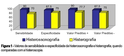
Purpose: to compare the methods used to investigate the endouterine cavity by testing the sensitivity and specificity of X-ray hysterography and sonohysterography compared with hysteroscopy (gold standard). Methods: we carried out a prospective study with 18 patients who, due to symptoms such as irregular menstrual cycles, unexplained postmenopausal uterine bleeding and ultrasound disturbance, were candidates for uterine cavity investigation by X-ray hysterography, sonohysterography and hysteroscopy. Results: sonohysterography sensitivity and specificity were 90 and 87.5%, respectively. Positive and negative predictive values were 90 and 87.5%. For X-ray hysterography, sensitivity, specificity, positive and negative predictive values were 70, 75, 77.7 and 75%, respectively. Conclusion: the use of saline instilation into the endometrial cavity in order to enhance the acuracy of the vaginal ultrasonography seems reliable as a mean to distinguish lesions in the uterine cavity, thereby facilitating the identification of candidates for diagnostic or operative hysteroscopy. X-ray hysterography produces results inferior to hysterosonography.
Summary
Rev Bras Ginecol Obstet. 2000;22(10):619-625
DOI 10.1590/S0100-72032000001000004
Purpose: to compare the methods used to investigate the endouterine cavity by testing the sensitivity and specificity of X-ray hysterography and sonohysterography compared with hysteroscopy (gold standard). Methods: we carried out a prospective study with 18 patients who, due to symptoms such as irregular menstrual cycles, unexplained postmenopausal uterine bleeding and ultrasound disturbance, were candidates for uterine cavity investigation by X-ray hysterography, sonohysterography and hysteroscopy. Results: sonohysterography sensitivity and specificity were 90 and 87.5%, respectively. Positive and negative predictive values were 90 and 87.5%. For X-ray hysterography, sensitivity, specificity, positive and negative predictive values were 70, 75, 77.7 and 75%, respectively. Conclusion: the use of saline instilation into the endometrial cavity in order to enhance the acuracy of the vaginal ultrasonography seems reliable as a mean to distinguish lesions in the uterine cavity, thereby facilitating the identification of candidates for diagnostic or operative hysteroscopy. X-ray hysterography produces results inferior to hysterosonography.
Summary
Rev Bras Ginecol Obstet. 2007;29(12):619-624
DOI 10.1590/S0100-72032007001200004
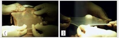
PURPOSE: to evaluate the results of neovaginoplasty with the use of a human amniotic graft in patients with the Mayer-Rokitansky-Küster-Hauser (MRKH) syndrome. METHODS: the study was a retrospective analysis of a series of 28 patients with the MRKH syndrome conducted from 1990 to 2003. The patients were attended and treated at the Ambulatório de Ginecologia Infanto-Puberal (AGIP) of the Hospital Universitário of the Faculdade de Medicina de Ribeirão Preto of the Universidade de São Paulo (FMRP-USP), being submitted to neovaginoplasty by the technique of McIndoe and Bannister, modified by the use of a human amniotic membrane graft. Epithelization, amplitude and depth of the neovaginas were evaluated 7 and 40 days after the procedure. Patient satisfaction was determined during the late postoperative period in terms of the presence of discomfort and dyspareunia during sexual relations. RESULTS: postoperatively, seven patients (25%) presented vaginal stenosis and six of them were submitted to a new surgical intervention, one had shortening of the neovagina, corrected with the use of exercises with a vaginal mold, three (10.7%) developed a rectovaginal fistula, one (3.6%) a uterovesical fistula, and one (3.6%) excess skin in the vaginal introitus - all successfully corrected with surgery. Four patients (14.3%) presented urinary tract infection. Two months after surgery, 11/19 patients (57.8%) presented satisfactory sexual activity and 42% dyspareunia, and within a maximum period of four years, 20/21 patients (95.2%) had satisfactory sexual activity and 4.8% dyspareunia. CONCLUSIONS: an amniotic membrane graft is a good option for the treatment of vaginal agenesis. Perioperative follow-up involves educational guidance regarding the use of the mold and regarding patient sexuality in order to reduce the complaints of dysfunctional coitus in the presence of a favorable surgical evolution and a neovagina of adequate aspect.
Summary
Rev Bras Ginecol Obstet. 2007;29(12):619-624
DOI 10.1590/S0100-72032007001200004
PURPOSE: to evaluate the results of neovaginoplasty with the use of a human amniotic graft in patients with the Mayer-Rokitansky-Küster-Hauser (MRKH) syndrome. METHODS: the study was a retrospective analysis of a series of 28 patients with the MRKH syndrome conducted from 1990 to 2003. The patients were attended and treated at the Ambulatório de Ginecologia Infanto-Puberal (AGIP) of the Hospital Universitário of the Faculdade de Medicina de Ribeirão Preto of the Universidade de São Paulo (FMRP-USP), being submitted to neovaginoplasty by the technique of McIndoe and Bannister, modified by the use of a human amniotic membrane graft. Epithelization, amplitude and depth of the neovaginas were evaluated 7 and 40 days after the procedure. Patient satisfaction was determined during the late postoperative period in terms of the presence of discomfort and dyspareunia during sexual relations. RESULTS: postoperatively, seven patients (25%) presented vaginal stenosis and six of them were submitted to a new surgical intervention, one had shortening of the neovagina, corrected with the use of exercises with a vaginal mold, three (10.7%) developed a rectovaginal fistula, one (3.6%) a uterovesical fistula, and one (3.6%) excess skin in the vaginal introitus - all successfully corrected with surgery. Four patients (14.3%) presented urinary tract infection. Two months after surgery, 11/19 patients (57.8%) presented satisfactory sexual activity and 42% dyspareunia, and within a maximum period of four years, 20/21 patients (95.2%) had satisfactory sexual activity and 4.8% dyspareunia. CONCLUSIONS: an amniotic membrane graft is a good option for the treatment of vaginal agenesis. Perioperative follow-up involves educational guidance regarding the use of the mold and regarding patient sexuality in order to reduce the complaints of dysfunctional coitus in the presence of a favorable surgical evolution and a neovagina of adequate aspect.
Summary
Rev Bras Ginecol Obstet. 2019;41(11):668-672
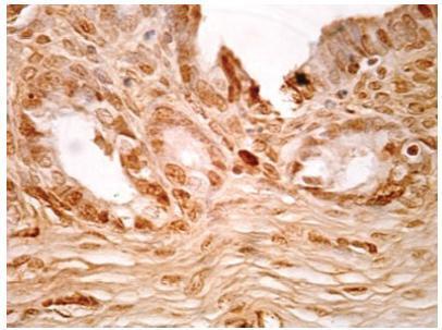
To analyze the effect of thalidomide on the progression of endometriotic lesions experimentally induced in rats and to characterize the pattern of cell proliferation by immunohistochemical Proliferating Cell Nuclear Antigen (PCNA) labeling of eutopic and ectopic endometrium.
Fifteen female Wistar rats underwent laparotomy for endometriosis induction by resection of one uterine horn, isolation of the endometrium and fixation of a tissue segment to the pelvic peritoneum. Four weeks after, the animals were divided into 3 groups: control (I), 10mg/kg/day (II) and 1mg/kg/day (III) intraperitoneal thalidomide for 10 days. The lesion was excised together with the opposite uterine horn for endometrial gland and stroma analysis. Eutopic and ectopic endometrial tissue was submitted to immunohistochemistry for analysis of cell proliferation by PCNA labeling and the cell proliferation index (CPI) was calculated as the number of labeled cells per 1,000 cells.
Group I showed a mean CPI of 0.248 ± 0.0513 in the gland and of 0.178 ± 0.046 in the stroma. In contrast, Groups II and III showed a significantly lower CPI, that is, 0.088 ± 0.009 and 0.080 ± 0.021 for the gland (p < 0.001) and 0.0945 ± 0.0066 and 0.075 ± 0.018 for the stroma (p < 0.001), respectively. Also, the mean lesion area of Group I was 69.2mm2, a significantly higher value compared with Group II (49.4mm2, p = 0.023) and Group III (48.6mm2, p = 0.006). No significant difference was observed between Groups II and III.
Thalidomide proved to be effective in reducing the lesion area and CPI of the experimental endometriosis implants both at the dose of 1mg/kg/day and at the dose of 10 mg/kg/day.
Summary
Rev Bras Ginecol Obstet. 2019;41(11):668-672
To analyze the effect of thalidomide on the progression of endometriotic lesions experimentally induced in rats and to characterize the pattern of cell proliferation by immunohistochemical Proliferating Cell Nuclear Antigen (PCNA) labeling of eutopic and ectopic endometrium.
Fifteen female Wistar rats underwent laparotomy for endometriosis induction by resection of one uterine horn, isolation of the endometrium and fixation of a tissue segment to the pelvic peritoneum. Four weeks after, the animals were divided into 3 groups: control (I), 10mg/kg/day (II) and 1mg/kg/day (III) intraperitoneal thalidomide for 10 days. The lesion was excised together with the opposite uterine horn for endometrial gland and stroma analysis. Eutopic and ectopic endometrial tissue was submitted to immunohistochemistry for analysis of cell proliferation by PCNA labeling and the cell proliferation index (CPI) was calculated as the number of labeled cells per 1,000 cells.
Group I showed a mean CPI of 0.248 ± 0.0513 in the gland and of 0.178 ± 0.046 in the stroma. In contrast, Groups II and III showed a significantly lower CPI, that is, 0.088 ± 0.009 and 0.080 ± 0.021 for the gland (p < 0.001) and 0.0945 ± 0.0066 and 0.075 ± 0.018 for the stroma (p < 0.001), respectively. Also, the mean lesion area of Group I was 69.2mm2, a significantly higher value compared with Group II (49.4mm2, p = 0.023) and Group III (48.6mm2, p = 0.006). No significant difference was observed between Groups II and III.
Thalidomide proved to be effective in reducing the lesion area and CPI of the experimental endometriosis implants both at the dose of 1mg/kg/day and at the dose of 10 mg/kg/day.