Summary
Revista Brasileira de Ginecologia e Obstetrícia. 2000;22(6):381-384
DOI 10.1590/S0100-72032000000600009
Parkinson's disease is characterized by tremor, stiffness of the musculature, bradykinesia, and postural and march abnormalities. It attacks all ethnic groups, with no sex preference, frequently in the 45-50-year range. The diagnosis is essentially clinical. The association with pregnancy is rare. The experience with that association is scarse, some questions remaining without answer. The authors describe a case of Parkinson's disease and gestation with satisfactory evolution, in spite of the clinical worsening during pregnancy. The mother presented elevation of blood pressure levels, alterations of the hepatic enzymes, and oligohydramnios. She used, independently, selegiline until the third month, and, later on, amantadine. The newborn presented low weight, respiratory distress and jaundice, being discharged from the hospital, with no other complications, on the fourth day of life.
Summary
Revista Brasileira de Ginecologia e Obstetrícia. 2012;34(8):381-385
DOI 10.1590/S0100-72032012000800007
PURPOSE: To assess perinatal factors associated with term newborns with pH<7.1 in the umbilical artery and 5th min Apgar score<7,0. METHODS: Retrospective case-control study carried out after reviewing the medical records of all births from September/1998 to March/2008, that occurred at the General Hospital of Caxias do Sul. The inclusion criterion was term newborns who presented a 5th min Apgar score <7.0 and umbilical artery pH<7.10. In the univariate analysis, we used the Student's t-test and the Mann-Whitney test for continuous variables, the c² test for dichotomous variables and risk estimation by the odds ratio (OR). The level of significance was set at p<0.05. RESULTS: Of a total of 15,495 consecutive births, 25 term neonates (0.16%) had pH<7.1 in the umbilical artery and a 5th min Apgar score <7.0. Breech presentation (OR=12.9, p<0.005), cesarean section (OR=3.5, p<0.01) and modified intrapartum cardiotocography (OR=7.8, p<0.02) presented a significant association with the acidosis event. Among the fetal characteristics, need for hospitalization in the neonatal intensive care unit (OR=79.7, p <0.0001), need for resuscitation (OR=12.2, p <0.0001) and base deficit were associated with the event (15.0 versus -4.5, p<0.0001). CONCLUSION: Low Apgar score at the 5th min of life associated with pH<7.1 in the umbilical artery can predict adverse neonatal outcomes.
Summary
Revista Brasileira de Ginecologia e Obstetrícia. 2016;38(8):381-390
The aims of the study were to evaluate, after pregnancy, the glycemic status of women with history of gestational diabetes mellitus (GDM) and to identify clinical variables associated with the development of type 2 diabetes mellitus (T2DM), impaired fasting glucose (IFG), and impaired glucose tolerance (IGT).
Retrospective cohort of 279 women with GDM who were reevaluated with an oral glucose tolerance test (OGTT) after pregnancy. Characteristics of the index pregnancy were analyzed as risk factors for the future development of prediabetes (IFG or IGT), and T2DM.
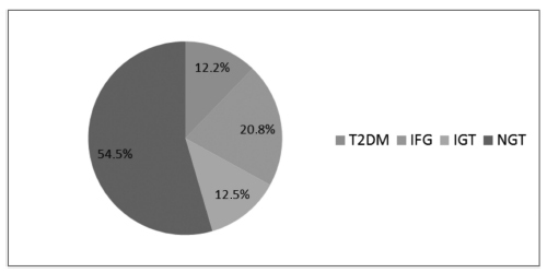
T2DM was diagnosed in 34 (12.2%) patients, IFG in 58 (20.8%), and IGT in 35 (12.5%). Women with postpartum T2DM showed more frequently a family history of T2DM, higher pre-pregnancy body mass index (BMI), lower gestational age, higher fasting and 2-hour plasma glucose levels on the OGTT at the diagnosis of GDM, higher levels of hemoglobin A1c, and a more frequent insulin requirement during pregnancy. Paternal history of T2DM (odds ratio [OR] =5.67; 95% confidence interval [95%CI] =1.64-19.59; p =0.006), first trimester fasting glucose value (OR =1.07; 95%CI =1.03-1.11; p =0.001), and insulin treatment during pregnancy (OR =15.92; 95%CI =5.54-45.71; p < 0.001) were significant independent risk factors for the development of T2DM.
A high rate of abnormal glucose tolerance was found in women with previous GDM. Family history of T2DM, higher pre-pregnancy BMI, early onset of GDM, higher glucose levels, and insulin requirement during pregnancy were important risk factors for the early identification of women at high risk of developing T2DM. These findings may be useful for developing preventive strategies.
Summary
Revista Brasileira de Ginecologia e Obstetrícia. 2016;38(8):381-390
The aims of the study were to evaluate, after pregnancy, the glycemic status of women with history of gestational diabetes mellitus (GDM) and to identify clinical variables associated with the development of type 2 diabetes mellitus (T2DM), impaired fasting glucose (IFG), and impaired glucose tolerance (IGT).
Retrospective cohort of 279 women with GDM who were reevaluated with an oral glucose tolerance test (OGTT) after pregnancy. Characteristics of the index pregnancy were analyzed as risk factors for the future development of prediabetes (IFG or IGT), and T2DM.
T2DM was diagnosed in 34 (12.2%) patients, IFG in 58 (20.8%), and IGT in 35 (12.5%). Women with postpartum T2DM showed more frequently a family history of T2DM, higher pre-pregnancy body mass index (BMI), lower gestational age, higher fasting and 2-hour plasma glucose levels on the OGTT at the diagnosis of GDM, higher levels of hemoglobin A1c, and a more frequent insulin requirement during pregnancy. Paternal history of T2DM (odds ratio [OR] =5.67; 95% confidence interval [95%CI] =1.64-19.59; p =0.006), first trimester fasting glucose value (OR =1.07; 95%CI =1.03-1.11; p =0.001), and insulin treatment during pregnancy (OR =15.92; 95%CI =5.54-45.71; p < 0.001) were significant independent risk factors for the development of T2DM.
A high rate of abnormal glucose tolerance was found in women with previous GDM. Family history of T2DM, higher pre-pregnancy BMI, early onset of GDM, higher glucose levels, and insulin requirement during pregnancy were important risk factors for the early identification of women at high risk of developing T2DM. These findings may be useful for developing preventive strategies.
Summary
Revista Brasileira de Ginecologia e Obstetrícia. 2015;37(8):381-387
DOI 10.1590/SO100-720320150005393
To evaluate the waiting times before obtaining the first colposcopic examination for women with abnormal Papanicolaou smears.
Retrospective cohort study conducted on patients who required a colposcopic examination to clarify an abnormal pap test, between 2002 January and 2008 August, in a metropolitan region of Brazil. The waiting times were defined as: Total Waiting Time (interval between the date of the pap test result and the date of the first colposcopic examination); Partial A Waiting Time (interval between the date of the pap test result and the date of referral); Partial B Waiting Time (interval between the date of referral and the date of the first colposcopic examination). Means, medians, relative and absolute frequencies were calculated. The Kruskal-Wallis test and Pearson's chi-square test were used to determine statistical significance.
A total of 1,544 women with mean of age of 34 years (SD=12.6 years) were analyzed. Most of them had access to colposcopic examination within 30 days (65.8%) or 60 days (92.8%) from referral. Mean Total Waiting Time, Partial A Waiting Time, and Partial B Waiting Time were 94.5 days (SD=96.8 days), 67.8 days (SD=95.3 days) and 29.2 days (SD=35.1 days), respectively.
A large part of the women studied had access to colposcopic examination within 60 days after referral, but Total waiting time was long. Measures to reduce the waiting time for obtaining the first colposcopic examination can help to improve the quality of care in the context of cervical cancer control in the region, and ought to be addressed at the phase between the date of the pap test results and the date of referral to the teaching hospital.
Summary
Revista Brasileira de Ginecologia e Obstetrícia. 2014;36(8):381-381
DOI 10.1590/SO100-72032014T0002
Summary
Revista Brasileira de Ginecologia e Obstetrícia. 2014;36(8):381-381
DOI 10.1590/SO100-72032014T0002
Summary
Revista Brasileira de Ginecologia e Obstetrícia. 2005;27(7):382-386
DOI 10.1590/S0100-72032005000700003
PURPOSE: to evaluate periodontal conditions and need for treatment supplied by Periodontal Screening and Recording (PSR) in mothers, in order to clarify the relationship between periodontal disease and low birth weight premature newborns. METHODS: PSR was used in a sample of 40 mothers, divided into: test group, consisting of mothers of premature newborns with weight less than 2,500 g (n=20), and the control group, consisting of mothers of term newborns with a weight equal to or over 2,500 g (n=20). The collected data were analyzed by descriptive statistics, and the results of PSR were submitted to statistical analysis in order to verify differences in periodontal condition and need for treatment of the mothers, using the Kolmogorov-Smirnov test, with a significance level of 5%. RESULTS: the presence of periodontal pocket of 3.5 to 5.5 mm was the most common finding among the mothers of newborns with low weight (39.2% of the cases), while the presence of bleeding at probing and absence of a periodontal pocket were the most frequent findings among the mothers of newborns with normal weight (37.5% of the cases). There was a statistically significant difference between periodontal conditions of the mothers of the groups (p=0.0494), but in relation to the need for treatment, there was no significant difference between the studied groups (p>0.05). CONCLUSION: the mothers of preterm newborns with low weight presented worse periodontal conditions, suggesting that periodontal infection may be related to preterm low birth weight newborns.
Summary
Revista Brasileira de Ginecologia e Obstetrícia. 2004;26(5):383-389
DOI 10.1590/S0100-72032004000500007
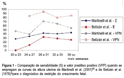
OBJECTIVE: to evaluate the measurement of uterine height in order to predict fetal growth restriction (FGR), according to a local curve. METHODS: from July 2000 to February 2003, 238 high-risk pregnant women were submitted to uterine height measurements between the 20th and the 42nd week of gestation. The gestational age of all the women was well known, confirmed by early ultrasound. Fifty (21%) women gave birth to infants considered small for their gestational age. The measures were performed by a single observer, who took 1617 uterine height measurements, from the upper border of the symphysis pubis to the fundus uteri, using tape measurement. The diagnosis of FGR was confirmed after birth according to the Ramos's curve. The women were divided into two groups according to their infant's birth weight and the data were statistically analyzed by the Fisher's exact test or Kruskal-Wallis's test. The sensitivity (SE), specificity (SP), positive predictive value (PPV), and negative predictive value (NPV) were calculated. The test for two proportions with normal approximation was performed to analyze the continuous variables. RESULTS: one measurement below the 10th percentile, according to gestational age, resulted in SE = 78.0%, SP = 77.1%, PPV = 47.6%, and NPV = 88.8% for the identification of FGR. If one measurement was below the 5th percentile, the SE, SP, PPV, and NPV were 64.0, 89.9, 62.7 and 90.4%, respectively. CONCLUSIONS: one measurement below the 10th percentile for the gestational age, according to the local curve, proved to be a good predictor of FGR.
Summary
Revista Brasileira de Ginecologia e Obstetrícia. 2004;26(5):383-389
DOI 10.1590/S0100-72032004000500007
OBJECTIVE: to evaluate the measurement of uterine height in order to predict fetal growth restriction (FGR), according to a local curve. METHODS: from July 2000 to February 2003, 238 high-risk pregnant women were submitted to uterine height measurements between the 20th and the 42nd week of gestation. The gestational age of all the women was well known, confirmed by early ultrasound. Fifty (21%) women gave birth to infants considered small for their gestational age. The measures were performed by a single observer, who took 1617 uterine height measurements, from the upper border of the symphysis pubis to the fundus uteri, using tape measurement. The diagnosis of FGR was confirmed after birth according to the Ramos's curve. The women were divided into two groups according to their infant's birth weight and the data were statistically analyzed by the Fisher's exact test or Kruskal-Wallis's test. The sensitivity (SE), specificity (SP), positive predictive value (PPV), and negative predictive value (NPV) were calculated. The test for two proportions with normal approximation was performed to analyze the continuous variables. RESULTS: one measurement below the 10th percentile, according to gestational age, resulted in SE = 78.0%, SP = 77.1%, PPV = 47.6%, and NPV = 88.8% for the identification of FGR. If one measurement was below the 5th percentile, the SE, SP, PPV, and NPV were 64.0, 89.9, 62.7 and 90.4%, respectively. CONCLUSIONS: one measurement below the 10th percentile for the gestational age, according to the local curve, proved to be a good predictor of FGR.
Summary
Revista Brasileira de Ginecologia e Obstetrícia. 2001;23(6):383-390
DOI 10.1590/S0100-72032001000600007
Purpose: to evaluate the ultrasonographic parameters associated with perinatal mortality increase in cases of fetal hydrocephalus. Method: 45 cases of fetal hydrocephalus were followed-up between January 1996 and December 1999. Fetal hydrocephalus was diagnosed when the ratio of lateral ventricles and the corresponding cerebral hemispheres was above 0.35 or when the measurement of the atrium of the lateral ventricles was above 10 mm. In all examinations the type of hydrocephalus, severity, symmetry, evolution and time of diagnosis were defined. The patients were submitted to morphologic ultrasound in the search of other anatomical abnormalities. The amniotic fluid index and fetal deaths were registered. The main ultrasonographic findings were correlated with perinatal mortality. For statistical analysis, chi² test and exact Fisher test were used. The value of p<0,05 was considered to be significant. Results: a total of 20 deaths were observed (44.4%), 6 occurred intra-uterus and 14 in the neonatal period. The diagnosis of hydrocephalus was established at a mean gestational age of 29 weeks. There was no association between perinatal mortality and alterations in the amniotic fluid volume, time of diagnosis, symmetry and type of hydrocephalus and the presence of other intra- or extracranial anomalies. On the other hand, the severity of the disease was associated significantly with perinatal death (p<0.0001). Conclusion: among all the analyzed ultrasonographic parameters, only the severity of hydrocephalus presented statistical association with perinatal death.