Summary
Rev Bras Ginecol Obstet. 1999;21(3):127-131
DOI 10.1590/S0100-72031999000300002
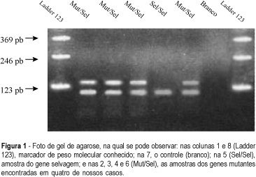
Purpose: the frequency of point mutation at codon 12 of the K¾ras gene was determined in paraffin blocks of surgical specimens from patients who had ductal invasive breast cancer. Material and Methods: Fifty surgical specimens blocked in paraffin from patients with ductal invasive breast cancer, with histological degree II and III, were used. The polymerase chain reaction (PCR) was used for amplification of DNA fragments studied. The material cleavage was obtained with restriction fragment length polymorphisms (RFLP). The electrophoresis in agarose gel, with Ladder 123 (GIBCO-BRL) marker, was employed to verify if some mutation had occurred. The results were shown using ultraviolet beam and recorded by photos. Results: mutations at codon 12 of K-ras gene were found in five samples (10%) and all of them were polymorphic for this caracter. The five patients whose tumors expressed mutation were in the postmenopausal period. Four patientes had tumors of histological degree II and one, III.
Summary
Rev Bras Ginecol Obstet. 1999;21(3):127-131
DOI 10.1590/S0100-72031999000300002
Purpose: the frequency of point mutation at codon 12 of the K¾ras gene was determined in paraffin blocks of surgical specimens from patients who had ductal invasive breast cancer. Material and Methods: Fifty surgical specimens blocked in paraffin from patients with ductal invasive breast cancer, with histological degree II and III, were used. The polymerase chain reaction (PCR) was used for amplification of DNA fragments studied. The material cleavage was obtained with restriction fragment length polymorphisms (RFLP). The electrophoresis in agarose gel, with Ladder 123 (GIBCO-BRL) marker, was employed to verify if some mutation had occurred. The results were shown using ultraviolet beam and recorded by photos. Results: mutations at codon 12 of K-ras gene were found in five samples (10%) and all of them were polymorphic for this caracter. The five patients whose tumors expressed mutation were in the postmenopausal period. Four patientes had tumors of histological degree II and one, III.
Summary
Rev Bras Ginecol Obstet. 1999;21(3):133-137
DOI 10.1590/S0100-72031999000300003
Purpose: to evaluate how knowledgeable medical students at the Universidade Federal de Goiás were concerning the basic diagnostic principles breast cancer. The study also aimed at promoting a debate among the students and at assessing the understanding of the students in the fifth year of medical school, who had already attended the Gynecology course. Methods: Through questionnaires given to 348 individuals, from the first to the fifth year, out of a total population of 550 students, the authors searched for information with regard to basic knowledge on the diagnosis of breast cancer. Of the 348 questionnaires, 55 (16%) were given to fifth-year students, who had already attended the Gynecology course. Furthermore, 43% of the students were women, 62% had medical doctors in their immediate family, and 17% had a family history of breast cancer. Results: in regard to the knowledge of diagnostic methods, 84% of the students were aware of the most frequent sign of breast cancer, 34% knew which was the best screening method, 49% knew when to refer asymptomatic women to mammography, 37% knew the recommended interval between mammography for women above the age of 50, and 24% knew when to associate ultrasound to mammography for the detection of breast cancer. The fifth-year students provided correct answers at a significantly higher rate, when compared to the others. Concerning gender, the only difference regarded the fact that women showed a better knowledge as to the best time for self-examination and when to recommend ultrasound associated with mammography. The presence of medical doctors in the family and a history of family members with breast cancer did not have any influence on the answers. Conclusion: The lack of information in regard to the diagnosis of breast cancer is very high, even among medical students. Nevertheless, the rate of information increases significantly after students are taught Gynecology, which is only offered during the fifth year of the medical school.
Summary
Rev Bras Ginecol Obstet. 1999;21(3):141-146
DOI 10.1590/S0100-72031999000300004
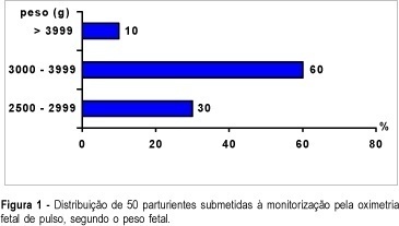
Purpose: to study fetal oxygen saturation (SpO2) levels during labor by continuous pulse oximetry tecnique, and its relation to umbilical artery (UA) pH. Patients and Methods: fetal SpO2 levels were measured during labor by the pulse oximetry technique in 50 subjects. Average values of SpO2 were compared between the first and second stage of labor, with the first stage further subdivided into phases, according to cervical dilatation of (<=4 cm, 5-7 cm and 8-9 cm). SpO2 values were studied in relation to umbilical artery pH at birth ( > or = 7.20 and <7.20). SpO2 > or = 30.0% was considered normal. Results: fetal SpO2 averages during the first stage were 53.0 ± 7.3% and 44.2 ± 6.8% (UA pH > or = 7.20 and <7.20, respectively; p<0.01). When the first stage was subdivided, the fetal SpO2 averages (UA pH > or = 7.20) were 55.1 ± 5.1% (<=4 cm), 52.3 ± 4.6% (5-7 cm) and 51.5 ± 7.2% (8-9 cm); for UA pH <7.20, the fetal SpO2 averages were 46.3 ± 5.1% (<=4 cm), 43.6 ± 6.7% (5-7 cm) and 42.8 ± 5.8% (8-9 cm). Considering the UA pH, these differences were statistically significant (p<0.01). Conclusion: a significant decrease of oxygen saturation values was observed during labor when fetal pulse oximetry was used.
Summary
Rev Bras Ginecol Obstet. 1999;21(3):141-146
DOI 10.1590/S0100-72031999000300004
Purpose: to study fetal oxygen saturation (SpO2) levels during labor by continuous pulse oximetry tecnique, and its relation to umbilical artery (UA) pH. Patients and Methods: fetal SpO2 levels were measured during labor by the pulse oximetry technique in 50 subjects. Average values of SpO2 were compared between the first and second stage of labor, with the first stage further subdivided into phases, according to cervical dilatation of (<=4 cm, 5-7 cm and 8-9 cm). SpO2 values were studied in relation to umbilical artery pH at birth ( > or = 7.20 and <7.20). SpO2 > or = 30.0% was considered normal. Results: fetal SpO2 averages during the first stage were 53.0 ± 7.3% and 44.2 ± 6.8% (UA pH > or = 7.20 and <7.20, respectively; p<0.01). When the first stage was subdivided, the fetal SpO2 averages (UA pH > or = 7.20) were 55.1 ± 5.1% (<=4 cm), 52.3 ± 4.6% (5-7 cm) and 51.5 ± 7.2% (8-9 cm); for UA pH <7.20, the fetal SpO2 averages were 46.3 ± 5.1% (<=4 cm), 43.6 ± 6.7% (5-7 cm) and 42.8 ± 5.8% (8-9 cm). Considering the UA pH, these differences were statistically significant (p<0.01). Conclusion: a significant decrease of oxygen saturation values was observed during labor when fetal pulse oximetry was used.
Summary
Rev Bras Ginecol Obstet. 1999;21(3):147-152
DOI 10.1590/S0100-72031999000300005
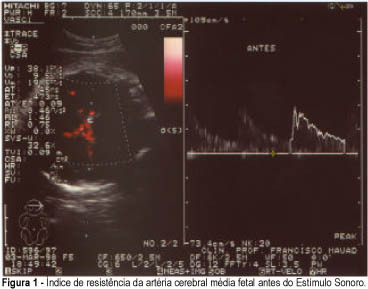
Purpose: to determine the possible occurrence of hemodynamic changes in the middle cerebral artery of the fetus (MCA) using color doppler after vibro-acoustic stimulation. Methods: thirty fetuses from pregnant women considered to be clinically normal, with a gestational age of 28 weeks or more were submitted to vibro-acoustic stimulation. We examined the changes in blood flow rate in the middle cerebral artery of the fetus on the basis of resistance index (RI) and fetal heart rate (FHR) by color doppler before and after the sound stimulus. Results: mean FHR before vibro-acoustic stimulation was 142.41 beats per minute (bpm) with a standard deviation of 9.01 and a range of 122 to 162 bpm. After stimulation, mean FHR was 159.44 bpm with a standard deviation of 15.49 and a range of 130 to 187 bpm (p<0.01). Mean RI in the MCA of the fetuses was 75.89% (range: 64 to 91%) before the experiment. After the vibro-acoustic stimulation, mean RI was 66.93% (range: 47 to 83%; p < 0.01). Conclusions: we observed that a sound stimulus provokes the well-known immediate and significant elevation of FHR and a decrease in cerebral vascular resistance when evaluated by the RI of the fetal middle cerebral artery.
Summary
Rev Bras Ginecol Obstet. 1999;21(3):147-152
DOI 10.1590/S0100-72031999000300005
Purpose: to determine the possible occurrence of hemodynamic changes in the middle cerebral artery of the fetus (MCA) using color doppler after vibro-acoustic stimulation. Methods: thirty fetuses from pregnant women considered to be clinically normal, with a gestational age of 28 weeks or more were submitted to vibro-acoustic stimulation. We examined the changes in blood flow rate in the middle cerebral artery of the fetus on the basis of resistance index (RI) and fetal heart rate (FHR) by color doppler before and after the sound stimulus. Results: mean FHR before vibro-acoustic stimulation was 142.41 beats per minute (bpm) with a standard deviation of 9.01 and a range of 122 to 162 bpm. After stimulation, mean FHR was 159.44 bpm with a standard deviation of 15.49 and a range of 130 to 187 bpm (p<0.01). Mean RI in the MCA of the fetuses was 75.89% (range: 64 to 91%) before the experiment. After the vibro-acoustic stimulation, mean RI was 66.93% (range: 47 to 83%; p < 0.01). Conclusions: we observed that a sound stimulus provokes the well-known immediate and significant elevation of FHR and a decrease in cerebral vascular resistance when evaluated by the RI of the fetal middle cerebral artery.
Summary
Rev Bras Ginecol Obstet. 1999;21(3):153-157
DOI 10.1590/S0100-72031999000300006
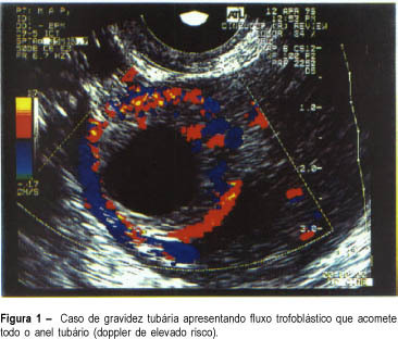
Purpose: to evaluate the efficacy of color Doppler in the prediction of results of the systemic treatment of unruptured ectopic pregnancy with a single dose of methotrexate. Methodology: twenty patients with a diagnosis of ectopic pregnancy were included in the study. The inclusion criteria were: hemodynamic stability, adnexal mass < 5.0 cm and decline of the titers of beta-hCG less than 15% in an interval of 24 h. The exclusion criteria were hepatic or renal disease and blood dyscrasias. Follow-up was by serial determinations of beta-hCG on days 4 and 7 after the beginning of the treatment, and weekly until the titers were negative. The patients were classified into 3 groups according to color Doppler: high risk (trophoblastic flow covering more than 2/3 of the mass), medium risk (when trophoblastic flow compromised 1/3 to 2/3 of tubal mass) and low risk (when trophoblastic flow covered less than 1/3 of the mass). Results: the success of the treatment with a single dose was 75% (15/20); when a second dose of MTX was used, the success rate was 85%. When comparing color Doppler with the results of the medical treatment, we had high risk in 4 patients and in all the treatment failed; medium and low risk in 16 patients, and in 15 the treatment was successful. Conclusion: color Doppler showing high risk indicated an unfavorable situation for the medical treatment with MTX, while medium and low risk in color doppler were favorable situations for the clinical treatment. However, these results should always be analyzed in association with the evolution curve of the beta-hCG titers.
Summary
Rev Bras Ginecol Obstet. 1999;21(3):153-157
DOI 10.1590/S0100-72031999000300006
Purpose: to evaluate the efficacy of color Doppler in the prediction of results of the systemic treatment of unruptured ectopic pregnancy with a single dose of methotrexate. Methodology: twenty patients with a diagnosis of ectopic pregnancy were included in the study. The inclusion criteria were: hemodynamic stability, adnexal mass < 5.0 cm and decline of the titers of beta-hCG less than 15% in an interval of 24 h. The exclusion criteria were hepatic or renal disease and blood dyscrasias. Follow-up was by serial determinations of beta-hCG on days 4 and 7 after the beginning of the treatment, and weekly until the titers were negative. The patients were classified into 3 groups according to color Doppler: high risk (trophoblastic flow covering more than 2/3 of the mass), medium risk (when trophoblastic flow compromised 1/3 to 2/3 of tubal mass) and low risk (when trophoblastic flow covered less than 1/3 of the mass). Results: the success of the treatment with a single dose was 75% (15/20); when a second dose of MTX was used, the success rate was 85%. When comparing color Doppler with the results of the medical treatment, we had high risk in 4 patients and in all the treatment failed; medium and low risk in 16 patients, and in 15 the treatment was successful. Conclusion: color Doppler showing high risk indicated an unfavorable situation for the medical treatment with MTX, while medium and low risk in color doppler were favorable situations for the clinical treatment. However, these results should always be analyzed in association with the evolution curve of the beta-hCG titers.
Summary
Rev Bras Ginecol Obstet. 1999;21(3):161-165
DOI 10.1590/S0100-72031999000300007
Purpose: to evaluate the social, demographic and obstetrical profile of adolescents as compared with adult women hospitalized for abortion complications. Material and methods: this is a descriptive study that evaluated 230 women with abortion complications. Among them, 59 were adolescents hospitalized at the IMIP Maternity (Recife, Brazil) from August 1994 to July 1995. The variables studied were: educational level, marital status, any paid activity, gestation age, number of pregnancies, desire to become pregnant, use of anticonceptive method, kind of relationship, reason for voluntary interruption, clinical classification of abortion and associated complications. The procedure for data analysis was the distribution of variables among adolescents and adults, the differences being evaluated through chi² and chi² for trend. Results: compared with the adult women who aborted, the adolescents showed a lower number of paid activity and multiparity and a higher number of pregnancies resulting from an unstable relationship. Conclusions: the results indicated that biologicallly the adolescents who were hospitalized for abortion have a similar profile to adult women. What differentiates them are the unfavorable social and demographic conditions that they are faced with at their generally unplanned first pregnancies.
Summary
Rev Bras Ginecol Obstet. 1999;21(3):167-169
DOI 10.1590/S0100-72031999000300008
Uterine torsion is an unusual pathology of difficult diagnosis and it is generally associated with an enlargement of uterine volume combined with other alterations of the pelvic organs. This report presents a case of a malnourished elderly woman with acute abdomen and intraoperative diagnosis of myoma and uterine torsion of 360 degrees to the right. The uterus showed signs of severe ischemia.
Summary
Rev Bras Ginecol Obstet. 1999;21(3):171-174
DOI 10.1590/S0100-72031999000300009
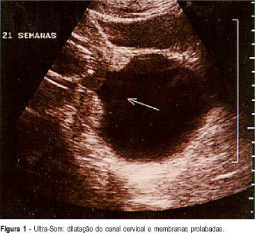
In pregnant women with cervical incompetence in whom there is also dilatation of the cervix and prolapsed membranes there are technical difficulties in performing cerclage in order to prolongate pregnancy until sufficient fetal maturity assures survival of the newborn. We describe a case of cervical incompetence with prolapsed membranes at 21 weeks of gestation, in which we caused the decrease of intrauterine pressure with drainage of amniotic fluid by amniocentesis, until reintroduction of membranes into the uterine cavity was possible. This procedure allowed traction of cervical lips and cerclage with less mechanical trauma, warranting the evolution of pregnancy for 12 weeks and fetal survival
Summary
Rev Bras Ginecol Obstet. 1999;21(3):171-174
DOI 10.1590/S0100-72031999000300009
In pregnant women with cervical incompetence in whom there is also dilatation of the cervix and prolapsed membranes there are technical difficulties in performing cerclage in order to prolongate pregnancy until sufficient fetal maturity assures survival of the newborn. We describe a case of cervical incompetence with prolapsed membranes at 21 weeks of gestation, in which we caused the decrease of intrauterine pressure with drainage of amniotic fluid by amniocentesis, until reintroduction of membranes into the uterine cavity was possible. This procedure allowed traction of cervical lips and cerclage with less mechanical trauma, warranting the evolution of pregnancy for 12 weeks and fetal survival