Summary
Revista Brasileira de Ginecologia e Obstetrícia. 2001;23(4):235-241
DOI 10.1590/S0100-72032001000400006
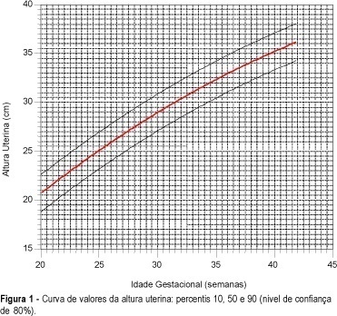
Purpose: to create a uterine height growth curve, according to gestational age, to verify differences among the existing curves and to evaluate the influence of color, parity and maternal weight on the variation of uterine height. Methods: during the period from July 1997 to July 1999, 100 normal pregnant women were submitted to uterine height measurements between the 20th and 42nd week of gestation. All the pregnant women had ultrasonically confirmed gestational age. A total of 726 measurements of uterine height were carried out by the same examiner, using a metric tape from the upper border of the symphysis pubis to the fundus uteri. Results: curves and tables of uterine height according to gestational age were obtained. The average uterine height growth was 0.7 cm/week. The study revealed different average uterine height values in relation to other uterine height growth curves. No statistically significant variations were found between the distributions of uterine heights according to color, parity and weight. Conclusion: the construction of a methodologically accepted uterine height growth curve aimed to detect, as a clinical method, the fetal growth disturbances. This should be analyzed in a posterior study.

Summary
Revista Brasileira de Ginecologia e Obstetrícia. 2001;23(4):225-232
DOI 10.1590/S0100-72032001000400005
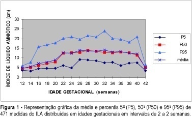
Purpose: to determine the amniotic fluid index (AFI) through ultrasound assessment in normal pregnancies and produce a curve of normalcy for the AFI from the 12th up to the 42nd week of pregnancy. Methods: the study involved 471 measurements on 256 pregnant women, all undergoing normal pregnancies. In pregnancies of more than 20 weeks an estimation was made of the sum of the largest vertical diameters of the amniotic fluid pockets in the four quadrants into which the uterus was divided. In the pregnancies of 20 weeks or less, the sum was obtained from the largest vertical diameters measured in the two halves into which the uterus was divided. Results were expressed in centimeters. Results: AFI was measured (471 measurements) and the results were stratified and grouped by weeks of pregnancy (every two weeks), except the 12th week which was analyzed alone. From an average of 4.7 cm (limits 3.8-5.9 for the 5th and the 95th percentiles) at the 12th week of pregnancy, the AFI grew progressively up to the maximum mean of 14.6 cm at the 32nd week (limits: 7.0-2.5 cm). AFI presented stable measurements from the 21st up to the 40th week. After that, AFI measurements suffered a sharp decrease. The AFI cutoff point occurred at the 21st week of pregnancy. The percent increase of AFI obtained at the 32nd week, when compared to the 12th was 197.7%, and 2.9% at the end of pregnancy when compared to the measurement of the week taken as reference. Conclusion: AFI varied during pregnancy. It increased progressively up to the 21st week and then stabilized up to the 40th week. After that, it experienced a sharp decline. The maximum measurement of the AFI occurred at the 32nd week. By establishing a normalcy curve for AFI it becomes easier to detect changes and allows for a better follow-up of the pregnancy period.

Summary
Revista Brasileira de Ginecologia e Obstetrícia. 2001;23(4):217-221
DOI 10.1590/S0100-72032001000400004
Purpose: to evaluate the influence of pregnancy, habit of smoking, and the contraceptive method in HPV infection and the frequency of cytologic findings in adolescent women with HPV infection. Methods: a total of 54,985 cytologic examinations of patients seen between July, 1993 and December, 1998 were retrospectively analyzed. Of this total, 6,498 (11.8%) examinations were from patients under 20 years old. Of the total of 6,498 cytologic examinations, 326 (5.9%) presented signs of HPV infection, with or without grade I cervical intraepithelial neoplasia (CIN). Patients with diagnosis of grade II and III CIN were excluded. The control group consisted of 333 patients paired by age, without cytological signs of HPV infection. Results: in adolescents, HPV infection was more frequent in oral contraceptive users (16.9% versus 13.8%, p<0.01) and in those who presented with clue cells in cytologic smears (22.4% versus 14.7%, p<0.001). The frequency of HPV infection in couples who used condom was 0% versus 2.1% in the control group (p<0.01). The difference in the number of pregnant women (41.1% versus 44.1%) and smokers (21.8% versus 16.5%) was not statistically significant. Conclusions: HPV infection is more frequent in adolescent women in use of oral contraceptive and with clue cells as cytologic finding. HPV infection did not occur in couples who used condom. Gestation and the habit of smoking did not influence the incidence of HPV infection.
Summary
Revista Brasileira de Ginecologia e Obstetrícia. 2001;23(4):209-215
DOI 10.1590/S0100-72032001000400003
Purpose: to evaluate a populational sample of the screening proposed by the National Program of Uterine Cervical Cancer Control (PNCC), regarding the following issues: frequency of unsatisfactory cytologic results, cytologic frequency of atypical squamous or glandular cells of undetermined significance (ASCUS, AGCUS), low- or high-grade squamous intraepithelial lesions (SIL), comparing the cytologic results with anatomicopathological results of colposcopically directed biopsies. Methods: through the written, broadcasting television and oral midia, women between 35 and 49 years were requested to have a preventive cytopathological test, to be collected by the authorized public health or other institutions accredited by SUS. The slides were analyzed by the program-authorized laboratories and all those patients from the populational sample from the municipality of Naviraí in the State of Mato Grosso do Sul with cellular alterations were submitted to colposcopy and directed biopsy. Results: the frequency of cytologic alterations of the ASCUS, AGCUS and SIL types was 3.3%, an index that is close to that predicted by the PNCC (4%); the percentage of samples that were unsatisfactory for evaluation was high (12.5%); among the ASCUS, AGCUS or low grade-SIL patients, 27.3% presented intraepithelial lesions of a high grade in the anatomicopathological study; while patients with cytology compatible with high grade-SIL, the directed biopsy revealed that 12.5% presented low-grade intraepithelial lesions. Conclusions: the choice of oncological cytology as the only method for the screening in the program allowed high indexes of false-negatives (27.3%) and of false-positives (12.5%). In the screening of cervical neoplasms, colposcopy has shown to be an important and indispensable method to guide the therapeutical management to be adopted.
Summary
Revista Brasileira de Ginecologia e Obstetrícia. 2001;23(4):205-208
DOI 10.1590/S0100-72032001000400002
Purpose: to evaluate the rate of lymphedema and its relation to the type of surgery, age and weight of the patient. Methods: one hundred and nine patients with breast cancer, submitted to modified radical mastectomy sparing the pectoralis major or both pectorales, were studied. Differences of more than 2.0 cm between the diameters of the upper members, measured above and below the elbow, were considered as due to lymphedema. Results: a total rate of 14% of this complication was observed (15 cases). In mastectomies sparing both pectoralis muscles, a smaller rate was observed (9%), when compared to 15% using Patey's technique. However, the difference was not significant. There was a significant relationship between the incidence of lymphedema and the patient's weight and age. The lymphedema was observed in only one of the 34 patients younger than 46 years old, and none of the 19 patients with up to 50 kg presented lymphedema. Conclusion: in the present series lymphedema of the upper limb was associated with the older and heavier patients.
Summary
Revista Brasileira de Ginecologia e Obstetrícia. 2001;23(2):83-86
DOI 10.1590/S0100-72032001000200004
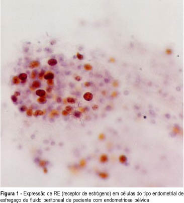
Purpose: to evaluate the expression of estrogen (ER) and progesterone (PR) receptors in smears of peritoneal fluid sediment from patients with and without endometriosis. Methods: immunocytochemical study of ER and PR in smears of peritoneal fluid sediment in 19 cases with endometriosis and 7 without (control group), observing their expression. The data were submitted to Student's t-test to evaluate statistical significance. Results: in 84.6% of the cases with endometriosis, endometrial-like cells expressed ER (mean = 4.1%). In cases without endometriosis there was ER expression in 42.9%, with a mean of 4.5% (p = 0.1706). PR was expressed in only one case of endometriosis, with an endometrioma rupture history. Conclusions: there was no difference of ER expression between cases with endometriosis and the control group, in contrast to tissue behavior. Further cases must be studied for a better evaluation of this enigmatic mechanism of hormonal receptors in exfoliated cells.

Summary
Revista Brasileira de Ginecologia e Obstetrícia. 2001;23(5):321-327
DOI 10.1590/S0100-72032001000500008
Purpose: to determine the relationship between fine needle aspiration cytology guided by ultrasound of nonpalpable breast lesions (cystic or solid masses) with the ultrasound and histopathological features of the biopsy lesions. Methods: a total of 617 nonpalpable lesions were analyzed by ultrasound. Fine needle aspiration cytology was guided by ultrasonography and the cysts were distinguished from the solid masses by comparing the biopsies. The cytologic results were compared with the histological results in the case surgical biopsy was carried out. Results: of the 617 nonpalpable lesions 471 were cysts (451 simple cysts with 100% negative cytology and 20 cases were considered complex cysts; 3 (15%) of these had a positive or suspected cytology and in 2 cases malignancy was confirmed. There were 105 solid masses, 63 of them with negative cytology. Fifty-nine cases had a negative biopsy, and 4 cases (0.3%) were false-negative but all of them presented disagreement between the cytological and image features; in 14 cases (13%) there was a suspected cytology and in 5 of them carcinoma was confirmed; in 14 cases (13%), the samples were insufficient, 1 case was carcinoma and in 51 cases, a triple diagnosis was concordant and the lesions were followed-up. Conclusion: cytological analysis of simple cysts is not required, but when they are complex, cytological analysis is mandatery. In the case of nonpalpable solid masses, cytology must be correlated with ultrasound and mammography features. If the results are discordant, the lesion should be followed-up.
Summary
Revista Brasileira de Ginecologia e Obstetrícia. 2001;23(5):313-319
DOI 10.1590/S0100-72032001000500007
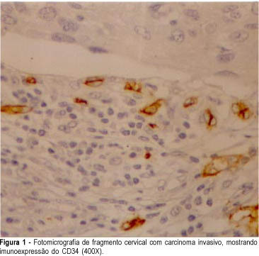
Purpose: to compare the efficiency of anti-factor VIII and anti-CD34 antibodies as vascular makers in cervical cancer, in cervical intraepithelial neoplasia and in normal cervix. Methods: using an immunohistochemical method, factor VIII-related antigen and leukocyte antigen CD34, we performed microvascular counts in 18 squamous cell carcinomas, in 15 cervical high-grade intraepithelial neoplasia, in 15 low-grade intraepithelial lesions and in 10 normal cervices. Using light microscopy we counted microvessels per 400X field in the most active areas of neovascularization with higher microvessel density in each case. Results: the average of microvessels stained with anti-CD34 in invasive carcinoma, high-grade intraepithelial lesions, low-grade intraepithelial lesions and in the normal cervices was 154, 134, 112 and 93, respectively. When we used anti-factor VIII the average was 56, 44, 33 and 30 vessels, following the same order. High-grade intraepithelial lesions and invasive carcinomas showed greater means number of vessels than normal tissue. Conclusions: the use of anti-CD34 allowed the detection of a greater number of vessels when compared to anti-factor VIII. However, we could observe that anti-factor VIII staining was able to significantly discriminate high-grade from low-grade lesions.
