Summary
Revista Brasileira de Ginecologia e Obstetrícia. 2002;24(9):601-608
DOI 10.1590/S0100-72032002000900006
Purpose: to appraise the value of ultrasonographic parameters for the diagnosis of fetal Down syndrome (T21), in order to permit its use in routine clinical practice. Methods: this is a prospective cohort study using various ultrasonographic parameters for the prediction of T21. A total of 1662 scans were evaluated in the cohort study and 289 examinations were analyzed as a differential sample to test the normality curve from October 1993 to November 2000. The statistical analysis was based on the calculation of intra- and interobserver variations, the construction of normality curves for the studied parameters, as well as their validity tests, and the calculation of sensitivity, specificity, relative risk, likelyhood ratio and posttest predictive values. Results: among 1662 cases, 22 fetuses (1.32%) with T21 were identified. The normality curves were built for nucal fold thickness, femur/foot ratio and nasal bone length. Renal pelvis had a semiquantitative distribution and the proposed cutoff level was 4.0 mm. Sensitivity, specificity, false positive rate, relative risk and likelyhood ratio for nucal fold measurements above the 95th percentile were 54.5%, 95.2%, 4.9%, 20.2 and 11, respectively. For nasal bone measurements below the 5th percentile, 59.0%, 90.1%, 9.0%, 13.4 and 6.5. For femur/foot ratio below the 5th percentile, 45.5%, 84.4%, 15.6%, 3.7 and 2,6. For renal pelvis greater than 4.0 mm, 36.4%, 89.2%, 10.9%, 4.5 and 3.4. For absent fifth finger middle phalanx, 22.7%, 98.1%, 1.9%, 13.2 and 11.9. For the presence of major malformations, 31.8%, 98.7%, 1.3%, 27.2 and 24,8. After calculating the probability rates and the incidence of T21 in different maternal ages, a table for posttest risk using ultrasonographic parameters was set up. Conclusions: normality curves and indices for the assessment of risk for fetal Down syndrome on a population basis were established by the utilization of different maternal ages and by multiplying factors proposed by the authors. It was not possible to establish a normality curve for renal pelvis measurements, because of their semiquantitative distribution.
Summary
Revista Brasileira de Ginecologia e Obstetrícia. 2002;24(9):585-591
DOI 10.1590/S0100-72032002000900004
Purpose: to report 15 breast cancer cases associated with pregnancy and to compare to a control group with breast ductal infiltrating carcinoma, evaluating clinical staging, metastatic axillary lymph node involvement, histopathologic aspects related to nuclear grade, histology grade and estrogen and progesterone hormonal receptors. Method: a retrospective study of 15 cases of patients with breast cancer associated with pregnancy, attended at Mastology Department in the Woman Health Reference Center, Pérola Byington Hospital, São Paulo, was done between September 1996 and April 2001. The evaluation of clinical staging, time of diagnosis and involved axillary lymph nodes was the main study basis. Also age, parity, histologic type, applied treatment, histologic characteristics regarding nuclear grade and histologic grade and the presence of hormonal receptors in the tumors were analyzed. Results: we observed that 7 patients (46.7%) presented a locally advanced breast cancer (clinical stage IIIA and IIIB) and that 3 patients (20%) presented a disseminated disease at the moment of diagnosis. The patients presented on average 2.4 involved axillary lymph nodes and in only one patient the lymph nodes were free of disease (6.6%). Regarding time of diagnosis, 40% of the tumors were diagnosed during the lactational period, 46.7% during the second trimester and 13.3% during the third trimester. The pregnant patients were compared to a control group of non-pregnant patients in the same age range, all of them with infiltrating breast carcinoma, and clinical staging, axillary lymph node involvement, nuclear grade, histologic grade and estrogen and progesterone hormonal receptors were evaluated. There was a statistically significant difference (p=0.0022) regarding clinical staging and axillary lymph node involvement (p=0.0017), and no statistically significant difference as concerns the remaining parameters. Conclusion: breast cancer associated with pregnancy is a neoplasia with a bad prognosis. There is no difference when comparing pregnant patients with non-pregnant patients in the same age range, the advanced clinical staging at the moment of diagnosis being the determinant factor for survival.
Summary
Revista Brasileira de Ginecologia e Obstetrícia. 2002;24(9):579-584
DOI 10.1590/S0100-72032002000900003
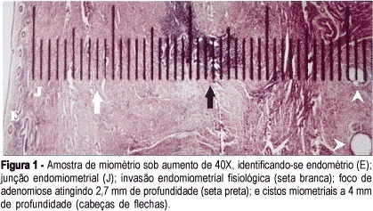
Purpose: to evaluate the sensitivity, specificity, positive and negative predictive values of a clinical and an ecographic method for adenomyosis diagnosis. Methods: a transversal study of validation of the diagnostic method was done, including 95 women in menacme submitted to hysterectomy for various causes. Adenomyosis was diagnosed through a clinical method in women aged 40 years or older, with 2 or more deliveries, increased menstrual bleeding associated with dysmenorrhea. The ecographic diagnosis was established if at least one myometrial ill defined area of abnormal ecotexture was found, which could be hypoechoic, hyperchoic, heterogeneous or cystic. Gold standard was histopathology, defined as the finding of endometrial glands or stroma more than 2.5 cm above the endomiometrial junction. Results: the clinical method had 68.2% sensitivity, 78.1% specificity, 48.4% positive predictive value and 89.1% negative predictive value. For the echographic method this figures were, respectively, 45.5%, 84.9%, 47.6% and 83.8%. Likelihood ratio was 3.11 for the clinical and 3.03 for the echographic method. Considering only those simultaneously positive cases by both methods, sensitivity was below 30% and specificity was near 100%. Considering all positive cases by one or the other method or concomitanty by both, the sensitivity reached 86% and specificity was 60%. Conclusion: the echographic method was not better than the clinical for the diagnosis of adenomyosis.

Summary
Revista Brasileira de Ginecologia e Obstetrícia. 2002;24(8):541-545
DOI 10.1590/S0100-72032002000800007
Purpose: to evaluate the diagnostic accuracy of sonohysterography as a diagnostic method for the evaluation of the uterine cavity in postmenopausal women with abnormal uterine cavity at conventional endovaginal sonography. Methods: this study consisted of the evaluation of 99 postmenopausal patients with abnormal uterine cavity on conventional endovaginal sonography, that was defined as endometrial thickness equal to or larger than 5 mm in a postmenopausal patient not on hormone replacement therapy, or endometrial thickness equal to or larger than 8 mm in patients on hormone replacement therapy, with irregular bleeding. These patients were subjected to sonohysterography, and specimens were obtained for pathologic examination by biopsy guided by histeroscopy in 92 patients, endometrial biopsy in four patientes and hysterectomy in three patients. The results of sonohysterography were compared with the pathologic findings, considered "gold standard". Results: there were eight cases of normal uterine cavity and 20 cases of atrophic endometrium and sonohysterography had high levels of specificity (97.8 and 97.5%) and low sensitivity (35 and 25%). There were high levels of sensitivity (92.3 and 75.0%) and specificity (94.1 and 97.9%) for polyps (65 cases) and submucous myomas (four cases). There were three cases of endometrial carcinoma and the sonohysterography had a sensitivity and specificity of 100%. Conclusions: sonohysterography showed to be accurate in the diagnostic of focal diseases (endometrial polyps and submucous myomas). There were three cases of endometrial cancer, and sonohysterography correctly diagnosed all of them. This method was also accurate to exclude endometrial abnormality. However, in the cases of diffusely thickened endometrium, the accuracy was low, because atrophic and normal endometrium on histopathology frequently appears as diffusely thickened endometrium at endovaginal sonography and sonohysterography. Sonohysterography did not lead to complications during and after the procedure.
Summary
Revista Brasileira de Ginecologia e Obstetrícia. 2002;24(8):535-539
DOI 10.1590/S0100-72032002000800006
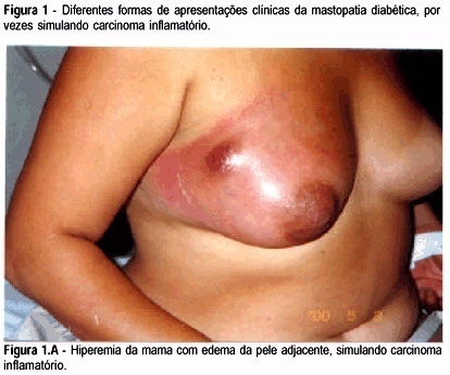
Purpose: to study the association between long-standing type 1 diabetes with bad glycemic control and breast inflammatory lesions which can simulate inflammatory carcinoma. Patients and Methods: eighteen patients were studied, retrospectively, in a mastology reference center from January 1998 to December 2001, presenting with breast inflammatory lesion with or without palpable mass. They were submitted to serum glucose and glycosylated hemoglobin determination, as well as image examination and histopathologic analysis, and diabetic mastopathy was diagnosed. Results: the patients' average age was 50.2 years, and all had insulin-dependent diabetes mellitus, with average disease time of 14.9 years. All patients, with no exception, had a bad glycemic control; the average blood glucose was 329.6 mg/dL and the glycosilated hemoglobin average was 9.7%. NPH insulin dose being applied per day was 37.2 units. Patients underwent a clinical treatment with antibiotics and control of the glycemic levels with NPH insulin and had resolution of the symptoms in about five weeks. Conclusion: the professionals involved in women health care must be aware of this inflammatory pathology of the breast and its benign characteristics to avoid unnecessary procedures sometimes with patient injury.

Summary
Revista Brasileira de Ginecologia e Obstetrícia. 2002;24(8):527-533
DOI 10.1590/S0100-72032002000800005
Purpose: to analyze the perinatal results of patients submitted to a 100 g oral glucose tolerance test (OGTT) during prenatal care at the Instituto Materno-Infantil de Pernambuco (IMIP), according to three different criteria. Methods: a cross-sectional study was conducted involving 210 pregnant patients attended at the IMIP, who were tested by a 100 g OGTT and had a singleton, topic pregnancy, without history of diabetes or glucose intolerance before pregnancy, and who delivered at the IMIP. The patients were classified into one of the following categories according to the levels found by OGTT: controls, mild hyperglycemia, Bertini's group, Carpenter's group and the National Diabetes Data Group (NDDG). These classes were then compared and association between the categories and preeclampsia, large for gestational age (LGA) newborns, rate of cesarean delivery, stillbirth, and mean birth weight was investigated. Results: the frequency of gestational diabetes was 48.1, 18.1, and 9% according to Bertini's, Carpenter and Coustan's and NDDG criteria, respectively, and mild hyperglycemia was present in 10.5%. Age of patients increased with a higher degree of carbohydrate intolerance. The groups did not differ regarding frequency of LGA, C-section, stillbirths, and birth weight. There was an increased frequency of preeclampsia among women with hyperglycemia and gestational diabetes according to Carpenter and Coustan's criteria. Conclusions: prevalence of gestational diabetes varied between 9 and 48% according to the different criteria, but maternal and perinatal results did not differ significantly among the groups. Strict diagnostic criteria can determine overdiagnosis without improvement of perinatal outcome.
Summary
Revista Brasileira de Ginecologia e Obstetrícia. 2002;24(8):521-526
DOI 10.1590/S0100-72032002000800004
Purpose: to assess the evolution of epileptic seizures during pregnancy and the occurrence of malformations in neonates born to epileptic mothers who used anticonvulsant drugs during pregnancy, as well as the perinatal characteristics of the newborns. Methods: a total of 126 medical records of epileptic patients seen at the high-risk pregnancy outpatient clinic were analyzed retrospectively in terms of the following variables: age, parity, diagnosis of the type of epileptic seizure, anticonvulsant drug used during the prenatal period, evolution of epileptic seizures during the prenatal period, type of delivery, gestational age at resolution, and perinatal characteristics of the newborns. Results: the incidence of pregnant women with epilepsy was 0.2% in relation to prenatal patients, with simple partial epilepsy being the most frequent type (40% of cases). Monotherapy was applied to 75% of the patients and carbamazepine was the most frequently used drug. Among the 111 patients evaluated in terms of course of the disease during pregnancy, 53% showed no change, 31% became worse and 16% improved. Normal delivery was performed in 62.5% of cases, with a satisfactory perinatal result in terms of Apgar score, and with a rate of low birth weight neonates above the values for low-risk populations. No fetal malformations were observed. Conclusion: epilepsy showed a favorable course during pregnancy and was not aggravated by the latter, with cases of worsening of signs and symptoms being associated with epilepsy of difficult control before pregnancy. Evaluation of the perinatal characteristics of the neonates showed satisfactory Apgar scores and evolution, indicating that epilepsy and anticonvulsant drugs do not cause severe impairment of intrapartum vitality. No cases of malformations or hemorrhagic complications were detected in the present study.