Summary
Revista Brasileira de Ginecologia e Obstetrícia. 2015;37(6):283-290
DOI 10.1590/SO100-720320150005292
To determine the basic expression of ABC transporters in an epithelial ovarian cancer cell line, and to investigate whether low concentrations of acetaminophen and ibuprofen inhibited the growth of this cell line in vitro.
TOV-21 G cells were exposed to different concentrations of acetaminophen (1.5 to 15 μg/mL) and ibuprofen (2.0 to 20 μg/mL) for 24 to 48 hours. The cellular growth was assessed using a cell viability assay. Cellular morphology was determined by fluorescence microscopy. The gene expression profile of ABC transporters was determined by assessing a panel including 42 genes of the ABC transporter superfamily.
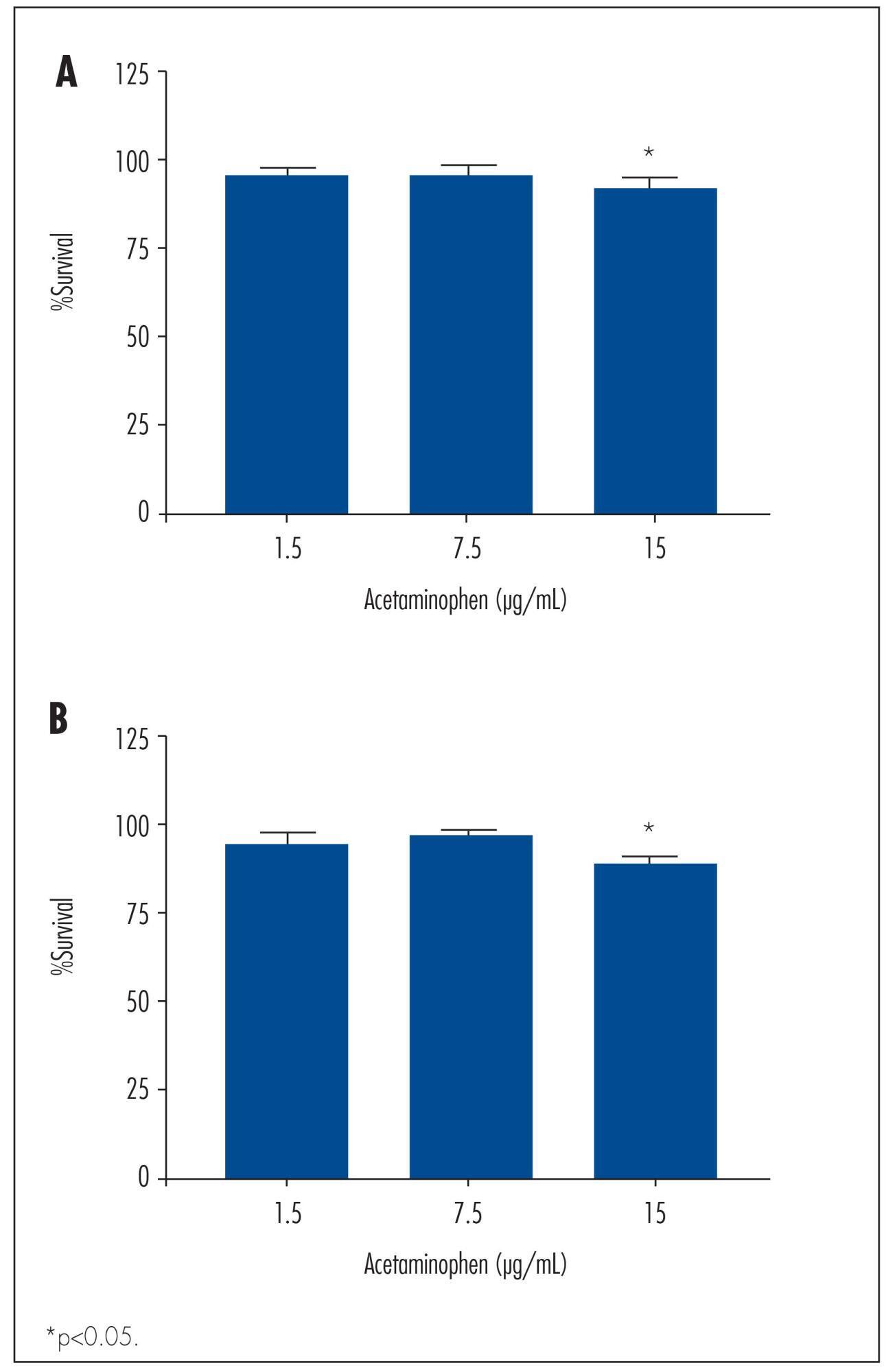
We observed a significant decrease in TOV-21 G cell growth after exposure to 15 μg/mL of acetaminophen for 24 (p=0.02) and 48 hours (p=0.01), or to 20 μg/mL of ibuprofen for 48 hours (p=0.04). Assessing the morphology of TOV-21 G cells did not reveal evidence of extensive apoptosis. TOV-21 G cells had a reduced expression of the genes ABCA1, ABCC3, ABCC4, ABCD3, ABCD4 and ABCE1 within the ABC transporter superfamily.
This study provides in vitro evidence of inhibitory effects of growth in therapeutic concentrations of acetaminophen and ibuprofen on TOV-21 G cells. Additionally, TOV-21 G cells presented a reduced expression of the ABCA1, ABCC3, ABCC4, ABCD3, ABCD4 and ABCE1 transporters.

Summary
Revista Brasileira de Ginecologia e Obstetrícia. 2014;36(3):113-117
DOI 10.1590/S0100-72032014000300004
To investigate the prevalence of chromosomal abnormalities in couples with two or more recurrent first trimester miscarriages of unknown cause.
The study was conducted on 151 women and 94 partners who had an obstetrical history of two or more consecutive first trimester abortions (1-12 weeks of gestation). The controls were 100 healthy women without a history of pregnancy loss. Chromosomal analysis was performed on peripheral blood lymphocytes cultured for 72 hours, using Trypsin-Giemsa (GTG) banding. In all cases, at least 30 metaphases were analyzed and 2 karyotypes were prepared, using light microscopy. The statistical analysis was performed using the Student t-test for normally distributed data and the Mann-Whitney test for non-parametric data. The Kruskal-Wallis test or Analysis of Variance was used to compare the mean values between three or more groups. The software used was Statistical Package for the Social Sciences (SPSS), version 17.0.
The frequency of chromosomal abnormalities in women with recurrent miscarriages was 7.3%, including 4.7% with X-chromosome mosaicism, 2% with reciprocal translocations and 0.6% with Robertsonian translocations. A total of 2.1% of the partners of women with recurrent miscarriages had chromosomal abnormalities, including 1% with X-chromosome mosaicism and 1% with inversions. Among the controls, 1% had mosaicism.
An association between chromosomal abnormalities and recurrent miscarriage in the first trimester of pregnancy (OR=7.7; 95%CI 1.2--170.5) was observed in the present study. Etiologic identification of genetic factors represents important clinical information for genetic counseling and orientation of the couple about the risk for future pregnancies and decreases the number of investigations needed to elucidate the possible causes of miscarriages.
Summary
Revista Brasileira de Ginecologia e Obstetrícia. 2014;36(2):72-78
DOI 10.1590/S0100-72032014000200005
To compare salivary and serum cortisol levels, salivary alpha-amylase (sAA), and unstimulated whole saliva (UWS) flow rate in pregnant and non-pregnant women.
A longitudinal study was conducted at a health promotion center of a university hospital. Nine pregnant and 12 non-pregnant women participated in the study. Serum and UWS were collected and analyzed every trimester and twice a month during the menstrual cycle. The salivary and serum cortisol levels were determined by chemiluminescence assay and the sAA was processed in an automated biochemistry analyzer.
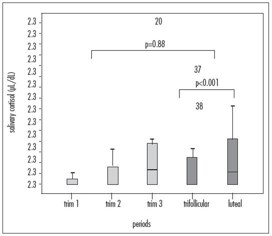
Significant differences between the pregnant and non-pregnant groups were found in median [interquartile range] levels of serum cortisol (23.8 µL/dL [19.4-29.4] versus 12.3 [9.6-16.8], p<0.001) and sAA (56.7 U/L [30.9-82.2] versus 31.8 [18.1-53.2], p<0.001). Differences in salivary and serum cortisol (µL/dL) and sAA levels in the follicular versus luteal phase were observed (p<0.001). Median UWS flow rates were similar in pregnant (0.26 [0.15-0.30] mL/min) and non-pregnant subjects (0.23 [0.20-0.32] mL/min). Significant correlations were found between salivary and serum cortisol (p=0.02) and between salivary cortisol and sAA (p=0.01).
Serum cortisol and sAA levels are increased during pregnancy. During the luteal phase of the ovarian cycle, salivary cortisol levels increase, whereas serum cortisol and sAA levels decline.

Summary
Revista Brasileira de Ginecologia e Obstetrícia. 2014;36(1):35-39
DOI 10.1590/S0100-72032014000100008
O objetivo do presente estudo longitudinal foi avaliar o valor da ultrassonografia Doppler das artérias uterinas no segundo e terceiro trimestres de gestação para a predição de desfecho adverso da gravidez em mulheres de baixo risco.
De julho de 2011 até agosto de 2012, 205 gestantes de feto único atendidas em nossa clínica de pré-natal foram incluídas no presente estudo prospectivo e avaliadas em termos de dados demográficos e obstétricos. As pacientes foram submetidas à avaliação de ultrassom durante o segundo e terceiro trimestres, incluindo avaliação Doppler das artérias uterinas bilaterais, visando determinar os valores do índice de pulsatilidade (IP) e do índice de resistência (IR), bem como a presença de incisura diastólica precoce. O desfecho do presente estudo foi a avaliação da sensibilidade, especificidade, valor preditivo positivo (VPP) e valor negativo preditivo (VNP) da ultrassonografia Doppler das artérias uterinas para a predição de desfechos adversos da gravidez, incluindo pré-eclâmpsia, natimortalidade, descolamento prematuro da placenta e trabalho de parto prematuro.
A média de idade das gestantes foi de 26,4±5,11 anos. Os valores de IP e IR das artérias uterinas para o primeiro (IP: 1,1±0,42 versus 1,53±0,59, p=0,002; IR: 0,55±0,09 versus 0.72±0.13, p=0,000, respectivamente) e para o terceiro trimestre (IP: 0,77±0,31 versus 1,09±0,46, p=0,000; IR: 0,46±0,10 versus 0,60±0,14, p=0,010, respectivamente) foram significativamente maiores em pacientes com desfecho adverso da gravidez em relação às mulheres com desfecho normal. A combinação de IP e IR > percentil 95 e a presença de incisura bilateral apresentou sensibilidade e especificidade de 36,1 e 97%, respectivamente, no segundo trimestre e de 57,5 e 98,2% no terceiro trimestre.
Com base no presente estudo, o Doppler das artérias uterinas parece ser ferramenta valiosa para a predição de uma variedade de desfechos adversos no segundo e terceiro trimestres de gestação.
Summary
Revista Brasileira de Ginecologia e Obstetrícia. 2014;36(1):35-39
DOI 10.1590/S0100-72032014000100008
The aim of this longitudinal study was to investigate the value of uterine artery Doppler sonography during the second and third trimesters in the prediction of adverse pregnancy outcome in low-risk women.
From July 2011 to August 2012, a total of 205 singleton pregnant women presenting at our antenatal clinic were enrolled in this prospective study and were assessed for baseline demographic and obstetric data. They underwent ultrasound evaluation at the time of second and third trimesters, both included Doppler assessment of bilateral uterine arteries to determine the values of the pulsatility index (PI) and resistance index (RI) and presence of early diastolic notch. The endpoint of this study was assessing the sensitivity, specificity, positive predictive value (PPV) and negative predictive value (NPV) of Doppler ultrasonography of the uterine artery, for the prediction of adverse pregnancy outcomes including preeclampsia, stillbirth, placental abruption and preterm labor.
The mean age of cases was 26.4±5.11. The uterine artery PI and RI values for both second (PI: 1.1±0.42 versus 1.53±0.59, p=0.002; RI: 0.55±0.09 versus 0.72±0.13, p=0.000 respectively) and third-trimester (PI: 0.77±0.31 versus 1.09±0.46, p=0.000; RI: 0.46±0.10 versus 0.60±0.14, p=0.010 respectively) evaluations were significantly higher in patients with adverse pregnancy outcome than in normal women. Combination of PI and RI >95th percentile and presence of bilateral notch in second trimester get sensitivity and specificity of 36.1 and 97% respectively, while these measures were 57.5 and 98.2% in third trimester.
According to our study, it seems that uterine artery Doppler may be a valuable tool for the prediction of a variety of adverse outcomes in second and third trimesters.
Summary
Revista Brasileira de Ginecologia e Obstetrícia. 2013;35(9):407-412
DOI 10.1590/S0100-72032013000900005
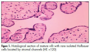
PURPOSE: In placentas from uncomplicated pregnancies, Hofbauer cells either disappear or become scanty after the fourth to fifth month of gestation. Immunohistochemistry though, reveals that a high percentage of stromal cells belong to Hofbauer cells. The aim of this study was to investigate the changes in morphology and density of Hofbauer cells in placentas from normal and pathological pregnancies. METHODS: Seventy placentas were examined: 16 specimens from normal term pregnancies, 10 from first trimester's miscarriages, 26 from cases diagnosed with chromosomal abnormality of the fetus, and placental tissue specimens complicated with intrauterine growth restriction (eight) or gestational diabetes mellitus (10). A histological study of hematoxylin-eosin (HE) sections was performed and immunohistochemical study was performed using the markers: CD 68, Lysozyme, A1 Antichymotrypsine, CK-7, vimentin, and Ki-67. RESULTS: In normal term pregnancies, HE study revealed Hofbauer cells in 37.5% of cases while immunohistochemistry revealed in 87.5% of cases. In first trimester's miscarriages and in cases with prenatal diagnosis of fetal chromosomal abnormalities, both basic and immunohistochemical study were positive for Hofbauer cells. In pregnancies complicated with intrauterine growth restriction or gestational diabetes mellitus, a positive immunoreaction was observed in 100 and 70% of cases, respectively. CONCLUSIONS: Hofbauer cells are present in placental villi during pregnancy, but with progressively reducing density. The most specific marker for their detection seems to be A1 Antichymotrypsine. It is remarkable that no mitotic activity of Hofbauer cells was noticed in our study, as the marker of cellular multiplication Ki-67 was negative in all examined specimens.

Summary
Revista Brasileira de Ginecologia e Obstetrícia. 2013;35(8):357-362
DOI 10.1590/S0100-72032013000800004
PURPOSE: To establish reference values for the first trimester uterine artery resistance index (UtA-RI) and pulsatility index (UtA-PI) in healthy singleton pregnant women from Northeast Brazil. METHODS: A prospective observational cohort study including 409 consecutive singleton pregnancies undergoing routine early ultrasound screening at 11 - 14 weeks of gestation was performed. The patients responded to a questionnaire to assess maternal epidemiological characteristics. The left and right UtA-PI and UtA-RI were examined by color and pulsed Doppler by transabdominal technique and the mean UtA-PI, mean UtA-RI and the presence of bilateral protodiastolic notching were recorded. Quartile regression was used to estimate reference values. RESULTS: The mean±standard deviation UtA-RI and UtA-PI were 0.7±0.1 and 1.5±0.5, respectively. When segregated for gestation age, mean UtA-PI was 1.6±0.5 at 11 weeks, 1.5±0.6 at 12 weeks, 1.4±0.4 at 13 weeks and 1.3±0.4 at 14 weeks' gestation and mean UtA-RI was 0.7±0.1 at 11 weeks, 0.7±0.1 at 12 weeks, 0.6±0.1 at 13 weeks and 0.6±0.1 at 14 weeks' gestation. Uterine artery bilateral notch was present in 261 (63.8%) patients. We observed that the 5th and 95th percentiles of the UtA-PI and UtA-RI uterine arteries were 0.7 and 2.3 and, 0.5 and 0.8, respectively. CONCLUSION: Normal reference range of uterine artery Doppler in healthy singleton pregnancies from Northeast Brazil was established. The 95th percentile of UtA-PI and UtA-RI values may serve as a cut-off for future prediction of pregnancy complications studies (i.e., pre-eclampsia) in Northeast Brazil.