-
Case Report03-01-2018
Postpartum Genital Melanoma – A Case Report
Revista Brasileira de Ginecologia e Obstetrícia. 2018;40(3):163-167
Abstract
Case ReportPostpartum Genital Melanoma – A Case Report
Revista Brasileira de Ginecologia e Obstetrícia. 2018;40(3):163-167
Views154See moreAbstract
Melanomas of the female genital tract may occur in the vulva, the vagina, the ovary or the cervix.Pregnancy has been considered an aggravating factor in the evolution and prognosis of melanoma. A 35-year-old female presented with vaginal bleeding 2 months after a term cesarean delivery. An endovaginal ultrasound revealed a lesion in the uterine cervix. The pathological report revealed a small round-cell neoplasm, and the immunohistochemistry confirmed the diagnosis of malignant melanoma. A positron emission tomography revealed an expansive hypermetabolic lesion centered on the cervix, and hypermetabolic lesions in the liver and right kidney. Non-surgical treatment was provided, with biochemotherapy followed by ipilimumab and nivolumab. The patient died one year later. Postpartum vaginal bleeding, even if late-onset, should be investigated, as it may be a pregnancy-associated malignant melanoma, which has a poor prognosis.
Views154

This is an Open Access article distributed under the terms of the Creative Commons Attribution License, which permits unrestricted use, distribution, and reproduction in any medium, provided the original work is properly cited. Abstract
Case ReportPostpartum Genital Melanoma – A Case Report
Revista Brasileira de Ginecologia e Obstetrícia. 2018;40(3):163-167
Views154See moreAbstract
Melanomas of the female genital tract may occur in the vulva, the vagina, the ovary or the cervix.Pregnancy has been considered an aggravating factor in the evolution and prognosis of melanoma. A 35-year-old female presented with vaginal bleeding 2 months after a term cesarean delivery. An endovaginal ultrasound revealed a lesion in the uterine cervix. The pathological report revealed a small round-cell neoplasm, and the immunohistochemistry confirmed the diagnosis of malignant melanoma. A positron emission tomography revealed an expansive hypermetabolic lesion centered on the cervix, and hypermetabolic lesions in the liver and right kidney. Non-surgical treatment was provided, with biochemotherapy followed by ipilimumab and nivolumab. The patient died one year later. Postpartum vaginal bleeding, even if late-onset, should be investigated, as it may be a pregnancy-associated malignant melanoma, which has a poor prognosis.
This is an Open Access article distributed under the terms of the Creative Commons Attribution License, which permits unrestricted use, distribution, and reproduction in any medium, provided the original work is properly cited. -
Case Report02-01-2018
Modified Pereira Suture as an Effective Option to Treat Postpartum Hemorrhage due to Uterine Atony
Revista Brasileira de Ginecologia e Obstetrícia. 2018;40(2):92-95
Abstract
Case ReportModified Pereira Suture as an Effective Option to Treat Postpartum Hemorrhage due to Uterine Atony
Revista Brasileira de Ginecologia e Obstetrícia. 2018;40(2):92-95
Views154See moreAbstract
Nowadays, postpartum hemorrhage is the major cause of maternal mortality and morbidity worldwide. Uterine atony is its main cause; thus, prophylactic measures, as well as medical and surgical fast approaches, have been developed to manage it. The uterine compression sutures are a possible treatment that preserves the uterus and, consequently, the fertility potential. Bearing that in mind, we report two cases of postpartum hemorrhage after caesarean section, successfully treated with a new modification of Pereira suture - longitudinal and transverse uterine sutures were applied after no response was registered to the first-line therapies. Both women recovered, and the postpartum evaluation revealed a normal uterus with an adequate blood supply, suggesting potential fertility, as described in the literature regarding this kind of therapeutic approach.
Views154This is an Open Access article distributed under the terms of the Creative Commons Attribution License, which permits unrestricted use, distribution, and reproduction in any medium, provided the original work is properly cited. Abstract
Case ReportModified Pereira Suture as an Effective Option to Treat Postpartum Hemorrhage due to Uterine Atony
Revista Brasileira de Ginecologia e Obstetrícia. 2018;40(2):92-95
Views154See moreAbstract
Nowadays, postpartum hemorrhage is the major cause of maternal mortality and morbidity worldwide. Uterine atony is its main cause; thus, prophylactic measures, as well as medical and surgical fast approaches, have been developed to manage it. The uterine compression sutures are a possible treatment that preserves the uterus and, consequently, the fertility potential. Bearing that in mind, we report two cases of postpartum hemorrhage after caesarean section, successfully treated with a new modification of Pereira suture - longitudinal and transverse uterine sutures were applied after no response was registered to the first-line therapies. Both women recovered, and the postpartum evaluation revealed a normal uterus with an adequate blood supply, suggesting potential fertility, as described in the literature regarding this kind of therapeutic approach.
This is an Open Access article distributed under the terms of the Creative Commons Attribution License, which permits unrestricted use, distribution, and reproduction in any medium, provided the original work is properly cited. -
Case Report01-01-2018
When an Unexpected Diagnosis Occurs: a Vaginal Premenopausal Sarcoma
Revista Brasileira de Ginecologia e Obstetrícia. 2018;40(1):47-52
Abstract
Case ReportWhen an Unexpected Diagnosis Occurs: a Vaginal Premenopausal Sarcoma
Revista Brasileira de Ginecologia e Obstetrícia. 2018;40(1):47-52
Views86See moreAbstract
Vaginal cancer is a rare entity. The evidence on its management resides mostly in clinical cases or small case series. Of the histological types, the most frequent is the squamous cell carcinoma, followed by adenocarcinoma. But what to do when identifying an even more infrequent sarcoma in a premenopausal woman? In this study, we describe the case of a 53-year-old woman presenting with metrorrhagia for two months, who was evaluated after an intense episode. A necrotic and ulcerative vaginal swelling was documented and then submitted to biopsy, which revealed a vaginal sarcoma. The patient was referred to radiation therapy with 50 Gy (aiming to control the symptoms and to cause tumor reduction for posterior pelvic exenteration with intraoperative radiotherapy) and developed an extra-pelvic metastization at the end of the treatment, which caused a fast negative outcome. Despite the initial poor prognosis, a chemo-irradiation or primary surgery regimen might have achieved (although with greater side effects) a better survival. This case-report entails a discussion about the strategies to manage vaginal sarcoma in advanced stage and in premenopausal women.
Views86This is an Open Access article distributed under the terms of the Creative Commons Attribution License, which permits unrestricted use, distribution, and reproduction in any medium, provided the original work is properly cited. Abstract
Case ReportWhen an Unexpected Diagnosis Occurs: a Vaginal Premenopausal Sarcoma
Revista Brasileira de Ginecologia e Obstetrícia. 2018;40(1):47-52
Views86See moreAbstract
Vaginal cancer is a rare entity. The evidence on its management resides mostly in clinical cases or small case series. Of the histological types, the most frequent is the squamous cell carcinoma, followed by adenocarcinoma. But what to do when identifying an even more infrequent sarcoma in a premenopausal woman? In this study, we describe the case of a 53-year-old woman presenting with metrorrhagia for two months, who was evaluated after an intense episode. A necrotic and ulcerative vaginal swelling was documented and then submitted to biopsy, which revealed a vaginal sarcoma. The patient was referred to radiation therapy with 50 Gy (aiming to control the symptoms and to cause tumor reduction for posterior pelvic exenteration with intraoperative radiotherapy) and developed an extra-pelvic metastization at the end of the treatment, which caused a fast negative outcome. Despite the initial poor prognosis, a chemo-irradiation or primary surgery regimen might have achieved (although with greater side effects) a better survival. This case-report entails a discussion about the strategies to manage vaginal sarcoma in advanced stage and in premenopausal women.
This is an Open Access article distributed under the terms of the Creative Commons Attribution License, which permits unrestricted use, distribution, and reproduction in any medium, provided the original work is properly cited. -
Case Report11-01-2017
Pregnancy in Non-Communicating Unicornuate Uterus: Diagnosis Difficulty and Outcomes – a Case Report
Revista Brasileira de Ginecologia e Obstetrícia. 2017;39(11):640-644
Abstract
Case ReportPregnancy in Non-Communicating Unicornuate Uterus: Diagnosis Difficulty and Outcomes – a Case Report
Revista Brasileira de Ginecologia e Obstetrícia. 2017;39(11):640-644
Views184See moreAbstract
Approximately 1 in every 76,000 pregnancies develops within a unicornuate uterus with a rudimentary horn.Müllerian uterus anomalies are often asymptomatic, thus, the diagnosis is a challenge, and it is usually made during the gestation or due to its complications, such as uterine rupture, pregnancy-induced hypertension, antepartum, postpartum bleeding and intrauterine growth restriction (IUGR). In order to avoid unnecessary cesarean sections and the risks they involve, the physicians should consider the several approaches and for how long it is feasible to perform labor induction in suspected cases of pregnancy in a unicornuate uterus with a rudimentary horn, despite the rarity of the anomaly. This report describes a case of a unicornuate uterus in which a pregnancy developed in the non-communicating rudimentary horn and the consequences of the delayed diagnosis.
Views184This is an Open Access article distributed under the terms of the Creative Commons Attribution License, which permits unrestricted use, distribution, and reproduction in any medium, provided the original work is properly cited. Abstract
Case ReportPregnancy in Non-Communicating Unicornuate Uterus: Diagnosis Difficulty and Outcomes – a Case Report
Revista Brasileira de Ginecologia e Obstetrícia. 2017;39(11):640-644
Views184See moreAbstract
Approximately 1 in every 76,000 pregnancies develops within a unicornuate uterus with a rudimentary horn.Müllerian uterus anomalies are often asymptomatic, thus, the diagnosis is a challenge, and it is usually made during the gestation or due to its complications, such as uterine rupture, pregnancy-induced hypertension, antepartum, postpartum bleeding and intrauterine growth restriction (IUGR). In order to avoid unnecessary cesarean sections and the risks they involve, the physicians should consider the several approaches and for how long it is feasible to perform labor induction in suspected cases of pregnancy in a unicornuate uterus with a rudimentary horn, despite the rarity of the anomaly. This report describes a case of a unicornuate uterus in which a pregnancy developed in the non-communicating rudimentary horn and the consequences of the delayed diagnosis.
This is an Open Access article distributed under the terms of the Creative Commons Attribution License, which permits unrestricted use, distribution, and reproduction in any medium, provided the original work is properly cited. -
Case Report10-01-2017
Fetal Skeletal Lethal Dysplasia: Case Report Displasia Esquelética Letal Fetal: Relato de Caso
Revista Brasileira de Ginecologia e Obstetrícia. 2017;39(10):576-582
Abstract
Case ReportFetal Skeletal Lethal Dysplasia: Case Report Displasia Esquelética Letal Fetal: Relato de Caso
Revista Brasileira de Ginecologia e Obstetrícia. 2017;39(10):576-582
Views192See moreAbstract
The clinical management and decision-making in pregnancies in which there is suspicion of lethal fetal malformations during the prenatal period, such as lethal skeletal dysplasia (SD), demand a multidisciplinary approach coordinated by an experienced physician. Based on the presentation of a case of osteogenesis imperfecta type IIA, we offer and discuss recommendations with the intention of organizing clinical and laboratory investigations aiming toward the clinical management, prognosis, and etiological diagnosis of these malformations, as well as genetic counselling to patients who wish to become pregnant.
Views192This is an Open Access article distributed under the terms of the Creative Commons Attribution License, which permits unrestricted use, distribution, and reproduction in any medium, provided the original work is properly cited. Abstract
Case ReportFetal Skeletal Lethal Dysplasia: Case Report Displasia Esquelética Letal Fetal: Relato de Caso
Revista Brasileira de Ginecologia e Obstetrícia. 2017;39(10):576-582
Views192See moreAbstract
The clinical management and decision-making in pregnancies in which there is suspicion of lethal fetal malformations during the prenatal period, such as lethal skeletal dysplasia (SD), demand a multidisciplinary approach coordinated by an experienced physician. Based on the presentation of a case of osteogenesis imperfecta type IIA, we offer and discuss recommendations with the intention of organizing clinical and laboratory investigations aiming toward the clinical management, prognosis, and etiological diagnosis of these malformations, as well as genetic counselling to patients who wish to become pregnant.
This is an Open Access article distributed under the terms of the Creative Commons Attribution License, which permits unrestricted use, distribution, and reproduction in any medium, provided the original work is properly cited. 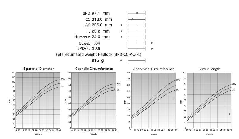
-
Case Report09-01-2017
Acute Abdomen Secondary to Ruptured Epithelial Ovarian Cancer during Pregnancy: The Relevance of Teamwork
Revista Brasileira de Ginecologia e Obstetrícia. 2017;39(9):513-515
Abstract
Case ReportAcute Abdomen Secondary to Ruptured Epithelial Ovarian Cancer during Pregnancy: The Relevance of Teamwork
Revista Brasileira de Ginecologia e Obstetrícia. 2017;39(9):513-515
Views181Abstract
Acute abdomen secondary to epithelial ovarian cancer rupture during pregnancy is a rare event. Our aim is to present how the work of a coordinated multidisciplinary team in a case of ruptured epithelial ovarian cancer during pregnancy is feasible to obtain the best results possible. A 34-year-old woman during the 37th week of her first gestation presented with an acute abdomen. During laparotomy, a ruptured 16.5-cm left ovarian tumor was detected; the tumor was extirpated and sent to pathologic evaluation. In the meantime, a Kerr cesarean section was performed, and a healthy female neonate was born. The tumor was diagnosed as a cystadenocarcinoma; therefore, the family and the combined surgical team (obstetricians and a surgical oncologist) decided to complete a definitive radical ovarian cancer surgery: hysterectomy, right salpingooophorectomy, lymphadenectomy, omentectomy and appendectomy. The patient’s postoperative evolution was uneventful, and she was sent to adjuvant chemotherapy.
Key-words Acute abdomenepithelial ovarian cancerOvarian cancerPregnancyruptured epithelial ovarian cancerSee moreViews181This is an Open Access article distributed under the terms of the Creative Commons Attribution License, which permits unrestricted use, distribution, and reproduction in any medium, provided the original work is properly cited. Abstract
Case ReportAcute Abdomen Secondary to Ruptured Epithelial Ovarian Cancer during Pregnancy: The Relevance of Teamwork
Revista Brasileira de Ginecologia e Obstetrícia. 2017;39(9):513-515
Views181Abstract
Acute abdomen secondary to epithelial ovarian cancer rupture during pregnancy is a rare event. Our aim is to present how the work of a coordinated multidisciplinary team in a case of ruptured epithelial ovarian cancer during pregnancy is feasible to obtain the best results possible. A 34-year-old woman during the 37th week of her first gestation presented with an acute abdomen. During laparotomy, a ruptured 16.5-cm left ovarian tumor was detected; the tumor was extirpated and sent to pathologic evaluation. In the meantime, a Kerr cesarean section was performed, and a healthy female neonate was born. The tumor was diagnosed as a cystadenocarcinoma; therefore, the family and the combined surgical team (obstetricians and a surgical oncologist) decided to complete a definitive radical ovarian cancer surgery: hysterectomy, right salpingooophorectomy, lymphadenectomy, omentectomy and appendectomy. The patient’s postoperative evolution was uneventful, and she was sent to adjuvant chemotherapy.
Key-words Acute abdomenepithelial ovarian cancerOvarian cancerPregnancyruptured epithelial ovarian cancerSee moreThis is an Open Access article distributed under the terms of the Creative Commons Attribution License, which permits unrestricted use, distribution, and reproduction in any medium, provided the original work is properly cited. 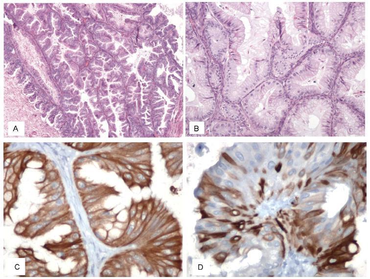
-
Case Report09-01-2017
Uterine Extramedullary Plasmacytoma as a Primary Manifestation of Multiple Myeloma
Revista Brasileira de Ginecologia e Obstetrícia. 2017;39(9):516-520
Abstract
Case ReportUterine Extramedullary Plasmacytoma as a Primary Manifestation of Multiple Myeloma
Revista Brasileira de Ginecologia e Obstetrícia. 2017;39(9):516-520
Views130Abstract
The association between plasmacytomas and multiple myeloma (MM) is well-described, and in about one third of the cases of plasmacytoma the additional study will lead to the diagnosis of MM. The finding of plasmacytomas in the genital tract is extremely rare, with sparse cases described in the literature, and these cases pose a challenge regarding the optimal guidance and treatment. This paper describes a case of uterine extramedullary plasmacytoma in a 79-year-old woman with complaints of postmenopausal abnormal uterine bleeding. The complementary study led to the diagnosis of uterine plasmacytoma and, subsequently, of MM. Despite the unfavorable outcome of this case, we consider pertinent to report it because it constitutes a differential diagnosis to be taken into account in the approach of pelvic masses.
Key-words multiple myelomaplasmacytomaSee moreViews130This is an Open Access article distributed under the terms of the Creative Commons Attribution License, which permits unrestricted use, distribution, and reproduction in any medium, provided the original work is properly cited. Abstract
Case ReportUterine Extramedullary Plasmacytoma as a Primary Manifestation of Multiple Myeloma
Revista Brasileira de Ginecologia e Obstetrícia. 2017;39(9):516-520
Views130Abstract
The association between plasmacytomas and multiple myeloma (MM) is well-described, and in about one third of the cases of plasmacytoma the additional study will lead to the diagnosis of MM. The finding of plasmacytomas in the genital tract is extremely rare, with sparse cases described in the literature, and these cases pose a challenge regarding the optimal guidance and treatment. This paper describes a case of uterine extramedullary plasmacytoma in a 79-year-old woman with complaints of postmenopausal abnormal uterine bleeding. The complementary study led to the diagnosis of uterine plasmacytoma and, subsequently, of MM. Despite the unfavorable outcome of this case, we consider pertinent to report it because it constitutes a differential diagnosis to be taken into account in the approach of pelvic masses.
Key-words multiple myelomaplasmacytomaSee moreThis is an Open Access article distributed under the terms of the Creative Commons Attribution License, which permits unrestricted use, distribution, and reproduction in any medium, provided the original work is properly cited. 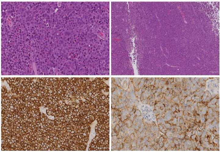
-
Case Report08-01-2017
Differential Diagnosis between Bartholin Cyst and Vulvar Leiomyoma: Case Report
Revista Brasileira de Ginecologia e Obstetrícia. 2017;39(8):433-435
Abstract
Case ReportDifferential Diagnosis between Bartholin Cyst and Vulvar Leiomyoma: Case Report
Revista Brasileira de Ginecologia e Obstetrícia. 2017;39(8):433-435
Views170See moreAbstract
Genital leiomyomas are rare tumors that can often be misdiagnosed as Bartholin cyst. We report a case of a 32-year-old patient who had a cystic nodulation in the left labium majus that was suggestive of Bartholin cyst. A resection surgery was performed, and the definitive histopathology diagnosis was vulvar leiomyoma. The macroscopic features of cystic lesions difficult the differential diagnosis between leiomyoma and Bartholin cyst; therefore, a histopathologic examination is often recommended.
Views170This is an Open Access article distributed under the terms of the Creative Commons Attribution License, which permits unrestricted use, distribution, and reproduction in any medium, provided the original work is properly cited. Abstract
Case ReportDifferential Diagnosis between Bartholin Cyst and Vulvar Leiomyoma: Case Report
Revista Brasileira de Ginecologia e Obstetrícia. 2017;39(8):433-435
Views170See moreAbstract
Genital leiomyomas are rare tumors that can often be misdiagnosed as Bartholin cyst. We report a case of a 32-year-old patient who had a cystic nodulation in the left labium majus that was suggestive of Bartholin cyst. A resection surgery was performed, and the definitive histopathology diagnosis was vulvar leiomyoma. The macroscopic features of cystic lesions difficult the differential diagnosis between leiomyoma and Bartholin cyst; therefore, a histopathologic examination is often recommended.
This is an Open Access article distributed under the terms of the Creative Commons Attribution License, which permits unrestricted use, distribution, and reproduction in any medium, provided the original work is properly cited.