Summary
Revista Brasileira de Ginecologia e Obstetrícia. 2012;34(5):221-227
DOI 10.1590/S0100-72032012000500006
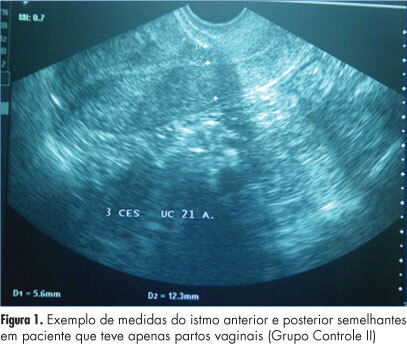
PURPOSE: To evaluate the thickness of the lower uterine segment by transvaginal ultrasound in a group of non-pregnant women and to describe the morphologic findings in the scar of those submitted to cesarean section. METHODS: A retrospective study of 155 transvaginal ultrasound images obtained from premenopausal and non-pregnant women, conducted between January 2008 and November 2011. the subjects were divided into three groups: women who were never pregnant (Control Group I), women with previous vaginal deliveries (Control Group II) and women with previous cesarean section (Observation Group). We excluded women with a retroverted uterus, intrauterine device users, pregnant women and those with less than one year of tsince the last obstetrical event. The data were analyzed statistically with Statistica®, version 8.0 software. ANOVA and LSD were used to compare the groups regarding quantitative variables and the Student's t-test was used to compare the thickness of the anterior and posterior isthmus. The Spearman correlation coefficient was calculated to estimate the association between quantitative variables. P values <0.05 were considered statistically significant. RESULTS: There was significant difference between the thickness of the anterior and posterior isthmus only in the group of women with previous cesarean section. Comparing the groups two by two, no significant differences between the thickness of the anterior and posterior isthmus were observed in the Control Groups, but this difference was significant when we compared the Observation Group with each Control Group. In the Observation Group, no correlation was found between the thickness of the isthmus and the number of previous cesarean deliveries or the time elapsed since the last birth. A niche was found in the cesarean scar in 30.6% of the women in the Observation Group, 93% of whom complained of post-menstrual bleeding. CONCLUSION: The relationship between the thickness of the anterior and posterior wall of the lower uterine segment by transvaginal ultrasound is a suitable method for the evaluation of the uterine lower segment in women with previous cesarean sections.

Summary
Revista Brasileira de Ginecologia e Obstetrícia. 2012;34(5):228-234
DOI 10.1590/S0100-72032012000500007
PURPOSE: To evaluate habits of sun exposure and sun protection of pregnant women in a public hospital, to assess orientation about photo protection during the prenatal care, and to detect the presence of melasma and its impact on their quality of life. METHODS: A descriptive cross sectional study conducted among women of 18 years old and older, after delivery, who participated in a program of prenatal care in the South Region of Brazil. The sample was non-probabilistic by convenience. Data collection occurred from July to August 2011 through direct interview using a structured questionnaire to obtain personal information and photo protection habits during pregnancy, skin assessment and photographic record of lesions through informed consent. The skin was classified per Fitzpatrick's phototypes and the melasma was diagnosed clinically. In the patients with melasma, the MELASQoL-PB version was applied. The analysis was performed using Statistica®, version 8.0, and the significance level of p<0.05. RESULTS: In the sample (109 mothers) predominated white women (60.6% phototype III), young (average age 24.4 years SD=6.1) and housewives (59.6%). The majority (80%) stayed exposed to sunlight for 1-2 hours per day between 10 am and 3 pm, and from those (72%) did not apply any photoprotection due to lack of sunscreen habit. Other physical means of sun protection were used by 15% of these patients. Information during prenatal care about the risks of sun exposure was reported by 34% of the mothers interviewed. There was a trend toward a significant association between prenatal guidance and daily use of sunscreen (p=0.088). About 20% of mothers had melasma. The average score MELASQol-PB (25) showed a negative impact on quality of life of these patients. CONCLUSION: In these women, sun exposure occurred at inappropriate times, without proper guidance and without the use of an effective sunscreen. The mothers with melasma complained about the appearance of their skin, frustration and embarrassment.
Summary
Revista Brasileira de Ginecologia e Obstetrícia. 2012;34(5):235-242
DOI 10.1590/S0100-72032012000500008
PURPOSE: To evaluate the survival and complications associated with prematurity of infants with less than 32 weeks of gestation. METHODS: It was done a prospective cohort study. All preterm infants with a gestational age between 25 and 31 weeks and 6 days, born alive without congenital anomalies and admitted to the NICU between August 1st, 2009 and October 31st, 2010 were included. Newborns were stratified into three groups: G25, 25 to 27 weeks and 6 days; G28, 28 to 29 weeks and 6 days; G30, 30 to 31 weeks and 6 days, and they were followed up to 28 days. Survival at 28 days and complications associated with prematurity were evaluated. Data were analyzed statistically by c² test, analysis of variance, Kruskal-Wallis test, odds ratio with confidence interval (CI) and multiple logistic regression, with significance set at 5%. RESULTS: The cohort comprised 198 preterm infants (G25=59, G28=43 and G30=96). The risk of death was significantly higher in G25 and G28 compared to G30 (RR=4.14, 95%CI 2.23-7.68 and RR=2.84, 95%CI: 1.41-5.74). Survival was 52.5%, 67.4% and 88.5%, respectively. Survival was greater than 50% in preterm >26 weeks and birth weight >700 g. Neonatal morbidity was inversely proportional to gestational age, except for necrotizing enterocolitis and leukomalacia, which did not differ among groups. Logistic regression showed that pulmonary hemorrhage (OR=3.3, 95%CI 1.4-7.9) and respiratory distress syndrome (OR=2.5, 95%CI 1.1-6.1) were independent risk factors for death. There was a predominance of severe hemorrhagic brain lesions in G25. CONCLUSION: Survival above 50% occurred in infants with a gestational age of more than 26 weeks and >700 g birth weight. Pulmonary hemorrhage and respiratory distress syndrome were independent predictors of neonatal death. It is necessary to identify the best practices to improve the survival of extreme preterm infants.
Summary
Revista Brasileira de Ginecologia e Obstetrícia. 2012;34(3):102-106
DOI 10.1590/S0100-72032012000300002
PURPOSE: To assess the prevalence of obstetric risk factors and their association with unfavorable outcomes for the mother and fetus. METHODS: A longitudinal, descriptive and analytical study was conducted on 204 pregnant women between May 2007 and December 2008. Clinical and laboratory assessments followed routine protocols. Risk factors included socio-demographic aspects; family, personal and obstetric history; high pre-gestational body mass index (BMI); excessive gestational weight gain and anemia. Adverse outcomes included pre-eclampsia (4.5%), gestational diabetes mellitus (3.4%), premature birth (4.4%), caesarian birth (40.1%), high birth weight (9.8%) and low birth weight (13.8%). RESULTS: The average age was 26±6.4 years; the mothers were predominantly non-white (84.8%), 51.8% had incomplete or complete secondary level schooling, 67.2% were in a stable marital relationship and 51.0% had a regular paid job; 63.7% were admitted to the prenatal clinic during the second trimester and 16.7% during the first, with 42.6% being primiparous. A past history of chronic hypertension was reported by 2.9%, pre-eclampsia by 9.8%, excessive gestational weight gain by 15.2% and former gestational diabetes mellitus by 1.0%. In the current pregnancy, elevated pre-gestational BMI was found in 34.6%; 45.5% presented with excessive gestational weight gain, 25.3% with anemia and 47.3% with dyslipidemia. Of the 17.5% of cases with altered blood glucose, gestational diabetes mellitus was confirmed in 3.4% and proteinuria occurred in 16.4% of all cases. Adverse maternal fetal outcomes included pre-eclampsia (4.5%), gestational diabetes mellitus (3.4%), premature birth (4.4%), caesarean birth (40.1%) and high and low birth weight (9.8% and 13.8%, respectively). Independent predictors of adverse maternal fetal outcomes were identified by Poisson multivariate regression analysis: pre-gestational BMI>25 kg/m² was a predictor for pre-eclampsia (RR=17.17; 95%CI 2.14-137.46) and caesarian operation (RR=1.79; 95%CI 1.13-2.85), previous caesarean was a predictor for present caesarean operation (RR=2.28; 95%CI 1.32-3.92) and anemia and high gestational weight gain were predictors for high birth weight (RR=3.38; 95%CI 1.41-8.14 and RR=4.68; 95%CI 1.56-14.01, respectively). CONCLUSION: Pre-gestational overweight/obesity, previous caesarean, excessive weight gain and anemia were major risk factors for pre-eclampsia, caesarean operations and high birth weight.
Summary
Revista Brasileira de Ginecologia e Obstetrícia. 2012;34(3):107-112
DOI 10.1590/S0100-72032012000300003
PURPOSE: To analyze the influence of maternal nutritional status, weight gain and energy consumption on fetal growth in high-risk pregnancies. METHODS: A prospective study from August 2009 to August 2010 with the following inclusion criteria: puerperae up to the 5th postpartum day; high-risk singleton pregnancies (characterized by medical or obstetrical complications during pregnancy); live fetus at labor onset; delivery at the institution; maternal weight measured on the day of delivery, and presence of medical and/or obstetrical complications characterizing pregnancy as high-risk. Nutritional status was assessed by pregestational body mass index and body mass index in late pregnancy, and the patients were classified as: underweight, adequate, overweight and obese. A food frequency questionnaire was applied to evaluate energy consumption. We investigated maternal weight gain, delivery data and perinatal outcomes, as well as fetal growth based on the occurrence of small for gestational age and large for gestational age neonates. RESULTS: We included 374 women who were divided into three study groups according to newborn birth weight: adequate for gestational age (270 cases, 72.2%), small for gestational age (91 cases, 24.3%), and large for gestational age (13 cases, 3.5%). Univaried analysis showed that women with small for gestational age neonates had a significantly lower mean pregestational body mass index (23.5 kg/m², p<0.001), mean index during late pregnancy (27.7 kg/m², p<0.001), and a higher proportion of maternal underweight at the end of pregnancy (25.3%, p<0.001). Women with large for gestational age neonates had a significantly higher mean pregestational body mass index (29.1 kg/m², p<0.001), mean index during late pregnancy (34.3 kg/m², p<0.001), and a higher proportion of overweight (30.8%, p=0.02) and obesity (38.5%, p=0.02) according to pregestational body mass index, and obesity at the end of pregnancy (53.8%, p<0.001). Multivariate analysis revealed the index value during late pregnancy (OR=0.9; CI95% 0.8-0.9, p<0.001) and the presence of hypertension (OR=2.6; 95%CI 1.5-4.5, p<0.001) as independent factors for small for gestational age. Independent predictors of large for gestational age infant were the presence of diabetes mellitus (OR=20.2; 95%CI 5.3-76.8, p<0.001) and obesity according to body mass index during late pregnancy (OR=3.6; 95%CI 1.1-11.7, p=0.04). CONCLUSION: The maternal nutritional status at the end of pregnancy in high-risk pregnancies is independently associated with fetal growth, the body mass index during late pregnancy is a protective factor against small for gestational age neonates, and maternal obesity is a risk factor for large for gestational age neonates.
Summary
Revista Brasileira de Ginecologia e Obstetrícia. 2012;34(3):113-117
DOI 10.1590/S0100-72032012000300004
PURPOSE: To determine the association between maternal complications and type of delivery in women with heart disease and to identify the possible clinical and obstetrical factors implicated in the determination of the route of delivery. METHODS: This was a retrospective and descriptive study of the medical records of pregnant women with heart disease admitted to a tertiary reference hospital in the municipality of Fortaleza, Ceará, from 2006 to 2007. The study population included all pregnant women with an antepartum diagnosis of heart disease admitted for delivery, while women who received a diagnosis of heart disease after delivery were excluded, regardless of age and gestational week. A semi-structured questionnaire regarding sociodemographic, clinical and obstetrical variables was used. A descriptive analysis was first performed based on simple frequencies and proportions of the sociodemographic variables. Next, possible associations between clinical and obstetrical aspects and type of delivery were analyzed, with the verification of association between maternal complications and type of delivery. The Fisher exact test was applied for this analysis, with the level of significance set at p<0.05. The collected data were processed and analyzed using the Epi-InfoTM software version 6.04 (Atlanta, USA). RESULTS: Seventy-three pregnant women with heart disease were included in the study. Interatrial communication was the condition most frequently observed among congenital diseases (11.0%) and mitral calcification among the acquired ones (24.6%). The proportion of cesarean deliveries was higher than the proportion of vaginal deliveries, except for women with acquired heart disease. An association was detected between type of heart disease and type of delivery (p=0.01). There were 13 cases of maternal complications (17.8%). Among them, ten (76.9%) occurred during cesarean section and three during vaginal delivery. No association mas detected between maternal complications and type of delivery in pregnant women with heart disease (p=0.74). CONCLUSIONS: There was no association between the occurrence of maternal complications and route of delivery among pregnant women with heart disease.
Summary
Revista Brasileira de Ginecologia e Obstetrícia. 2012;34(3):118-121
DOI 10.1590/S0100-72032012000300005
PURPOSE: To report the use of colpotomy for the treatment of ectopic pregnancies. METHODS: This was a retrospective cross-sectional study conducted on all women hospitalized with a clinical-laboratory suspicion of ectopic pregnancy who did not fulfill the criteria for drug treatment with methothrexate, during the period from February 2007 to August 2008. Demographic variables, gynecologic history and characteristics associated with treatment were obtained by reviewing the medical records. RESULTS: Eighteen women were included in the study. Mean age was 27±5.2 years. All patients presented ruptured ectopic pregnancy and all were submitted to partial salpingectomy. Surgical time ranged from 30 to 120 minutes (mean: 64.5 minutes) calculated from the moment when the patient entered the operating room to the moment when she left it. No patient presented postoperative infection. Mean time of hospitalization was 40±14.3 hours. The medications used during the postoperative period were similar in all cases, being based on nonsteroid anti-inflammatory drugs, dipyrone, paracetamol and meperidine, as needed. The diet was reintroduced 8 hours after the end of surgery. CONCLUSIONS: The use of colpotomy in the treatment of ectopic pregnancy showed good results, with the absence of important complications and a short hospitalization time. The basic surgical instruments needed for this procedure are relatively common to all hospitals, and the surgical technique is reproducible.
Summary
Revista Brasileira de Ginecologia e Obstetrícia. 2012;34(3):122-127
DOI 10.1590/S0100-72032012000300006
PURPOSE: To compare the diagnostic accuracy of sonohysterography (HSN) and conventional transvaginal ultrasound (USG) in assessing the uterine cavity of infertile women candidate to assisted reproduction techniques (ART). METHODS: Comparative cross-sectional study with 120 infertile women candidate to ART, assisted at Centro de Reprodução Assistida (CRA) of Hospital Regional da Asa Sul (HRAS), Brasília - DF, from August 2009 to November 2010. Sonohysterography was performed with saline solution infusion in a close system. The sonohysterography finding was compared to previous USG results. The uterine cavity was considered abnormal when the endometrium was found to be thicker than expected during the menstrual cycle and when an endometrial polyp, a submucous myoma and an abnormal shape of the uterine cavity were observed. The statistical analysis was done using absolute frequencies, percentage values and the χ², with the level of significance set at 5%. RESULTS: HSN revealed that 92 (76.7%) infertile women candidate to ART had a normal uterine cavity, while 28 (23.3%) had the following abnormalities: 15 polyps (12.5%), 9 cases of abnormal shape of the uterine cavity (7.5%), 6 submucous myomas (5%), 4 cases of inadequate endometrial thickness for the menstrual cycle phase (3.3%), and 2 cases of uterine septum (1.7%); 5 women presented more than one abnormality (4.2%). While USG showed alteration in the cavity only in 5 (4.2%) women, the sonohysterography confirmed 4 out of the 5 abnormalities shown by USG and detected an abnormal uterine cavity in 24 other women, who had not been detected by USG. This means that sonohysterography was able to detect more abnormalities in the uterine cavity than USG, with a statistically significant difference (p=0.002). CONCLUSION: The sonohysterography was more accurate than USG in the assessment of the uterine cavity of this cohort of infertile women candidate to ART. The sonohysterography can be easily incorporated into the investigation of these women and contribute to reducing embryo implantation failures.