Summary
Revista Brasileira de Ginecologia e Obstetrícia. 2008;30(8):406-412
DOI 10.1590/S0100-72032008000800006
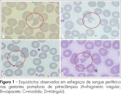
PURPOSE: to evaluate the significance of schizocytes presence in peripheral blood smear of pregnant women with pre-eclampsia, identifying and correlating them with other markers of hemolysis and of the disease severity. METHODS: Seventh six glass slides of peripheral blood smear of pregnant women with pre-eclampsia have been evaluated. After the smear, the slides have been stained with Leishman's dye and stored till they were examined with a Leica, model DLMB microscope, provided with the Qwin Lite 2.5 software that made it possible to record the images of selected fields in CD-ROM. Ten fields with approximately 100 erythrocytes were counted in each glass slide. Schizocytes (irregular fragment or helmet-shaped, bite-shaped or triangular) were considered as present, when their percentage was equal or higher than 0.2%, their presence being correlated with other hemolysis markers (hemoglobin, total bilirubin, lactic desidrogenasis and reticulocytes), pre-eclampsia markers (proteinuria and platelet number). The Statistical Package in Social Science for Windows (SPSS), 10.0 version has been used for statistical analysis, at p<0.05. RESULTS: schizocytes have been present in 31.6% of the pregnant women with pre-eclampsia. In most (75%) of the blood smears there have been three or four schizocytes. There has been no correlation between schizocyte presence and any other hemolysis marker, any pre-eclampsia marker or disease severity. CONCLUSIONS: schizocytes have been identified in a small number and in less than a third of the pregnant women with pre-eclampsia. There has been no correlation with other hemolysis marker parameters or with the disease severity. This way, the presence of schizocytes is not a marker of the clinical evolution of pre-eclampsia.

Summary
Revista Brasileira de Ginecologia e Obstetrícia. 2008;30(8):413-419
DOI 10.1590/S0100-72032008000800007
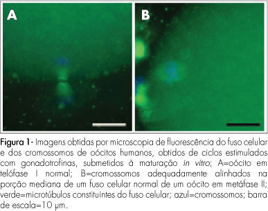
PURPOSE: to evaluate the meiotic spindle and the chromosome distribution of in vitro mature oocytes from stimulated cycles of infertile women with endometriosis, and with male and/or tubal infertility factors (Control Group), comparing the rates of in vitro maturation (IVM) between the two groups evaluated. METHODS: fourteen patients with endometriosis and eight with male and/or tubal infertility factors, submitted to ovarian stimulation for intracytoplasmatic sperm injection have been prospectively and consecutively selected, and formed a Study and Control Group, respectively. Immature oocytes (46 and 22, respectively, from the Endometriosis and Control Groups) were submitted to IVM. Oocytes presenting extrusion of the first polar corpuscle were fixed and stained for microtubules and chromatin evaluation through immunofluorescence technique. Statistical analysis has been done by the Fisher's exact test, with statistical significance at p<0.05. RESULTS: there was no significant difference in the IVM rates between the two groups evaluated (45.6 and 54.5% for the Endometriosis and Control Groups, respectively). The chromosome and meiotic spindle organization was observed in 18 and 11 oocytes from the Endometriosis and Control Groups, respectively. In the Endometriosis Group, eight oocytes (44.4%) presented themselves as normal metaphase II (MII), three (16.7%) as abnormal MII, five (27.8%) were in telophase stage I and two (11.1%) underwent parthenogenetic activation. In the Control Group, five oocytes (45.4%) presented themselves as normal MII, three (27.3%) as abnormal MII, one (9.1%) was in telophase stage I and two (18.2%) underwent parthenogenetic activation. There was no significant difference in meiotic anomaly rate between the oocytes in MII from both groups. CONCLUSIONS: the present study data did not show significant differences in the IVM or in the meiotic anomalies rate between the IVM oocytes from stimulated cycles of patients with endometriosis, as compared with controls. Nevertheless, they have suggested a delay in the outcome of oocyte meiosis I from patients with endometriosis, shown by the higher proportion of oocytes in telophase I observed in this group.

Summary
Revista Brasileira de Ginecologia e Obstetrícia. 2008;30(7):328-334
DOI 10.1590/S0100-72032008000700002
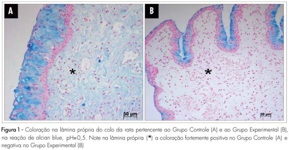
PURPOSE: to study the histochemical changes related to the uterine cervix glycosaminoglycan of the albino female rat, after local ministration of hyaluronidases at the end of pregnancy. METHODS: ten female rats with positive pregnancy tests were randomly distributed in two numerically equal groups. The control group (Cg) was built up with rats that received a single dose of 1 mL of distilled water in the uterine cervix, under anesthesia, at the 18th pregnancy day. In the experimental group (Exg), the rats received 0.02 mL of hyaluronidase, diluted in 0.98 mL of distilled water (1 mL as a total), under the same conditions as the Cg. At the 20th pregnancy day, the rats were anesthetized once again and submitted to dissection, and the cervix prepared for histochemical study with alcian blue dye and its blockades (pH=0.5, pH=2.5, methylation and saponification). RESULTS: strongly positive reaction in the lamina propria (+3) has been seen in the Cg, and negative reaction in the Exg, with pH=0.5 alcian blue staining. With pH=2.5, staining has also been strongly positive (+4) in the Cg, and weakly positive (+1) in the Exg slide. After methylation, both groups have shown negative reaction, with pH=2.5 alcian blue staining. The lamina propria staining became negative after methylation in both groups, followed by saponification and enzymatic digestion on slide. CONCLUSIONS: there is clear predominance of sulphated glycosaminoglycans in the Cg as compared to the Exg and a small amount of identified carboxylated glycosaminoglycans in the Exg. The changes evidenced in the extracellular matrix have suggested that the hyaluronidase injected in the uterine cervix has promoted biochemical changes compatible with cervix maturation.

Summary
Revista Brasileira de Ginecologia e Obstetrícia. 2008;30(7):335-340
DOI 10.1590/S0100-72032008000700003
PURPOSE: to evaluate the effect of exposure of female rats to therapeutic ultrasound in the pre-implantation phase. METHODS: pregnant Wistar female rats have been exposed to 3 MHz, 0.6 W/cm² ultrasound, pulsatile ultrasound (PUS) or continuous ultrasound (CUS), and controls, unplugged ultrasound (UUS), for five minutes. The rats were sacrificed at the 20th day post-insemination. Biochemical and hematological analyses have been done. Animals have been submitted to necropsy in order to identify lesions of internal organs, and to remove and weight the liver, kidneys and ovaries. Alive, malformed, dead and reabsorbed fetuses have been counted. RESULTS: the rats have not presented changes in their body and organs weight, and neither in their reproductive capacity, but there has been an increase in triglycerides in the PUS and CUS groups, when compared to the UUS group. The fetuses' relative weights of the heart (0.7 ± 0.9), liver (9.8 ± 0.8), kidneys (6.2 ± 0.8) and lungs (3.8 ± 0.4) increased in the CUS, when compared to the heart (0.7 ± 0.9), liver (9.8 ± 0.8), kidneys (6.2 ± 0.8) e lungs (3.8 ± 0.4) of the UUS. CONCLUSIONS: in the experimental model, the therapeutic ultrasound used has not caused meaningful maternal toxicity. Pulsatile waves have not changed fetal morphology, but continuous waves have caused increase in the relative weight of the fetuses' heart, liver, lungs and kidneys.
Summary
Revista Brasileira de Ginecologia e Obstetrícia. 2008;30(7):341-348
DOI 10.1590/S0100-72032008000700004
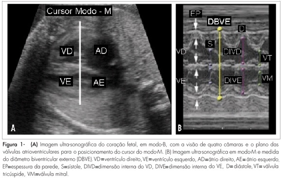
PURPOSE: to verify the correlation between ultrasonography heart measures and hemoglobin deficit in fetuses of alloimmunized pregnant women. METHODS: a transversal study, including 60 fetuses, with 21 to 35 weeks of gestational age, from 56 isoimmunized pregnant women. A number of 139 procedures were performed. Before cordocentesis for the collection of fetal blood, cardiac measures and femur length (FL) were assessed by ultrasonography. The external biventricular diameter (EBVD) was obtained by measuring the distance between the epicardic external parts at the end of the diastole, with the M-mode cursor perpendicular to the interventricular septum, in the atrioventricular valves. The measure of the atrioventricular diameter (AVD) was obtained by positioning the same cursor along the interventricular septum, evaluating the distance between the heart basis and apex. The FL was determined from the trochanter major to the distal metaphysis. The cardiac circumference (CC) was also calculated. To adjust the cardiac measure to the gestational age, each of these measures were divided by the FL measure. Hemoglobin concentration has been determined by spectrophotometry with the Hemocue® system. Hemoglobin deficit calculation was based in the Nicolaides's normality curve. RESULTS: direct and significant correlations were observed between the cardiac measures evaluated and the hemoglobin deficit. To predict moderate and severe anemia, the sensitivity and specificity found were 71.7 and 66.3% for EBVD and FL, 65.8 and 62.4% for AVD and FL, and 73.7 and 60.4% for CC and FL, respectively. CONCLUSIONS: ultrasonography cardiac measures assessed from fetuses of isoimmunized pregnant women correlate directly with hemoglobin deficit.

Summary
Revista Brasileira de Ginecologia e Obstetrícia. 2008;30(7):349-354
DOI 10.1590/S0100-72032008000700005
PURPOSE: to describe the prevalence and behavioral profile of genital infections in women attended at a Primary Health Unit in Vitoria, ES. METHODS: a transversal study including 14 to 49-year-old women attended by the Family Health Program (FHP). Exclusion criteria were: having been submitted to gynecological examination in less than one year before, and history of recent treatment (in the last three months) for genital infections. An interview including socio-demographic, clinical and behavioral data was applied. Genital specimens were collected for cytology, GRAM bacterioscopy and culture, and urine sample for molecular biological test for Chlamydia trachomatis. RESULTS: two hundred and ninety-nine women took part in the study. The median age was 30.0 (interquartile interval: 24;38) years old; the average age of the first intercourse was 17.3 (sd=3.6) years old. The first pregnancy average age was 19.2 (3.9) years old. About 70% reported up to 8 years of schooling; 5% reported previous Sexually Transmitted Diseases (STD), and 8%, the use of illicit drugs. Only 23.7% reported consistent use of condoms. Clinical complaints were: genital ulcer (3%); dysuria (7.7%); vaginal discharge (46.6%): pruritus (20%) and pelvic pain (18%). Prevalence rates were: Chlamydia trachomatis 7.4%; gonorrhea 2%; trichomoniasis 2%; bacterial vaginosis 21.3%; candidiasis 9.3%; and cytological changes suggestive of HPV 3.3%. In the final logistic regression model, the factors independently associated to genital infections were: abnormal cervical mucus, OR=9.7 (CI95%=5.6-13.7), previous HIV testing, OR=6.5 (CI95%=4.0-8.9), having more than one partner during the previous year, OR=3.9 (CI95%=2.7-5.0), and having more than one partner in life, OR=4.7 (CI95%=2.4-6.8). CONCLUSIONS: results show a high rate of genital infections and the need of preventive measures, such as STD surveys and risk reduction programs for women that look for routine gynecological service.