Summary
Revista Brasileira de Ginecologia e Obstetrícia. 2009;31(3):111-116
DOI 10.1590/S0100-72032009000300002
PURPOSE: to evaluate whether the presence of insulin resistance (IR) alters cardiovascular risk factors in women with polycystic ovary syndrome (POS). METHODS: transversal study where 60 POS women with ages from 18 to 35 years old, with no hormone intake, were evaluated. IR was assessed through the quantitative insulin sensitivity check index (QUICKI) and defined as QUICKI <0.33. The following variables have been compared between the groups with or without IR: anthropometric (weight, height, waist circumference, arterial blood pressure, cardiac frequency), laboratorial (homocysteine, interleucines-6, factor of tumoral-α necrosis, testosterone, fraction of free androgen, total cholesterol and fractions, triglycerides, C reactive protein, insulin, glucose), and ultrasonographical (distensibility and carotid intima-media thickness, dilation mediated by the brachial artery flux). RESULTS: Eighteen women (30%) presented IR and showed significant differences in the following anthropometric markers, as compared to the women without IR (POS with and without IR respectively): body mass index (35.56±5.69 kg/m² versus 23.90±4.88 kg/m², p<0.01), waist (108.17±11.53 versus 79.54±11.12 cm, p<0.01), systolic blood pressure (128.00±10.80 mmHg versus 114.07±8.97 mmHg, p<0.01), diastolic blood pressure (83.67±9.63 mmHg versus 77.07±7.59 mmHg, p=0.01). It has also been observed significant differences in the following laboratorial markers: triglycerides (120.00±56.53 mg/dL versus 77.79±53.46 mg/dL, p=0.01), HDL (43.06±6.30 mg/dL versus 40.45±10.82 mg/dL, p=0.01), reactive C protein (7.98±10.54 mg/L versus 2.61±3.21 mg/L, p<0.01), insulin (28.01±18.18 µU/mL versus 5.38±2.48 µU/mL, p<0.01), glucose (93.56±10.00 mg/dL versus 87.52±8.75 mg/dL, p=0.02). Additionally, two out of the three ultrasonographical markers of cardiovascular risk were also different between the groups: carotid distensibility (0.24±0.05 mmHg-1 versus 0.30±0.08 mmHg-1, p<0.01) and carotid intima-media thickness (0.52±0.08 mm versus 0.43±0.09, p<0.01). Besides, the metabolic syndrome ratio was higher in women with IR (nine cases=50% versus three cases=7.1%, p<0.01). CONCLUSIONS: POS and IR women present significant differences in several ultrasonographical, seric and anthropometric markers, which point out to higher cardiovascular risk, as compared to women without POS and IR. In face of that, the systematic IR evaluation in POS women may help to identify patients with cardiovascular risk.
Summary
Revista Brasileira de Ginecologia e Obstetrícia. 2009;31(3):131-137
DOI 10.1590/S0100-72032009000300005
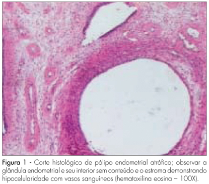
PURPOSE: to evaluate the effects of tamoxifen on the expression of TGF-β and p27 proteins in polyps and adjacent endometrium of women after menopause. METHODS: prospective study with 30 post-menopausal women with diagnosis of breast cancer, taking tamoxifen (20 mg/day), presenting diagnosis of suspect endometrial polyps through transvaginal ultrasonography, and submitted to diagnostic and surgical hysterectomy to withdraw the polyps and adjacent endometrium. A immunohistochemical study has been done to verify the expression of the TGF-β and p27 proteins in the polyps and adjacent endometrium. These proteins' quantification has been done by morphometry. RESULTS: the patients' average age was 61.7 years old; their average age at the menopause onset was 49.5; and the average of using tamoxifen was 25.3 months. The average concentration of positive cells for TGF-β protein in the glandular and stroma polyp epithelium was 62.6±4.5 cells/mm². For the p27, in the glandular polyp epithelium, it was 24.2±18.6 cells/mm² and for the stroma, 19.2±15.2 cells/mm². There was no significant difference between the expression of TGF-β and p27 in the glandular epithelial form the polyps and the adjacent endometrium. The expression of proteins in the polyp and adjacent endometrium with its respective glandular and stroma epithelium showed a significant difference for the p27 protein (r=0.9, p<0.05). CONCLUSIONS: we have concluded that the TGF-β expression is not related to the effect of tamoxifen on the growing of endometrial polyps, but the absence of polyps' malignization by tamoxifen may be explained by the high expression of p27 protein in its glandular epithelium.
Summary
Revista Brasileira de Ginecologia e Obstetrícia. 2009;31(3):142-147
DOI 10.1590/S0100-72032009000300007
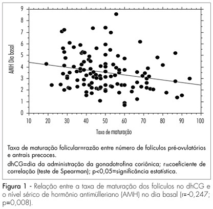
PURPOSE: to test the hypothesis that the anti-müllerian hormone (AMH) serum level reflects the antral follicles' response to the administration of FSH. METHODS: prospective study, including 116 normo-ovulatory infertile patients submitted to controlled ovarian hyperstimulation with GnRH and FSH agonists. The AMH serum level was measured after reaching the pituitary suppression and before the FSH administration (basal day). The number of antral follicles was determined by ultrasonography at the basal day (precocious antral follicles; 2 to 8 mm) and at the day of hCG administration (dhCG; pre-ovulatory follicles; >16 mm). The follicle response to FSH was determined by the percentage of precocious antral follicles which reached pre-ovulatory stage in response to FSH (maturation rate). The correlation of AMH with the patients' age, the total number of precocious antral and pre-ovulatory follicles, collected oocytes, total dose of FHS in the controlled ovarian stimulation and the rate of follicular maturation was studied. For the statistical analysis, it simple regression analysis and the Spearman's test were used, at a 5% significance level. RESULTS: The serum level of AMH was positively correlated with the number of precocious antral follicles at the basal day (r=0.64; p<0.0001) and pre-ovulatory follicles in dhCG (r=0.23; p=0.01). Exceptionally, the serum level of AMH was negatively correlated with the maturation ratio (r=-0.24; p<0.008). CONCLUSIONS: AMH attenuates the follicular development caused by FSH administration.
Summary
Revista Brasileira de Ginecologia e Obstetrícia. 2009;31(2):90-93
DOI 10.1590/S0100-72032009000200007
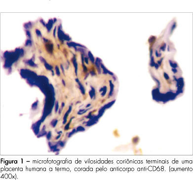
PURPOSE: to verify the amount of CD68+ cells in chorionic villosities in placentae from gestations submitted or not to labor. METHODS: transversal study with healthy near-term pregnant women, among whose placentae, 31 have been examined by immunohistochemical technique. Twenty placentae were obtained after vaginal delivery (VAGG) and eleven after elective cesarean sections (CESG). Slides were prepared with chorionic villosities samples and labeled with anti-CD68 antibody, specific for macrophages. Labeled and nonlabeled cells were counted inside the villosities. Non-parametric statistical tests were used for the analysis. RESULTS: among the 6,424 cells counted in the villosities' stroma from the 31 placentae, 1,135 cells (17.6%) were stained by the CD68+. The mean of cells labeled by the anti-CD68 was 22±18 for the VAGG group and 20±16 for the CESG, in each placentary sample. CONCLUSIONS: there were no significant differences in the percentage of macrophages (CD68+) in association with labor.
Summary
Revista Brasileira de Ginecologia e Obstetrícia. 2009;31(2):54-60
DOI 10.1590/S0100-72032009000200002
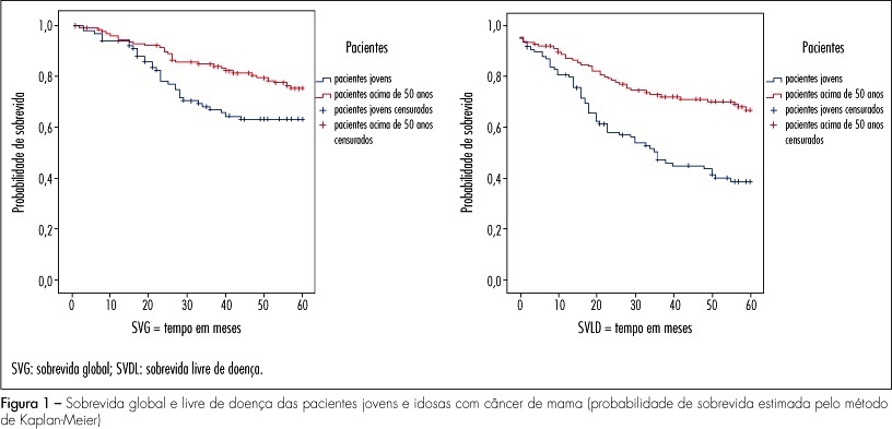
PURPOSE: the objective of this study was to evaluate the clinical, pathological and molecular characteristics in very young women and postmenopausal women with breast cancer. METHODS: we selected 106 cases of breast cancer of very young women (<35 years) and 130 cases of postmenopausal women. We evaluated clinical characteristics of patients (age at diagnosis, ethnic group, family history of breast cancer, staging, presence of distant metastases, overall and disease-free survival), pathological characteristics of tumors (tumor size, histological type and grade, axillary lymph nodes status) and expression of molecular markers (hormone receptors, HER2, p53, p63, cytokeratins 5 and 14, and EGFR), using immunohistochemistry and tissue microarray. RESULTS: when comparing clinicopathologic variables between the age groups, younger women demonstrated greater frequency of nulliparity (p=0.03), larger tumors (p<0.000), higher stage disease (p=0.01), lymph node positivity (p=0.001), and higher grade tumors (p=0.004). Most of the young patients received chemotherapy (90.8%) and radiotherapy (85.2%) and less tamoxifen therapy (31.5%) comparing with postmenopausal women. Lower estrogen receptor positivity 49.1% (p=0.01) and higher HER2 overexpression 28.7% (p=0.03) were observed in young women. In 32 young patients (29.6%) and in 20% of the posmenopausal women, the breast carcinomas were of the triple-negative phenotype (p=0.034). In 16 young women (50%) and in 10 postmenopausal women (7.7%), the tumors expressed positivity for cytokeratin 5 and/or 14, basal phenotype (p=0.064). Systemic metastases were detected in 55.3% of the young women and in 39.2% of the postmenopausal women. Breast cancer overall survival and disease-free survival in five years were, respectively, 63 and 39% for young women and 75 and 67% for postmenopausal women. CONCLUSIONS: breast cancer arising in very young women showed negative clinicobiological characteristics and more aggressive tumors.
Summary
Revista Brasileira de Ginecologia e Obstetrícia. 2009;31(2):61-67
DOI 10.1590/S0100-72032009000200003
PURPOSE: to evaluate the quality of life and sexuality features of women with breast cancer, according to the type of surgery they underwent and their sociodemographic characteristics. METHODS: transversal study with 110 women treated for breast cancer, for at least one year in the Centro de Atenção Integral à Saúde da Mulher of UNICAMP. The quality of life was assessed by the WHOQOL-bref questionnaire, and the issues on sexuality, by a specific questionnaire in which Cronbach's Alpha coefficient was used to validate the concordance of responses (alpha=0.72) and the technique of factor analysis, with the criterion of self value and variance maximum rotation, resulting in two components: intrinsic or intimacy ( how the woman sees herself sexually) and extrinsic or attractiveness (how the woman believes the others see her sexually). Sociodemographic variables have been assessed according to the WHO questionnaire, and the sexuality components, through the Kruskal-Wallis followed by the Mann-Whitney's test and Spearman correlation test. RESULTS: age, schooling, type of surgery and lapse of time from the surgery did not influence the quality of life concerning physical, environmental, and psychological aspects, as well as the social relationships. Women with a stable marital relationship got higher scores in the psychological area (p=0.04) and in the area of social relationships (p=0.02). Higher socioeconomic level influenced the quality of life concerning physical appearance (p=0.01) and environment (p=0.002). Regarding the sexuality, age had influence in the extrinsic component (p=0.0158). Women with a stable marital relationship had higher scores of quality of life in both components of sexuality. Higher schooling influenced in a positive way the intrinsic factor. Women submitted to quadrantectomy or mastectomy with immediate breast reconstruction showed higher scores relating to attractiveness in comparison to mastectomized women without reconstruction. CONCLUSIONS: better socioeconomic level and better schooling, stable marital relationship and surgery with breast conservation are linked to better rates of quality of life, including sexuality.
Summary
Revista Brasileira de Ginecologia e Obstetrícia. 2009;31(2):68-74
DOI 10.1590/S0100-72032009000200004
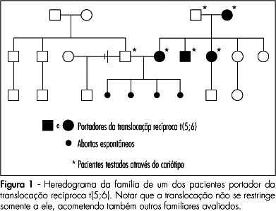
PURPOSE: to asses the prevalence and clinical characteristics of couples with history of recurrent spontaneous abortion and chromosome abnormality, attended at the present service. METHODS: all the couples referred to our service due to history of recurrent spontaneous abortion, from January 1975 to June 2008, were evaluated. Only the ones whose chromosome karyotype analysis by GTG bands has been successfully made were included in the study. Clinical data on their age, as well as on the number of abortions, stillbirth, multiple malformations, livebirth per couple, and the result of the karyotype exam were collected. Fisher's exact test (p<0.05) has been used to compare the incidence of chromosome alterations found in our study, with data in the literature. RESULTS: there were 108 couples in the sample. Their ages varied from 21 to 58 years old among the men (average of 31.4 years old), and from 19 to 43 among the women (average of 29.9 years old). In ten couples, one of the mates (9.3%) presented chromosome alterations, which corresponded respectively to three cases (30%) of reciprocal translocation [two of t(5;6) and one of t(2;13)], two (20%) of Robertsonian translocation [two of der(13;14) and one of der(13;15)], five(50%) of mosaicism (mos) [two cases of mos 45,X/46,XX, one of mos 46,XX/47,XXX, one of mos 46,XY/47,XXY and one of mos 46,XY/47,XYY] and one (10%) of chromosome inversion [inv(10)]. In one of the couples, the female presented two concomitant alterations: t(2;13) and der(13;14). Chromosome abnormalities were found in 5% of the couples with a history of two abortions, in 10.3% with three abortions, and in 14.3% with four or more abortions. CONCLUSIONS: the incidence of chromosome abnormalities seen in our study (9.3%) was similar to most of the studies carried out in the last 20 years, varying from 4.8 to 10.8%. Nevertheless the high percentage of patients with mosaicism in our sample, has called our attention. It is believed that this fact may be associated to the high number of metaphases ordinarily analyzed in the present service.
Summary
Revista Brasileira de Ginecologia e Obstetrícia. 2009;31(2):75-81
DOI 10.1590/S0100-72032009000200005
PURPOSE: to evaluate the factors leading to delays in obtaining definitive diagnosis of suspicious lesions for breast cancer. METHODS: a cross-sectional, observational study was carried out with 104 women attending a cancer hospital with a diagnosis or suspected diagnosis of breast cancer. A semistructured questionnaire on the patients' demographic, clinical characteristics and the use of services was applied.Variables were compared using t-Student test, Mann-Whitney test, Pearson's χ2 test or Fisher's exact test, as appropriate. In order to identify the variables associated with delays in breast cancer diagnosis, the Odds Ratio (OR) were calculated together with their respective 95% confidence intervals (95%CI) and a logistic regression model was constructed. RESULTS: age of patients was 54±12.6 years (mean±standard deviation). Most of the women were white (48.1%), married (63.5%), living in the city of Rio de Janeiro (57.7%) and poorly educated (60.6%). The median time between the first sign or symptom of the disease and first consultation was one month and the mean time between first consultation and confirmation of diagnosis was 6.5 months. In 51% of the women, diagnosis was late (stages II-IV). Symptomatic presentation and longer delay between symptom onset and the first evaluation and between symptom onset and the diagnosis were found to be significant factors (p<0.05) for delays in obtaining definitive diagnosis of suspicious lesions. CONCLUSIONS: the results of this study suggest that efforts must be made to reduce the time needed to get an appointment with a doctor and to confirm a diagnosis of suspicious lesions, as well as to educate physicians and the women themselves regarding the importance of breast symptoms and the value of prompt evaluation, diagnosis, and treatment.