Summary
Revista Brasileira de Ginecologia e Obstetrícia. 2008;30(10):499-503
DOI 10.1590/S0100-72032008001000004
PURPOSE: to evaluate the embryo's volume (EV) between the seventh and the tenth gestational week, through tridimensional ultrasonography. METHODS: a transversal study with 63 normal pregnant women between the seventh and the tenth gestational week. The ultrasonographical exams have been performed with a volumetric abdominal transducer. Virtual Organ Computer-aided Analysis (VOCAL) has been used to calculate EV, with a rotation angle of 12º and a delimitation of 15 sequential slides. The average, median, standard deviation and maximum and minimum values have been calculated for the EV in all the gestational ages. A dispersion graphic has been drawn to assess the correlation between EV and the craniogluteal length (CGL), the adjustment being done by the determination coefficient (R²). To determine EV's reference intervals as a function of the CGL, the following formula was used: percentile=EV+K versus SD, with K=1.96. RESULTS: CGL has varied from 9.0 to 39.7 mm, with an average of 23.9 mm (±7.9 mm), while EV has varied from 0.1 to 7.6 cm³, with an average of 2.7 cm³ (±3.2 cm³). EV was highly correlated to CGL, the best adjustment being obtained with quadratic regression (EV=0.2-0.055 versus CGL+0.005 versus CGL²; R²=0.8). The average EV has varied from 0.1 (-0.3 to 0.5 cm³) to 6.7 cm³ (3.8 to 9.7 cm³) within the interval of 9 to 40 mm of CGL. EV has increased 67 times in this interval, while CGL, only 4.4 times. CONCLUSIONS: EV is a more sensitive parameter than CGL to evaluate embryo growth between the seventh and the tenth week of gestation.
Summary
Revista Brasileira de Ginecologia e Obstetrícia. 2008;30(10):494-498
DOI 10.1590/S0100-72032008001000003
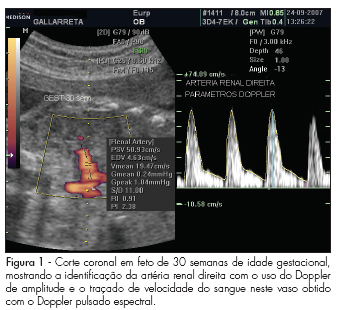
PURPOSE: to describe values found for the resistance index (RI), pulsatility index (PI) and the systole/diastole (S/D) ratio of fetal renal arteries in non-complicated gestations between the 22nd and the 38th week, and to evaluate whether those values vary along that period. METHODS: observational study, where 45 fetuses from non-complicated gestations have been evaluated in the 22nd, 26th, 30th and 38th weeks of gestational age. Doppler ultrasonography has been performed by the same observer, using a device with 4 to 7 MHz transducer. For the acquisition of the renal arteries velocity record, a 1 mm to 2 mm probe has been placed in the mean third of the renal artery for the evaluation through pulsed Doppler ultrasonography. The measurement of RI, PI and S/D ratio from three consecutive waves was performed with the automatic mode. To detect significant differences in the indexes' values along gestation, we have compared values obtained at the different gestational ages, through repeated measures ANOVA, followed by Tukey's post-hoc test. RESULTS: There were no significant differences between the right and left renal arteries, when the RI, IP and S/D ratio were compared. Nevertheless, a change in the values of these parameters has been observed between the 22nd week (RI=0.9 ± 0.02; PI=2.4 ± 0.02; S/D ratio=11.6 ± 2.2; mean ± standard deviation of the combined mean values of the right and left renal artery) and the 38th week (RI=0.8 ± 0.03; PI=2.1 ± 0.2; S/D ratio=8.7 ± 2.3) of gestation. CONCLUSIONS: the parameters evaluated (RI, PI and S/D ratio) have presented decreasing values between the 22nd and 38th, with no difference between the fetus's right and left sides.

Summary
Revista Brasileira de Ginecologia e Obstetrícia. 2008;30(10):486-493
DOI 10.1590/S0100-72032008001000002
PURPOSE: to investigate factors accountable for macrosomia incidence in a study with mothers and progeny attended at a Basic Unity of Health in Rio de Janeiro, Brazil. METHODS: a prospective study, with 195 pairs of mothers and progeny, in which the dependent variable was macrosomia (weight at delivery >4,000 g - independent of the gestational age or of other demographic variables), and socioeconomic, previous pregnancies/gestation course, biochemical, behavioral and anthropometric, the independent variables. Statistical analysis has been done by multiple logistic regression. Relative risk (RR) values have been estimated, based on the simple form: RR=OR/ (1 - I0) + (I0 versus OR), in which I0 is the macrosomia incidence in non-exposed people. RESULTS: Macrosomia incidence was 6.7%, the highest value being found in the progeny of women >30 years old (12.8%), white (10.4%), with two or more children (16.7%), with male newborns (9.6%), with height >1,6 m (12.5%), with overweight or obesity as a nutritional pre-gestational state (13.6%), and with excessive gestational gain of weight (12.7%). The final model has shown that having two or more children (RR=3.7; CI95%=1.1-9.9), and having a male newborn (RR=7.5; CI95%=1.0-37.6) were the variables linked to the macrosomia occurrence. CONCLUSIONS: macrosomia incidence was higher than the one observed in Brazil as a whole, but inferior to the one reported in studies from developed countries. Having two or more children and a newborn male were the factors accountable for the occurrence of macrosomia.
Summary
Revista Brasileira de Ginecologia e Obstetrícia. 2008;30(9):459-465
DOI 10.1590/S0100-72032008000900006
PURPOSE: to evaluate the effect of maternal, socioeconomic and obstetric variables, as well the presence of artery incisions in the 20th and 24th weeks on the fetal weight estimated at the end of pregnancy (36th week) in pregnant women attended by Programa Saúde da Família, in an inland town of the northeast of Brazil. METHODS: a longitudinal study including 137 pregnant women, who have been followed up every four weeks in order to assess clinical, socioeconomic and obstetric conditions, including their weight. The uterine arteries were evaluated by Doppler in the 20th and 24th weeks, the fetal weight and the amniotic fluid index (AFI), determined in the 36th week. The initial maternal nutritional state has been determined by the body mass index (BMI), the pregnant women being classified as low weight, eutrophic, over weight and obese. Weight gain during gestation has been evaluated, according to the initial nutritional state, being classified at the end of the second and third trimester as insufficient, adequate and excessive weight gain. Analysis of variance was performed to evaluate the association of the fetal weight in the 36th week with the predictor variables, adjusted by multiple linear regression. RESULTS: an association between the fetal weight estimated in the 36th week and the mother's age (p=0.02), mother's job (p=0.02), initial nutritional state (p=0.04), weight gain in the second trimester (p=0.01), presence of incisions in the uterine arteries (p=0.02), and AFI (p=0.007) has been observed. The main factors associated to the fetal weight estimated in the 36th week, after the multiple regression analysis were: BMI at the pregnancy onset, weight gain in the second trimester, AFI and tabagism. CONCLUSIONS: in the present study, the fetal weight is positively associated with the initial maternal nutritional state, the weight gain in the second trimester and the volume of amniotic fluid, and negatively, to tabagism.
Summary
Revista Brasileira de Ginecologia e Obstetrícia. 2008;30(9):452-458
DOI 10.1590/S0100-72032008000900005
PURPOSE: to evaluate the experience of Hospital das Clínicas da Faculdade de Medicina de Botucatu da Universidade Estadual Paulista "Júlio de Mesquita Filho", in the follow-up of pregnant women with hyperthyroidism. METHODS: Sixty patients, divided in groups with compensated hyperthyroidism (CHG=24) and with uncompensated hyperthyroidism (UHG=36) were retrospectively studied and compared concerning clinical-laboratorial characteristics and intercurrences. The t-Student test, contingency tables, multiple linear regression and multiple logistic regression with significance level at 5.0% were used. RESULTS: propylthiouracil (PTU) was used by 94.0% of UHG and by 42.0% of CHG (p<0.0001); maternal complications close to delivery have occurred in 20.6% of UHG and in 11.8% of CHG, and UHG presented three fetal deaths, influenced by the mother age, higher level of T4L (lT4L) and of PTU dose (PTUd) in the third trimester (p=0.007); restriction of intra-uterine growth, influenced by lT4L and PTUd in the third trimester has occurred in nine UHG and in three CHG cases, and oligoamnios has occurred in 12 patients (83.3% of UGH and 16.7% of CGH), influenced by age and lT4L in the third trimester (p=0.04); the gestational age at delivery was 34.4±4.6 weeks in UHG and 37.0±2.5 in CHG, influenced by the T4Ll in the third trimester (p<0.05). CONCLUSIONS: the UHG has presented less satisfactory results than CHG, influenced by high lT4L and PTUd in the third trimester, and by more advanced age of some pregnant women.
Summary
Revista Brasileira de Ginecologia e Obstetrícia. 2008;30(9):445-451
DOI 10.1590/S0100-72032008000900004
PURPOSE: to determine the prevalence and risk factors associated to anemia in pregnant women from the semiarid region of Alagoas, Brazil. METHODS: transversal study comprising a sample (n=150) obtained taking into consideration the prevalence estimated by World Health Organization of 52%, an error of 8% and a confidence interval of 95%. Sampling has been done in three stages: 15 towns among the 38 in the region, four census sectors by town and 24 residences by sector. All the resident pregnant women were eligible, and their socio-economic, demographic, anthropometric and health data have been collected. Anemia was identified at the <11 g/dL hemoglobin level (Hemocue®), and its association with risk factors, tested by multiple linear regression analysis. RESULTS: anemia prevalence was 50%. Seventy eight per cent of the pregnant women were under pre-natal care. From those, 79.3% were in the second or third trimester of gestation. Nevertheless, only 21.2% of them were taking iron supplementation. Variables (p<0.05) independently associated with anemia (anemic versus not-anemic pregnant women) were: larger number of family members (4.5±2.3 versus 4,3±2.3; p=0.02), lower age group of the pregnant woman (23.9±6.3 versus 24.7±6.7; p=0.04), or of her partner (34.5±15.8 versus 36±17.5; p=0.03), no toilet in the house (30.7 versus 24%; p<0.001), history of child abortion and/or death (32.4 versus 16.4%; p<0.001), living in the country (60 versus 46.7%; p=0.03), average per capita income
Summary
Revista Brasileira de Ginecologia e Obstetrícia. 2008;30(9):437-444
DOI 10.1590/S0100-72032008000900003
PURPOSE: to verify the accuracy of uterine cervix cytology for HPV diagnosis, as compared to polymerase chain reaction (PCR) in samples of women with HIV. METHODS: 158 patients who had undergone a first collection of material from the uterine cervix with Ayre's spatula for PCR were included in the study. Then, another collection with Ayre's spatula and brush for oncotic cytology was performed. Only 109 slides were reviewed, as 49 of them had already been destructed for have being filed for over two years. RESULTS: the prevalence of HPV was 11% in the cytological exam and 69.7% in the PCR. Age varied from 20 to 61 years old, median 35 years. The HIV contagious route was heterosexual in 91.8% of the cases, and 79.1% of the patients had had from one to five sexual partners along their lives. The most frequent complaint was pelvic mass (5.1%), and 75.3% of the women had looked for the service for a routine medical appointment. The categorical variable comparison was done through contingency tables, using the χ2 test with Yates's correction to compare the ratios. The Fisher's test was used when one of the expected rates was lower than five. In the comparison of diagnostic tests, sensitivity, specificity and similarity ratios have been calculated. Among the 76 patients with HPV, detected by PCR, only 12 had the diagnosis confirmed by cytology (sensitivity=15.8%), which on the other hand did not present any false-positive results (specificity=100%). Concerning the HPV presence, the cytological prediction for positive results was 100% and 33.3% for negative, when both results were compared. Among the 12 patients with HPV positive cytology, four (33.3%) presented cervical intraepithelial neoplasia (OR=56; positive similarity ratio=positive infinity; negative similarity ratio=0.83). CONCLUSIONS: As the cytology specificity is quite high, it is possible to rely on the positive result, which means that a positive result will surely indicate the presence of HPV. The low sensitivity of cytology does not qualify it as a survey exam for HPV detection in this female group.