-
Case Report
High-Grade Transformation in Adenoid Cystic Carcinoma of the Bartholin Gland: Case Report
- Aline Evangelista Santiago
 ,
, - Nicky Teunissen ,
- Bernardo Ferreira de Paula Ricardo ,
- Eduardo Batista Cândido ,
- Rafaela de Souza Furtado , [ ... ],
- Agnaldo Lopes da Silva Filho
01-24-2021
Summary
Case ReportHigh-Grade Transformation in Adenoid Cystic Carcinoma of the Bartholin Gland: Case Report
Revista Brasileira de Ginecologia e Obstetrícia. 2021;43(12):980-984
01-24-2021- Aline Evangelista Santiago ,
- Nicky Teunissen ,
- Bernardo Ferreira de Paula Ricardo ,
- Eduardo Batista Cândido ,
- Rafaela de Souza Furtado ,
- Agnaldo Lopes da Silva Filho
Views111See moreAbstract
Introduction
In the present study, we report a case of primary adenoid cystic carcinoma (ACC) of the Bartholin gland with high-grade transformation (HGT). Adenoid cystic carcinoma of the Bartholin gland is a rare tumor and HGT has only been reported in head and neck tumors.
Case Report
A 77-year-old woman with a non-ulcerated vulvar lesion on the topography of the right Bartholin gland. The patient was submitted to tumor resection followed by V–Y island flap and adjuvant radiotherapy. The histopathological examination revealed primary ACC of the Bartholin gland, with areas of HGT and extensive perineural invasion. The immunohistochemical study with p53 showed a diffuse and strong positive reaction in areas with HGT. After 24 months of follow-up, the patient presented distant metastases and died, despite having undergone to chemotherapy.
Conclusion
As far as we know, this case is the first description in the literature of HGT in ACC of the Bartholin gland, and HGT appears to be associated with tumor aggressiveness.
Views111

This is an Open Access article distributed under the terms of the Creative Commons Attribution License, which permits unrestricted use, distribution, and reproduction in any medium, provided the original work is properly cited. Summary
Case ReportHigh-Grade Transformation in Adenoid Cystic Carcinoma of the Bartholin Gland: Case Report
Revista Brasileira de Ginecologia e Obstetrícia. 2021;43(12):980-984
01-24-2021- Aline Evangelista Santiago ,
- Nicky Teunissen ,
- Bernardo Ferreira de Paula Ricardo ,
- Eduardo Batista Cândido ,
- Rafaela de Souza Furtado ,
- Agnaldo Lopes da Silva Filho
Views111See moreAbstract
Introduction
In the present study, we report a case of primary adenoid cystic carcinoma (ACC) of the Bartholin gland with high-grade transformation (HGT). Adenoid cystic carcinoma of the Bartholin gland is a rare tumor and HGT has only been reported in head and neck tumors.
Case Report
A 77-year-old woman with a non-ulcerated vulvar lesion on the topography of the right Bartholin gland. The patient was submitted to tumor resection followed by V–Y island flap and adjuvant radiotherapy. The histopathological examination revealed primary ACC of the Bartholin gland, with areas of HGT and extensive perineural invasion. The immunohistochemical study with p53 showed a diffuse and strong positive reaction in areas with HGT. After 24 months of follow-up, the patient presented distant metastases and died, despite having undergone to chemotherapy.
Conclusion
As far as we know, this case is the first description in the literature of HGT in ACC of the Bartholin gland, and HGT appears to be associated with tumor aggressiveness.
This is an Open Access article distributed under the terms of the Creative Commons Attribution License, which permits unrestricted use, distribution, and reproduction in any medium, provided the original work is properly cited. 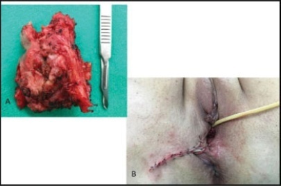
- Aline Evangelista Santiago
-
Case Report
Vulvar Hemangioma: Case Report
Revista Brasileira de Ginecologia e Obstetrícia. 2018;40(6):369-371
06-01-2018
Summary
Case ReportVulvar Hemangioma: Case Report
Revista Brasileira de Ginecologia e Obstetrícia. 2018;40(6):369-371
06-01-2018Views134See moreAbstract
Hemangioma is a benign neoplasm that may affect the vulva, and it can cause functional or emotional disability. This article reports the case of a 52-year-old female patient with a history of a genital ulcer for the past 3 years and who had undergone various treatments with creams and ointments. The patient was biopsied and diagnosed with vulvar hemangioma and was subsequently submitted to surgical excision of the lesion. We emphasize the importance of following the steps of the differential diagnosis and proceeding with a surgical approach only if necessary.
Views134This is an Open Access article distributed under the terms of the Creative Commons Attribution License, which permits unrestricted use, distribution, and reproduction in any medium, provided the original work is properly cited. Summary
Case ReportVulvar Hemangioma: Case Report
Revista Brasileira de Ginecologia e Obstetrícia. 2018;40(6):369-371
06-01-2018Views134See moreAbstract
Hemangioma is a benign neoplasm that may affect the vulva, and it can cause functional or emotional disability. This article reports the case of a 52-year-old female patient with a history of a genital ulcer for the past 3 years and who had undergone various treatments with creams and ointments. The patient was biopsied and diagnosed with vulvar hemangioma and was subsequently submitted to surgical excision of the lesion. We emphasize the importance of following the steps of the differential diagnosis and proceeding with a surgical approach only if necessary.
This is an Open Access article distributed under the terms of the Creative Commons Attribution License, which permits unrestricted use, distribution, and reproduction in any medium, provided the original work is properly cited. 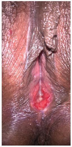
-
Case Report
Differential Diagnosis between Bartholin Cyst and Vulvar Leiomyoma: Case Report
Revista Brasileira de Ginecologia e Obstetrícia. 2017;39(8):433-435
08-01-2017
Summary
Case ReportDifferential Diagnosis between Bartholin Cyst and Vulvar Leiomyoma: Case Report
Revista Brasileira de Ginecologia e Obstetrícia. 2017;39(8):433-435
08-01-2017Views129See moreAbstract
Genital leiomyomas are rare tumors that can often be misdiagnosed as Bartholin cyst. We report a case of a 32-year-old patient who had a cystic nodulation in the left labium majus that was suggestive of Bartholin cyst. A resection surgery was performed, and the definitive histopathology diagnosis was vulvar leiomyoma. The macroscopic features of cystic lesions difficult the differential diagnosis between leiomyoma and Bartholin cyst; therefore, a histopathologic examination is often recommended.
Views129This is an Open Access article distributed under the terms of the Creative Commons Attribution License, which permits unrestricted use, distribution, and reproduction in any medium, provided the original work is properly cited. Summary
Case ReportDifferential Diagnosis between Bartholin Cyst and Vulvar Leiomyoma: Case Report
Revista Brasileira de Ginecologia e Obstetrícia. 2017;39(8):433-435
08-01-2017Views129See moreAbstract
Genital leiomyomas are rare tumors that can often be misdiagnosed as Bartholin cyst. We report a case of a 32-year-old patient who had a cystic nodulation in the left labium majus that was suggestive of Bartholin cyst. A resection surgery was performed, and the definitive histopathology diagnosis was vulvar leiomyoma. The macroscopic features of cystic lesions difficult the differential diagnosis between leiomyoma and Bartholin cyst; therefore, a histopathologic examination is often recommended.
This is an Open Access article distributed under the terms of the Creative Commons Attribution License, which permits unrestricted use, distribution, and reproduction in any medium, provided the original work is properly cited. -
Case Report
Extramammary Paget Disease of the Vulva – Case Report
Revista Brasileira de Ginecologia e Obstetrícia. 2016;38(10):524-528
10-01-2016
Summary
Case ReportExtramammary Paget Disease of the Vulva – Case Report
Revista Brasileira de Ginecologia e Obstetrícia. 2016;38(10):524-528
10-01-2016Views85See moreAbstract
Extramammary Paget disease (EPD) is a rare malign neoplasm that may affect the vulva and has manifestations common to benign diseases such as itching, pain and eczema. This leads to delay in diagnosis and consequent worse prognosis. The definitive diagnosis is obtained by biopsy of the vulva, which shows Paget cells. The treatment of choice is wide excision with margins, which leads to sequelae, functional and aesthetic. Recurrence is common. This article reports the case of a 48-year-old female patient with history of vulvar itching for the past 2 years, who had been submitted to various treatments for benign pathologies. The patient was biopsied and was diagnosed with extensive EPD, being submitted to vulvectomy. This article aims to draw attention to the need for biopsy of pruritic vulvar lesions that do not respond to usual treatment.
Views85This is an Open Access article distributed under the terms of the Creative Commons Attribution License, which permits unrestricted use, distribution, and reproduction in any medium, provided the original work is properly cited. Summary
Case ReportExtramammary Paget Disease of the Vulva – Case Report
Revista Brasileira de Ginecologia e Obstetrícia. 2016;38(10):524-528
10-01-2016Views85See moreAbstract
Extramammary Paget disease (EPD) is a rare malign neoplasm that may affect the vulva and has manifestations common to benign diseases such as itching, pain and eczema. This leads to delay in diagnosis and consequent worse prognosis. The definitive diagnosis is obtained by biopsy of the vulva, which shows Paget cells. The treatment of choice is wide excision with margins, which leads to sequelae, functional and aesthetic. Recurrence is common. This article reports the case of a 48-year-old female patient with history of vulvar itching for the past 2 years, who had been submitted to various treatments for benign pathologies. The patient was biopsied and was diagnosed with extensive EPD, being submitted to vulvectomy. This article aims to draw attention to the need for biopsy of pruritic vulvar lesions that do not respond to usual treatment.
This is an Open Access article distributed under the terms of the Creative Commons Attribution License, which permits unrestricted use, distribution, and reproduction in any medium, provided the original work is properly cited. 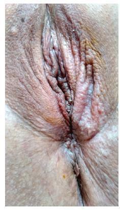
-
Article
Aggressive angiomyxoma of the vagina: a case report
Revista Brasileira de Ginecologia e Obstetrícia. 2013;35(12):575-582
02-03-2013
Summary
ArticleAggressive angiomyxoma of the vagina: a case report
Revista Brasileira de Ginecologia e Obstetrícia. 2013;35(12):575-582
02-03-2013DOI 10.1590/S0100-72032013001200008
Views112See moreAggressive angiomyxoma is a rare, slow-growing soft tissue tumor that usually arises in the pelvis and perineal regions of women in reproductive age, with a marked tendency to local recurrence. Because of its rarity, it is often initially misdiagnosed. Surgical resection is the main treatment modality of aggressive angiomyxoma. We describe a case of a vaginal aggressive angiomyxoma in a 47-year-old woman in which the diagnosis was only made after histological examination. The etiology, presentation, diagnosis and management of this rare tumor are outlined. Angiomyxoma of vulva and vagina refers to a rare disease. Pre-operative diagnosis is difficult due to rarity and absence of diagnostic features, but it should be considered in every mass in genital, perianal and pelvic region in a woman in the reproductive age. Thus, these cases should have complete radiological workup before excision, as pre-diagnosis can change the treatment modality and patient prognosis'.
Views112This is an Open Access article distributed under the terms of the Creative Commons Attribution License, which permits unrestricted use, distribution, and reproduction in any medium, provided the original work is properly cited. Summary
ArticleAggressive angiomyxoma of the vagina: a case report
Revista Brasileira de Ginecologia e Obstetrícia. 2013;35(12):575-582
02-03-2013DOI 10.1590/S0100-72032013001200008
Views112See moreAggressive angiomyxoma is a rare, slow-growing soft tissue tumor that usually arises in the pelvis and perineal regions of women in reproductive age, with a marked tendency to local recurrence. Because of its rarity, it is often initially misdiagnosed. Surgical resection is the main treatment modality of aggressive angiomyxoma. We describe a case of a vaginal aggressive angiomyxoma in a 47-year-old woman in which the diagnosis was only made after histological examination. The etiology, presentation, diagnosis and management of this rare tumor are outlined. Angiomyxoma of vulva and vagina refers to a rare disease. Pre-operative diagnosis is difficult due to rarity and absence of diagnostic features, but it should be considered in every mass in genital, perianal and pelvic region in a woman in the reproductive age. Thus, these cases should have complete radiological workup before excision, as pre-diagnosis can change the treatment modality and patient prognosis'.
This is an Open Access article distributed under the terms of the Creative Commons Attribution License, which permits unrestricted use, distribution, and reproduction in any medium, provided the original work is properly cited. -
Artigo de Revisão
Vulvar intraepithelial neoplasia: a current problem
Revista Brasileira de Ginecologia e Obstetrícia. 2008;30(8):420-426
10-16-2008
Summary
Artigo de RevisãoVulvar intraepithelial neoplasia: a current problem
Revista Brasileira de Ginecologia e Obstetrícia. 2008;30(8):420-426
10-16-2008DOI 10.1590/S0100-72032008000800008
Views42See moreVulvar intraepithelial neoplasia (VIN) is a pathological denomination coined by the International Society for Study of Vulvo-vaginal Diseases (ISSVD) and adopted by the International Society of Gynaecological Pathology (ISGYP) and by the World Health Organization. VIN is a heterogeneous pathological entity with a usual type (warty, basaloid and mixed) and a differentiated type. The incidence of the disease is increasing, especially in young women. The high-risk human papilomavirus (HR-HPV) infection, human immunodeficiency virus (HIV) infection, smoking, cervical, vaginal and rectal intraepithelial neoplasia are considered to be high risk factors for development of VIN. There are no specific symptoms or vulvar macroscopic aspects of VIN. However, a clinical lesion is always present. Liberal vulvar biopsies under colposcopy guidance should be done. Patients with diagnosis of VIN harbor an increased risk for vulvar invasive cancer. Surgical excision and laser CO2 vaporization are the most popular therapeutic modalities for VIN treatment, both with high rates of recurrence. A close follow-up of the patients is advised. Topical imiquimod seems to be a promising treatment option. Probably, prophylactic vaccination against HR-HPV will be an important tool for VIN prevention.
Views42This is an Open Access article distributed under the terms of the Creative Commons Attribution License, which permits unrestricted use, distribution, and reproduction in any medium, provided the original work is properly cited. Summary
Artigo de RevisãoVulvar intraepithelial neoplasia: a current problem
Revista Brasileira de Ginecologia e Obstetrícia. 2008;30(8):420-426
10-16-2008DOI 10.1590/S0100-72032008000800008
Views42See moreVulvar intraepithelial neoplasia (VIN) is a pathological denomination coined by the International Society for Study of Vulvo-vaginal Diseases (ISSVD) and adopted by the International Society of Gynaecological Pathology (ISGYP) and by the World Health Organization. VIN is a heterogeneous pathological entity with a usual type (warty, basaloid and mixed) and a differentiated type. The incidence of the disease is increasing, especially in young women. The high-risk human papilomavirus (HR-HPV) infection, human immunodeficiency virus (HIV) infection, smoking, cervical, vaginal and rectal intraepithelial neoplasia are considered to be high risk factors for development of VIN. There are no specific symptoms or vulvar macroscopic aspects of VIN. However, a clinical lesion is always present. Liberal vulvar biopsies under colposcopy guidance should be done. Patients with diagnosis of VIN harbor an increased risk for vulvar invasive cancer. Surgical excision and laser CO2 vaporization are the most popular therapeutic modalities for VIN treatment, both with high rates of recurrence. A close follow-up of the patients is advised. Topical imiquimod seems to be a promising treatment option. Probably, prophylactic vaccination against HR-HPV will be an important tool for VIN prevention.
This is an Open Access article distributed under the terms of the Creative Commons Attribution License, which permits unrestricted use, distribution, and reproduction in any medium, provided the original work is properly cited. -
Artigos Originais
Vulval intraepithelial lesions in HIV-infected patients
Revista Brasileira de Ginecologia e Obstetrícia. 2005;27(7):407-414
11-22-2005
Summary
Artigos OriginaisVulval intraepithelial lesions in HIV-infected patients
Revista Brasileira de Ginecologia e Obstetrícia. 2005;27(7):407-414
11-22-2005DOI 10.1590/S0100-72032005000700007
Views109See morePURPOSE: to evaluate the prevalence of vulval squamous intraepithelial lesions and associated factors in HIV-infected patients attended at the public health services of Rio de Janeiro city. METHOD: a total of 374 HIV-infected patients were attended at public services in Rio de Janeiro city and submitted to gynecological examination, Pap smear and colposcopic examination of the cervix and vulva. The association of vulval intraepithelial lesion was analyzed according to the results of clinical (age and cervical lesions), laboratorial (CD4 count) and behavioral (number of partners and smoking habit) variables. The study (independent) variables were the epidemiological data, the immunologic status and the results of gynecological propaedeutic. Thus, age, the smoking habit, number of sexual partners, count of T CD4 lymphocites, and cervical intraepithelial lesion were selected. In the beginning, a bivariate analysis was performed, aiming at assessing the association between the presence of vulval intraepithelial lesion (ultimate variable) and the independent variables (age, smoking habits, number of sexual partners, cytology, colposcopy and CD4 count). Thereafter, the results with statistical significance (p<0.05) were submitted to a multiple logistic regression, and the probability ratio with the respective 95% confidence interval was established. RESULTS: the prevalence of vulval intraepithelial lesions was 40%. In the multivariate analysis CD4 count below 500 cells/mm³ OR=2.69 [IC 95%: 1.61-4.52], abnormal colposcopy OR=1.64 [IC 95%: 1.01-2.67] and age under 26 OR=1.98 [IC 95%: 1.18-3.30] were significant. In the vulval and cervical simultaneous lesion subgroup, age under 26 OR=3.30 [IC 95%: 1.65-6.59] and CD4 count below 500 cells/mm³ OR=4.15 [IC 95%: 1.92-8.96], were significant on analysis. CONCLUSIONS: the prevalence of vulval squamous intraepithelial lesions in HIV-infected patients is high. Immunodeficiency, presence of cervical intraepithelial lesions and age under 26 were associated with the presence of vulval intraepithelial lesions.
Views109This is an Open Access article distributed under the terms of the Creative Commons Attribution License, which permits unrestricted use, distribution, and reproduction in any medium, provided the original work is properly cited. Summary
Artigos OriginaisVulval intraepithelial lesions in HIV-infected patients
Revista Brasileira de Ginecologia e Obstetrícia. 2005;27(7):407-414
11-22-2005DOI 10.1590/S0100-72032005000700007
Views109See morePURPOSE: to evaluate the prevalence of vulval squamous intraepithelial lesions and associated factors in HIV-infected patients attended at the public health services of Rio de Janeiro city. METHOD: a total of 374 HIV-infected patients were attended at public services in Rio de Janeiro city and submitted to gynecological examination, Pap smear and colposcopic examination of the cervix and vulva. The association of vulval intraepithelial lesion was analyzed according to the results of clinical (age and cervical lesions), laboratorial (CD4 count) and behavioral (number of partners and smoking habit) variables. The study (independent) variables were the epidemiological data, the immunologic status and the results of gynecological propaedeutic. Thus, age, the smoking habit, number of sexual partners, count of T CD4 lymphocites, and cervical intraepithelial lesion were selected. In the beginning, a bivariate analysis was performed, aiming at assessing the association between the presence of vulval intraepithelial lesion (ultimate variable) and the independent variables (age, smoking habits, number of sexual partners, cytology, colposcopy and CD4 count). Thereafter, the results with statistical significance (p<0.05) were submitted to a multiple logistic regression, and the probability ratio with the respective 95% confidence interval was established. RESULTS: the prevalence of vulval intraepithelial lesions was 40%. In the multivariate analysis CD4 count below 500 cells/mm³ OR=2.69 [IC 95%: 1.61-4.52], abnormal colposcopy OR=1.64 [IC 95%: 1.01-2.67] and age under 26 OR=1.98 [IC 95%: 1.18-3.30] were significant. In the vulval and cervical simultaneous lesion subgroup, age under 26 OR=3.30 [IC 95%: 1.65-6.59] and CD4 count below 500 cells/mm³ OR=4.15 [IC 95%: 1.92-8.96], were significant on analysis. CONCLUSIONS: the prevalence of vulval squamous intraepithelial lesions in HIV-infected patients is high. Immunodeficiency, presence of cervical intraepithelial lesions and age under 26 were associated with the presence of vulval intraepithelial lesions.
This is an Open Access article distributed under the terms of the Creative Commons Attribution License, which permits unrestricted use, distribution, and reproduction in any medium, provided the original work is properly cited.