-
Case Report07-17-2020
Cellular Angiofibroma: A Rare Vulvar Tumor Case Report Angiofibroma celular: Um caso raro de tumor vulvar
- Ana Helena Barbosa Fachada
 ,
, - Cátia Sofia Guilherme Ferreira Pais ,
- Marta Andrea Ferreira Fernandes ,
- Nuno Jorge Lopes Dias ,
- António Manuel Leitão Loureiro Pipa
Abstract
Case ReportCellular Angiofibroma: A Rare Vulvar Tumor Case Report Angiofibroma celular: Um caso raro de tumor vulvar
Revista Brasileira de Ginecologia e Obstetrícia. 2020;42(6):365-368
- Ana Helena Barbosa Fachada ,
- Cátia Sofia Guilherme Ferreira Pais ,
- Marta Andrea Ferreira Fernandes ,
- Nuno Jorge Lopes Dias ,
- António Manuel Leitão Loureiro Pipa
Views180See moreAbstract
Cellular angiofibroma (CA)is a rare benign mesenchymal tumor. In women, it occurs mainly in the vulvovaginal region, with vulvar location in 70% of the cases. Its clinical presentation is nonspecific and similar to several other vulvar tumors of different cellular origins. Thus, its histological and immunohistochemical features allow distinction fromother tumors. Cellular angiofibromas have good prognosis, despite some risk of relapse. The authors present the case of a 49-year-old woman with a bulky right vulvar lesion, for which the preoperative diagnosis was a Bartholin cyst, but the histological and immunohistochemical evaluation yielded a CA.
Views180

This is an Open Access article distributed under the terms of the Creative Commons Attribution License, which permits unrestricted use, distribution, and reproduction in any medium, provided the original work is properly cited. Abstract
Case ReportCellular Angiofibroma: A Rare Vulvar Tumor Case Report Angiofibroma celular: Um caso raro de tumor vulvar
Revista Brasileira de Ginecologia e Obstetrícia. 2020;42(6):365-368
- Ana Helena Barbosa Fachada ,
- Cátia Sofia Guilherme Ferreira Pais ,
- Marta Andrea Ferreira Fernandes ,
- Nuno Jorge Lopes Dias ,
- António Manuel Leitão Loureiro Pipa
Views180See moreAbstract
Cellular angiofibroma (CA)is a rare benign mesenchymal tumor. In women, it occurs mainly in the vulvovaginal region, with vulvar location in 70% of the cases. Its clinical presentation is nonspecific and similar to several other vulvar tumors of different cellular origins. Thus, its histological and immunohistochemical features allow distinction fromother tumors. Cellular angiofibromas have good prognosis, despite some risk of relapse. The authors present the case of a 49-year-old woman with a bulky right vulvar lesion, for which the preoperative diagnosis was a Bartholin cyst, but the histological and immunohistochemical evaluation yielded a CA.
This is an Open Access article distributed under the terms of the Creative Commons Attribution License, which permits unrestricted use, distribution, and reproduction in any medium, provided the original work is properly cited. - Ana Helena Barbosa Fachada
-
Case Report10-01-2018
Vulvar Lipoma: A Case Report
Revista Brasileira de Ginecologia e Obstetrícia. 2018;40(10):647-649
Abstract
Case ReportVulvar Lipoma: A Case Report
Revista Brasileira de Ginecologia e Obstetrícia. 2018;40(10):647-649
Views159See moreAbstract
The present study is a case report of vulvar lipoma. The vulva is a rare site for the development of lipomas, and the aim of the study is to determine if the current imaging modalities can diagnose lipomas correctly. A 43-year-old patient presented with a painless, slowly progressive, oval, mobile and non-tender right vulvar mass compressing the vagina and totally covering the introitus. Both the ultrasonography and magnetic resonance imaging (MRI) exams suggested the diagnosis of lipoma. Surgical excision was performed, and the histopathological examination of the mass confirmed a lipoma.
Views159This is an Open Access article distributed under the terms of the Creative Commons Attribution License, which permits unrestricted use, distribution, and reproduction in any medium, provided the original work is properly cited. Abstract
Case ReportVulvar Lipoma: A Case Report
Revista Brasileira de Ginecologia e Obstetrícia. 2018;40(10):647-649
Views159See moreAbstract
The present study is a case report of vulvar lipoma. The vulva is a rare site for the development of lipomas, and the aim of the study is to determine if the current imaging modalities can diagnose lipomas correctly. A 43-year-old patient presented with a painless, slowly progressive, oval, mobile and non-tender right vulvar mass compressing the vagina and totally covering the introitus. Both the ultrasonography and magnetic resonance imaging (MRI) exams suggested the diagnosis of lipoma. Surgical excision was performed, and the histopathological examination of the mass confirmed a lipoma.
This is an Open Access article distributed under the terms of the Creative Commons Attribution License, which permits unrestricted use, distribution, and reproduction in any medium, provided the original work is properly cited. 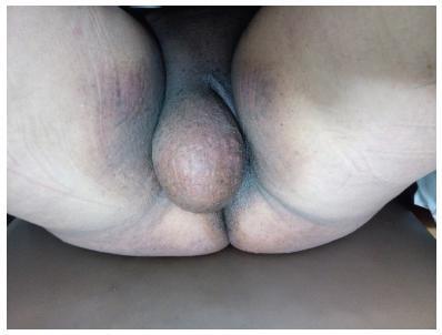
-
Case Report07-20-2004
Invasive Paget’s disease of the vulva and perianal region: a case report
Revista Brasileira de Ginecologia e Obstetrícia. 2004;26(4):329-335
Abstract
Case ReportInvasive Paget’s disease of the vulva and perianal region: a case report
Revista Brasileira de Ginecologia e Obstetrícia. 2004;26(4):329-335
DOI 10.1590/S0100-72032004000400011
Views152See moreExtramammary Paget's disease (EPD) is an uncommon neoplasic condition observed mostly in areas with numerous apocrine and or eccrine glands. In the woman it is most commonly seen on the vulva, although it can occur in other locations. Vulvar Paget's disease (VPD) can be classified into primary, of cutaneous origin, and secondary, of extracutaneous origin, with significant clinical e prognostic implications. Clinically VPD begins insidiously with pruritus and burning sensation. The lesion appears as a solitary patch with an eczematous, erythematous and squamous surface. This is a report of a case of a 72-year-old patient with an erythematous slightly thickened patch lesion with spots of erosion involving both the right and the left majus and minus labia, the clitoris, the pubic region, and the perineal and perianal regions. The operation performed included radical vulvectomy and bilateral inguinal lymphadenectomy. The histopathology revealed invasive Paget's disease. Immunohistochemical methods showed positive Paget cells for CEA, EMA, and cytokeratin pan. Pathogenesis and diagnosis of EPD is discussed, with differential diagnosis and reference to immunohistochemical methods. Recurrence rate is 30%, even with margin control. Experience with EPD is limited and long-term follow-up is required to exclude recurrence of the disease and development of an associated cancer.
Views152This is an Open Access article distributed under the terms of the Creative Commons Attribution License, which permits unrestricted use, distribution, and reproduction in any medium, provided the original work is properly cited. Abstract
Case ReportInvasive Paget’s disease of the vulva and perianal region: a case report
Revista Brasileira de Ginecologia e Obstetrícia. 2004;26(4):329-335
DOI 10.1590/S0100-72032004000400011
Views152See moreExtramammary Paget's disease (EPD) is an uncommon neoplasic condition observed mostly in areas with numerous apocrine and or eccrine glands. In the woman it is most commonly seen on the vulva, although it can occur in other locations. Vulvar Paget's disease (VPD) can be classified into primary, of cutaneous origin, and secondary, of extracutaneous origin, with significant clinical e prognostic implications. Clinically VPD begins insidiously with pruritus and burning sensation. The lesion appears as a solitary patch with an eczematous, erythematous and squamous surface. This is a report of a case of a 72-year-old patient with an erythematous slightly thickened patch lesion with spots of erosion involving both the right and the left majus and minus labia, the clitoris, the pubic region, and the perineal and perianal regions. The operation performed included radical vulvectomy and bilateral inguinal lymphadenectomy. The histopathology revealed invasive Paget's disease. Immunohistochemical methods showed positive Paget cells for CEA, EMA, and cytokeratin pan. Pathogenesis and diagnosis of EPD is discussed, with differential diagnosis and reference to immunohistochemical methods. Recurrence rate is 30%, even with margin control. Experience with EPD is limited and long-term follow-up is required to exclude recurrence of the disease and development of an associated cancer.
This is an Open Access article distributed under the terms of the Creative Commons Attribution License, which permits unrestricted use, distribution, and reproduction in any medium, provided the original work is properly cited. -
Case Report06-26-2001
Vulvar Fibroma: Case Report
Revista Brasileira de Ginecologia e Obstetrícia. 2001;23(3):187-190
Abstract
Case ReportVulvar Fibroma: Case Report
Revista Brasileira de Ginecologia e Obstetrícia. 2001;23(3):187-190
DOI 10.1590/S0100-72032001000300009
Views84See moreA vulvar fibroma, of the molluscum pendulum type, was present in a 20-year-old patient. The tumor began to develop slowly after her menarche, when she was 14 years of age. The physical examination revealed a mass with considerable volume, painless, located at the upper third of the greator left lip, elastic consistency, greater diameter at its distal portion measuring 12 cm by 23 cm in length. The treatment was exeresis from the base of the pedicle, under local anesthesia. The tumor weighed 950 g. A literature review is included.
Views84This is an Open Access article distributed under the terms of the Creative Commons Attribution License, which permits unrestricted use, distribution, and reproduction in any medium, provided the original work is properly cited. Abstract
Case ReportVulvar Fibroma: Case Report
Revista Brasileira de Ginecologia e Obstetrícia. 2001;23(3):187-190
DOI 10.1590/S0100-72032001000300009
Views84See moreA vulvar fibroma, of the molluscum pendulum type, was present in a 20-year-old patient. The tumor began to develop slowly after her menarche, when she was 14 years of age. The physical examination revealed a mass with considerable volume, painless, located at the upper third of the greator left lip, elastic consistency, greater diameter at its distal portion measuring 12 cm by 23 cm in length. The treatment was exeresis from the base of the pedicle, under local anesthesia. The tumor weighed 950 g. A literature review is included.
This is an Open Access article distributed under the terms of the Creative Commons Attribution License, which permits unrestricted use, distribution, and reproduction in any medium, provided the original work is properly cited.