Summary
Revista Brasileira de Ginecologia e Obstetrícia. 2022;44(5):519-531
To provide a survey of relevant literature on umbilical artery Doppler ultrasound use in clinical practice, technical considerations and limitations, and future perspectives.
Literature searches were conducted in PubMed and Medline, restricted to articles written in English. Additionally, the references of all analyzed studies were searched to obtain necessary information.
The use of this technique as a routine surveillance method is only recommended for high-risk pregnancies with impaired placentation. Meta-analyses of randomized trials have established that obstetric management guided by umbilical artery Doppler findings can improve perinatal mortality and morbidity. The values of the indices of Umbilical artery Doppler decrease with advancing gestational age; however, a lack of consensus on reference ranges prevails.
Important clinical decisions are based on the information obtained with umbilical artery Doppler ultrasound. Future efforts in research are imperative to overcome the current limitations of the technique.
Summary
Revista Brasileira de Ginecologia e Obstetrícia. 2006;28(12):693-699
DOI 10.1590/S0100-72032006001200002
PURPOSE: to analyze the effect of glucose in the materno-fetal hemodynamics through dopplervelocimetric assessment of the materno-fetal and fetoplacentary circulation. METHODS: the study was carried out by a single observer on 31 clinically healthy pregnant women from the 28th to the 36th gestational week. Parameters were assessed immediately before or 60 minutes after the ingestion of 50 g of glucose. The including criteria comprised normal clinical and laboratorial evaluation, the presence of only one fetus, gestational age between 28 and 36 weeks confirmed by ultrasonography and/or the date of the last menstruation, fasting glycemia less or equal to 110 mg/dL and less than 140 mg/dL after 50 g of glucose overload. The excluding criteria consisted of the presence of fetal malformation or development alterations, labor, diabetes as a family predisposition, pathologies due to or underlying gestation and use of tobacco, alcohol and/or other substances. The mother´s common carotid artery and uterine arteries, the umbilical artery and the fetal medial cerebral artery and abdominal aorta were evaluated. In each blood vessel, the following parameters were analyzed: resistance index, pulsatility index, maximum systolic speed, final diastolic speed and acceleration time. The fetal heart rate was evaluated by M Mode ultrasonography. For the statistical analysis, the Student's t test was used when the variable presented normal distribution in Kolmogorov-Smirnov's test. When normality was rejected, the Wilcoxon's non-parametric test was used, with the significance level always established at p<0.05. RESULTS: the maternal glycemia increased after the ingestion of 50 g of glucose (before: 68.0±10.1 mg/dL and after: 104.6±28.2 mg/dL; p<0.001), and fetal heart rate decreased after the glucose ingestion (before: 137.9±6.1 bpm and after: 134.5±6.9 bpm; p<0.001). The umbilical artery presented an increase in the pulsatility index (before: 0.8±0.1 and after: 0.9±0.2; p=0.03). Significant velocimetric alterations were not found in the other vessels or in the other indexes investigated. CONCLUSIONS: in spite of the variation in the levels of maternal glycemia and in the fetal heart rate following glucose ingestion, no significant flow alteration occurred in the following vessels: umbilical artery, fetal medial cerebral artery and aorta; nor in the carotid and uterine maternal arteries. We conclude that the glucose concentration used was released without hemodynamic interference in the materno-fetal compartment.
Summary
Revista Brasileira de Ginecologia e Obstetrícia. 2001;23(5):291-298
DOI 10.1590/S0100-72032001000500004
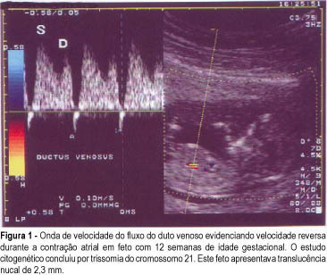
Objective: to study the value of Doppler velocimetry of the ductus venosus and of the umbilical artery and vein, in the screening for chromosomal abnormalities at 10-14 weeks of gestation. Patients and Methods: a total of 314 fetuses were studied consecutively. In 112 cases a cytogenetic study was performed on material obtained from a biopsy of the chorionic villus, and in 202 cases the postnatal phenotype was used as a basis for the result. In addition to the routine ultrasonographic examination, all the fetuses were submitted to measurement of the nuchal translucency thickness and to Doppler velocimetry of the umbilical artery and vein, particularly of the ductus venosus. For statistical analysis the Fisher exact test and the Mann-Whitney test were used. Results: twenty-three cases of chromosomal abnormalities occurred. Of these abnormal cases, the ductus venosus blood flow during atrial contraction was absent (1 case) and reverse (22 cases), sensitivity was 92%. In the group of normal fetuses (289 cases), 6 evaluations demonstrated alterations in the Doppler of the ductus venosus (specificity of 97.6%, positive and negative predictive values of 76.7% and 93.3%, respectively); the false-positive rate was 2.4%. In reference to the umbilical vein and umbilical artery, there was no statistically significant difference between the abnormal and the normal group. Conclusion: The only parameter of Doppler velocimetry of the umbilical artery and vein which contributed to the detection of aneuploidies was the accidental discovery of the reverse blood flow in both vessels. Although our favorable results demonstrated that the Doppler velocimetry of the ductus venosus is effective in detecting aneuploidies, this conclusion, however, is preliminary and needs further investigation.
Summary
Revista Brasileira de Ginecologia e Obstetrícia. 2001;23(5):291-298
DOI 10.1590/S0100-72032001000500004
Objective: to study the value of Doppler velocimetry of the ductus venosus and of the umbilical artery and vein, in the screening for chromosomal abnormalities at 10-14 weeks of gestation. Patients and Methods: a total of 314 fetuses were studied consecutively. In 112 cases a cytogenetic study was performed on material obtained from a biopsy of the chorionic villus, and in 202 cases the postnatal phenotype was used as a basis for the result. In addition to the routine ultrasonographic examination, all the fetuses were submitted to measurement of the nuchal translucency thickness and to Doppler velocimetry of the umbilical artery and vein, particularly of the ductus venosus. For statistical analysis the Fisher exact test and the Mann-Whitney test were used. Results: twenty-three cases of chromosomal abnormalities occurred. Of these abnormal cases, the ductus venosus blood flow during atrial contraction was absent (1 case) and reverse (22 cases), sensitivity was 92%. In the group of normal fetuses (289 cases), 6 evaluations demonstrated alterations in the Doppler of the ductus venosus (specificity of 97.6%, positive and negative predictive values of 76.7% and 93.3%, respectively); the false-positive rate was 2.4%. In reference to the umbilical vein and umbilical artery, there was no statistically significant difference between the abnormal and the normal group. Conclusion: The only parameter of Doppler velocimetry of the umbilical artery and vein which contributed to the detection of aneuploidies was the accidental discovery of the reverse blood flow in both vessels. Although our favorable results demonstrated that the Doppler velocimetry of the ductus venosus is effective in detecting aneuploidies, this conclusion, however, is preliminary and needs further investigation.
Summary
Revista Brasileira de Ginecologia e Obstetrícia. 2002;24(2):113-120
DOI 10.1590/S0100-72032002000200007
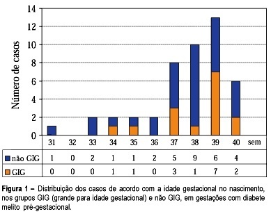
Purpose: to study fetal surveillance examinations in pregnancies complicated by pregestational diabetes mellitus, and to correlate them with large for gestational age (LGA) newborns. Methods: Between March 1999 and June 2001, 46 singleton pregnancies with pregestational diabetes mellitus without fetal anomalies were followed prospectively. From the 28th gestational week on, the following examinations were performed weekly: fetal biophysical profile, amniotic fluid index (AFI), and dopplervelocimetry of umbilical and middle cerebral arteries. The newborns with birthweight above the 90th percentile according to local standard values were characterized as LGA infants. Fisher's exact test and Student's t test were used for statistical analysis. Results: The mean gestational age at delivery was 37.6 weeks and 15 (32.6%) newborns were LGA. LGA fetuses showed significant increase in the AFI mean performed in the 32nd (16.5 cm, p=0.02), 33rd (16.7 cm, p=0.03), 34th (17.0 cm, p=0.02), 35th (17.9 cm, p=0.000), 36th (15.8 cm, p=0.03) and 37th (17.5 cm, p=0.003) weeks. Non-LGA fetuses presented the following mean AFI values: 13.5cm (32nd week), 13.1cm (33th week), 13.4 (34th week), 12.8 (35th week), 12.5 (36th week) and 12.8cm (37th week). AFI values equal to or above 18.0 cm were associated with the occurrence of LGA infants, when detected at the following gestational ages: 34th (60%, p=0.03), 35th (71.4%, p=0.01), 36th (80%, p=0.02) and 37th (66.7%, p=0.04) week. Non-LGA infants presented the following proportion of AFI values equal to or above 18.0 cm: 40.0% (34th week), 28.6% (35th week), 20.0% (36th week), and 33.3% (37th week). Conclusions: abnormal increase in AFI, mainly with values equal to or above 18.0 cm, is related to LGA infants at delivery. The maternal treatment should be adjusted to achieve the best result for maternal-fetal control, according to the AFI values during pregnancy.
Summary
Revista Brasileira de Ginecologia e Obstetrícia. 2002;24(2):113-120
DOI 10.1590/S0100-72032002000200007
Purpose: to study fetal surveillance examinations in pregnancies complicated by pregestational diabetes mellitus, and to correlate them with large for gestational age (LGA) newborns. Methods: Between March 1999 and June 2001, 46 singleton pregnancies with pregestational diabetes mellitus without fetal anomalies were followed prospectively. From the 28th gestational week on, the following examinations were performed weekly: fetal biophysical profile, amniotic fluid index (AFI), and dopplervelocimetry of umbilical and middle cerebral arteries. The newborns with birthweight above the 90th percentile according to local standard values were characterized as LGA infants. Fisher's exact test and Student's t test were used for statistical analysis. Results: The mean gestational age at delivery was 37.6 weeks and 15 (32.6%) newborns were LGA. LGA fetuses showed significant increase in the AFI mean performed in the 32nd (16.5 cm, p=0.02), 33rd (16.7 cm, p=0.03), 34th (17.0 cm, p=0.02), 35th (17.9 cm, p=0.000), 36th (15.8 cm, p=0.03) and 37th (17.5 cm, p=0.003) weeks. Non-LGA fetuses presented the following mean AFI values: 13.5cm (32nd week), 13.1cm (33th week), 13.4 (34th week), 12.8 (35th week), 12.5 (36th week) and 12.8cm (37th week). AFI values equal to or above 18.0 cm were associated with the occurrence of LGA infants, when detected at the following gestational ages: 34th (60%, p=0.03), 35th (71.4%, p=0.01), 36th (80%, p=0.02) and 37th (66.7%, p=0.04) week. Non-LGA infants presented the following proportion of AFI values equal to or above 18.0 cm: 40.0% (34th week), 28.6% (35th week), 20.0% (36th week), and 33.3% (37th week). Conclusions: abnormal increase in AFI, mainly with values equal to or above 18.0 cm, is related to LGA infants at delivery. The maternal treatment should be adjusted to achieve the best result for maternal-fetal control, according to the AFI values during pregnancy.